Category Archives: All Updates

No Picnic? Really?

They say emergency medicine and critical care are no picnic, but I’ve been trying to change that. There’s something about sitting down on a blanket and sharing protected time for conversation that makes for good team building and effective communication. If you have snacks, it’s even better.
In the emergency department or intensive care unit one sometimes has to be opportunistic regarding finding time for teaching, debriefing a resuscitation case, or even eating. We end up doing these things (if at all) on the fly, in a rushed manner, and often standing up. Do we really have to? All you need for a picnic is a blanket, a floor, and some people. Hospitals have these. If you don’t want to be seen, pop outside or use a bed space with a curtain round it.


Picnic Debrief
Here’s an example of an impromptu picnic. It was late in the evening, early 2013. After two busy resus cases, my senior registrar and I debrief picnic-style, with potato chips from a vending machine and a nice pot of tea. We’re still in the ED and available to our team, but anyone can clearly see we’re in the ‘picnic zone’ and so we’re left alone for ten minutes to gather our thoughts and identify any learning points. The ED is usually a factory of interruption, but no-one wants to interrupt a picnic.


Picnic Teaching
Here’s resident teaching. We don’t have time to leave the ED, but there’s always time for a picnic, during which we cover a surprising number of critical care topics. People won’t fall asleep while picnicking.


Picnic Picnic
And here’s a picnic with the intensive care trainees outside the unit. This is actually lunch, but why shouldn’t lunch be a picnic once in a while?

We’re encouraged to practice mindfulness and take mental time out as a way to prevent or manage stress in the critical care environment. I think this is enhanced with an accompanying brief physical time out too. One person sitting on a blanket on the floor might be a weirdo. Get two or more people, and you have a picnic. Everyone loves a picnic.

Spot the WOBBLER in syncope!

Syncope is a common ED presentation. An ECG is a critical investigation in syncope to identify the cause, including rare conditions associated with risk of sudden cardiac death.
So we should be really grateful when we are invited to interpret an ECG while we’re in the middle of six other tasks.

The problem with syncope is that some of the important life-threatening causes have fairly obscure ECG features that might be hard to remember. Some of these disorders and their ECG features are not entirely familiar to the clinicians who first screen the ECG.

When you’re busy and cognitively stretched you can save time and reduce the risk of missing important findings by having a structured, memorable checklist. I use the acronym WOBBLER, because I don’t want these people to wobble and kiss the dirt again.

The nice thing about WOBBLER is that it uses the sequence that you follow when you look at an ECG, ie from left to right, or from P wave to T wave.

The key is that this is for ECGs without obvious ischaemia or dysrhythmia. If you see something like this (STEMI):

or this (VT):

you don’t need WOBBLER, you need to be treating that patient.

So we apply WOBBLER to screen the ECG of well-looking syncope patients without obvious ischaemia or dysrhythmia.

Here goes…

W is Wolff-Parkinson-White syndrome – look for a short PR interval or delta wave:

O is obstructed AV pathway – look for 2nd or 3rd degree block:

or axis deviation:

…which is the first step in looking for B bifascicular block, or the combination of axis deviation and right bundle branch block:

the second B is Brugada, looking for characteristic morphology of the ST segment, so called coved ST elevation:

Now syncope, especially exertional syncope, can be caused by left ventricular outflow tract obstruction. Two conditions not to be missed associated with LVOTO (and exertional syncope) are hypertrophic cardiomyopathy and aortic stenosis. These both characteristically cause Lleft ventricular hypertrophy:

This example of Hypertrophic Cardiomyopathy has voltage criteria for LVH in the precordial leads as well as narrow, “dagger-like” Q waves in lateral and inferior leads

E– stands for epsilon wave, a feature of arrythmogenic right ventricular dysplasia, a rare disorder associated with sudden cardiac death. The epsilon wave looks a bit like the J wave of hypothermia and may be associated with other T wave abnormalities in V1-V3:

Epsilon wave in more detail

Finally, R stands for Repolarisation abnormality, particularly delayed Repolarisation as found in long QT syndrome:

but remember there is also a short QT syndrome too:

So WOBBLER may help you find the important and rare abnormalities not to be missed in the syncope patient, going from left to right from P wave through to T wave, in the patient that does not have obvious dysrhythmia or ischaemia. I hope you find it a helpful cognitive forcing tool.

All ECGs reproduced with kind permission of Life in the Fast Lane

Das SMACC in Berlin

street-art-1499524_1280
Berlin graffiti art depicting Oli Flower (left) and Roger Harris (right) on stage at the close of the last SMACC conference


Don’t miss your chance to register for the best emergency/prehospital/critical care conference out there

Tickets will be released on the following dates:das-smacc-tix-schedule-oct

Here are a few ‘rules of the game’ from the course organisers:

  • There will be 3 separate ticket releases: the major release will be as above on Wednesday, 26th October, a smaller allocation will be released on Wednesday, 7th December and a final limited release on Wednesday, 1st February
  • Your best chance will be with the first release, but if you really need to wait until you have leave confirmed then you can chance your hand on the February release
  • All prior delegates will receive an email reminder the week before tickets go on sale, but there is no other preference (first in best dressed!)
  • Owing to the limited number of spots there will be no DAY ONLY registrations issue
  • Workshop registration also opens on Wednesday, 26th October and like last year will be on a first come first served basis
  • If you miss out on a ticket there will be a waiting list
  • If you miss your preferred workshop there will also be a waiting list

You can check out the program here and registration fees here

Don’t worry if you can’t make it – all smacc talks are published free on line and you’ll find talks from the last four conferences at the smacc site

Good Luck!

It’s Tamponade – Now What?

You ultrasound the chest of your shocked patient in resus with fluid refractory hypotension. You see fluid around the heart. The right ventricle keeps bowing inwards, which you recall being described as ‘a little invisible man jumping up and down using the RV as a trampoline’, and you know this is in fact a sign of right ventricular diastolic collapse.


The collapse of the right side of the heart during diastole is the mechanism for shock and cardiac arrest due to tamponade, because the high pericardial pressures prevent the right heart from filling in diastole. This patient therefore has ‘tamponade physiology’ on ultrasound. A quick scan of the IVC shows it is dilated and does not collapse with respiration. This confirms a high central venous pressure (as do the patient’s distended neck veins), also consistent with tamponade physiology.


A formal echo done in resus confirms your suspicion of a dliated aortic root and visible dissection flap, so the diagnosis is now clear. This is type A aortic dissection with tamponade. The patient remains hypotensive and mottled with increasing drowsiness. Cardiothoracic surgery is based at another hospital site 30 minutes away by ambulance.


As the critical care clinician responsible for, or assisting with this patient’s care (emergency physician, intensivist, anaesthetist, rural GP, physician’s assistant, etc.), how do we get this patient to definitive care and mitigate the risk of deterioration en route? Let’s discuss the options using real life case examples, and consider the physiology, the evidence, and the dogma.


Here are four key questions to consider:
1. To drain or not to drain the pericardium?
2. To intubate or not to intubate?
3. If they arrest – CPR or no CPR?
4. How to transfer – physician escort or just send in an ambulance on lights and sirens?


Here are three scenarios that follow the intial assessment of the above patient. They are based on similar cases shared with me by participants on the Critical Care in the Emergency Department course.


Case 1

The patient is obtunded with profound shock and too unstable for transfer. The resus team undertakes pericardiocentesis and aspirates 30 ml of blood. The patient becomes conscious and cooperative and the systolic blood pressure (SBP) is 95 mmHg. The patient is transferred by paramedic ambulance to the cardothoracic centre where he is successfully operated on, resulting in a full recovery.


Case 2

As the patient is unconscious and requires interhospital transfer, the decision is made to intubate him for airway protection. He undergoes rapid sequence induction with ketamine, fentanyl, and rocuronium in the resus room. After capnographic confirmation of tracheal intubation he is manually ventilated via a self-inflating bag. The ED nurse reports a loss of palpable pulse and CPR is started. A team member suggests pericardiocentesis but a senior critical care physician says there is no point because ‘it won’t fix the underlying problem of aortic dissection’ and ’the blood will be clotted anyway’. After a brief attempt at standard ACLS, resuscitation efforts are discontinued and the patient is declared dead.


Case 3

The patient is hypotensive with a SBP of 90mmHg and drowsy but cooperative. The receiving centre has accepted the referral and an ambulance has been requested. The critical care physician responsible for patient transfers is requested to accompany the patient but declines, on the basis that ‘these cases are just like abdominal aortic aneurysms – they just need to get there asap. If they deteriorate en route we’re not going to do anything.’


The patient is transferred but 15 minutes into the journey he becomes unresponsive and loses his cardiac output. The transporting paramedics provide chest compressions and adrenaline/epinephrine but are unable to resuscitate him.
These cases illustrate some of the pitfalls and fallacies associated with tamponade due to type A dissection.


Pericardiocentesis

Pericardiocentesis can definitely be life-saving, restoring vital organ perfusion and buying time to get the patient to definitive surgery. Numerous case reports and case series provide evidence of its utility, even in patients in PEA cardiac arrest(1). The authors of the two largest cases series both used 8F pigtail drainage catheters(1,2).


Reports in the literature regarding the pericardiocentesis of critical cardiac tamponade complicating aortic dissection from Cruz et al


One key component of procedural success was controlled pericardial drainage, removing small volumes and reassessing the blood pressure, aiming for a SBP of 90 mmHg. The danger is overshooting, resulting in hypertension and extending the underlying aortic dissection which can be fatal (3).


Those still unconvinced by the evidence may be swayed by guidelines. The 2015 European Society of Cardiology Guidelines for the diagnosis and management of pericardial diseases (4) state:


“In the setting of aortic dissection with haemopericardium and suspicion of cardiac tamponade, emergency transthoracic echocardiography or a CT scan should be performed to confirm the diagnosis. In such a scenario, controlled pericardial drainage of very small amounts of the haemopericardium can be attempted to temporarily stabilize the patient in order to maintain blood pressure at 90 mmHg. (Class IIa, Level C)”


Intubation

Deterioration of tamponade patients following intubation is well described in the literature and the risk is well appreciated by cardiothoracic anaesthetists(5).


Once positive pressure ventilation is started, positive pleural pressure is transmitted to the pericardium, where pressures can exceed right ventricular diastolic pressure and prevent cardiac filling. The result is a fall in and possible loss of cardiac output. This is further exacerbated by the addition of PEEP(6).


One suggested approach if the patient must be intubated for airway protection but is not yet in the operating room with a surgeon ready to cut, is to consider intubation under local anaesthesia and allow the patient to breathe spontaneously (maintaining negative pleural pressure) through the tube until the surgeon is ready to open the chest(5).


Alternatively preload with fluid, use cautious doses of induction agent, and ventilate with low tidal volumes and zero PEEP. However the patient can still crash, so remember that these effects of ventilation on cardiac output in tamponade can be mitigated by the removal of a relatively small volume of pericardial fluid(6).


Cardiac Arrest

In cardiac arrest, external chest compressions are unlikely to be of benefit. In a study on baboons subjected to cardiac tamponade, closed chest massage resulted in an increase in intrapericardial pressure. There was an increase in systolic pressure, but a marked decrease in diastolic pressure, with an overall decrease in mean arterial pressure(7).

Pressure changes from CPR during tamponade in baboons. From Luna et al (7)
This would lead to impaired coronary perfusion and would be very unlikely to result in return of spontaneous circulation (ROSC). In the clinical situation described above, it is only relief of tamponade that is going to provide an arrested patient with a chance of recovery.


Transport

For patients with cardiac tamponade requiring interhospital (or intrahospital) transfer, it would seem vital therefore that the patient is accompanied by a clinician willing and capable to perform pericardiocentesis in the event of severe deterioriation or arrest en route. This simple life-saving intervention to deliver the patient alive to the operating room should be made available should the need arise.


Summary

  • Patients presenting in shock from cardiac tamponade often have treatable underlying causes and represent a situation where the planning and actions of the resuscitationist can be truly life-saving.
  • Pericardiocentesis is recommended in profound shock to buy time for definitive intervention. Controlled pericardiocentesis should be performed paying strict attention to SBP to avoid ‘overshooting’ to a hypertensive state in type A aortic dissection. In cardiac arrest, chest compressions are likely to be ineffective and pericardiocentesis is mandatory for ROSC.
  • The institution of positive pressure ventilation often results in worsened shock or cardiac arrest, and this is exacerbated by PEEP. Where possible, avoid intubation until the patient is in the operating room, or use low tidal volumes and no PEEP. Even then pericardiocentesis may be necessary to maintain or restore cardiac output.
  • Patients requiring transport who have tamponade should be accompanied by a clinician able to perform pericardiocentesis in the event of en route deterioration.


References

  1. Cruz I, Stuart B, Caldeira D, Morgado G, Gomes AC, Almeida AR, et al. Controlled pericardiocentesis in patients with cardiac tamponade complicating aortic dissection: Experience of a centre without cardiothoracic surgery. European Heart Journal: Acute Cardiovascular Care. 2015 Mar 19;4(2):124–8.
  2. Hayashi T, Tsukube T, Yamashita T, Haraguchi T, Matsukawa R, Kozawa S, et al. Impact of Controlled Pericardial Drainage on Critical Cardiac Tamponade With Acute Type A Aortic Dissection. Circulation. 2012 Sep 10;126(11_suppl_1):S97–S101.
  3. Isselbacher EM, Cigarroa JE, Eagle KA. Cardiac tamponade complicating proximal aortic dissection. Is pericardiocentesis harmful? Circulation. 1994 Nov 1;90(5):2375–8.
  4. Adler Y, Charron P, Imazio M, Badano L, Barón-Esquivias G, Bogaert J, et al. 2015 ESC Guidelines for the diagnosis and management of pericardial diseases. European Heart Journal. 2015 Nov 7;36(42):2921–64.
  5. Ho AMH, Graham CA, Ng CSH, Yeung JHH, Dion PW, Critchley LAH, et al. Timing of tracheal intubation in traumatic cardiac tamponade: A word of caution. Resuscitation. 2009 Feb;80(2):272–4.
  6. Möller CT, Schoonbee CG, Rosendorff C. Haemodynamics of cardiac tamponade during various modes of ventilation. Br J Anaesth. 1979 May;51(5):409–15.
  7. Luna GK, Pavlin EG, Kirkman T, Copass MK, Rice CL. Hemodynamic effects of external cardiac massage in trauma shock. The Journal of Trauma: Injury, Infection, and Critical Care. 1989 Oct;29(10):1430–3.

Difficult Airway? What Kind Exactly?

crazed-nutter-sm
Mention the term ‘difficult airway’ and many of us will conjure mental images of some kind of distorted anatomy. However, airway management may be ‘difficult’ for a number of reasons, and no internationally agreed definition of the term exists. Given the wrong staff and circumstances, an ‘easy’ airway in your or my hands may indeed become very difficult. In their editorial “The myth of the difficult airway: airway management revisited” (1) Huitink & Bouwman state:

“In our opinion, the ‘difficult airway’ does not exist. It is a complex situational interplay of patient, practitioner, equipment, expertise and circumstances.”

Airways that are anatomically difficult (eg. limited mouth opening, short thyromental distance, large tongue, neck immobility, etc.) and physiologically difficult (hypoxaemia, hypotension, acidosis) are well described among FOAM resources (2-4). In addition to these, a third category of difficulty is well worth considering.

At the smaccDUB conference, intensivist and human factors legend Peter Brindley described three types of difficult airway:

  1. Anatomically difficult
  2. Physiologically difficult
  3. Situationally difficult
    brindleysm
    Brindley = Legend

This last category probably surfaces more commonly than realised, particularly outside the operating room.

Imagine attending a cardiac arrest call on a medical ward. The patient is a 70 year old 120 kg male. The nurses have flattened the bed and discarded the pillow to optimise supine position for CPR. Gobs of vomitus splash from the patient’s pharynx with each compression. The wall suction system is disconnected. There is no bougie in the crash cart’s airway drawer. The nearest capnograph is on another floor of the hospital. In this scenario, no matter how excellent the critical care practitioner’s airway skills, this is a damned difficult airway.

I think Brindley’s third category is a term that should catch on, as a way of helping analyse cases that progress suboptimally and to identify factors during pre-intubation checks that can be addressed. It is terminology that I have added to my own Resuscitese Lexicon, particularly for case discussions during morbidity & mortality and airway audit meetings.

I would like to hear the ‘Situationally Difficult Airway‘ become more widely used, as it fills a gap in how we describe this important area of resuscitation practice.


1. Huitink JM, Bouwman RA. The myth of the difficult airway: airway management revisited. Anaesthesia. 2015 Mar;70(3):244–9. (Full text)

2. LITFL: Airway Assessment

3. EMCrit: HOP Killers

4. PulmCCM: The Physiologically Difficult Airway

The Course I Made For Me

My whole career has been about finding ways to optimise resuscitation. Many others also have the bug. The ‘resuscitationist movement’ is sweeping across Europe, with Katrin Hruska and Femke Geijsel about to run amazing courses for emergency teams in Sweden and The Netherlands. I have the honour of joining Clare Richmond in helping them do that. But first Critical Care in the Emergency Department is going to be run in London one more time.

This course contains the stuff I wish someone had told me as a registrar. A synthesis of my learning points in intensive care, prehospital & retrieval medicine, paediatric critical care, and being a front line ED doc for 20 years.

I’ve been running the course for over a decade, including in London, Birmingham, Basingstoke, Dublin, Stockholm, Sydney and Maribor. Each time I try to improve it, and try to squeeze one or two more learning points in. It’s a tough day – just me and fifty or so critical care cases to talk about. But no-one goes to sleep – guaranteed! Everyone has to work – to talk, think, and interact.

It is of course primarily a clinical course, focusing on optimal clinical practice. But consistent feedback from participants is that they get far more from it: a reassurance that they’re not crazy wanting to do more for their patients, and a way forward for remaining inspired and motivated to make changes to their practice and to their departments.

Next course Friday 9 December 2022, London, UK

You can register here or read more here

Humbling Reminders of the Power of Educators

the teaching hospitalvsmI’ve always had strong feelings about education. I was an uninspired and underachieving medical student, exasperated at the fact that the preclinical course at my medical school consisted of lengthy lectures about detailed aspects of basic sciences like histology and embryology. To make it worse, the teaching was delivered by basic science PhD students who were required to teach medical students as part of their contract. They taught because they had to, not because they were good at it. In other words, the best way to summarise how I was initially taught to be a doctor is this: my medical training consisted of being taught stuff I didn’t need to know, by people who weren’t doctors, and who didn’t know how to teach.

This frustrated me enormously. It wasn’t until I hit the wards as a senior medical student and then junior doctor that I would occasionally run into enthusiastic and supportive clinicians who were keen to share what they knew. They seemed to be few and far between, but the crumbs they dropped were enough to leave a trail that led me to be determined to become a doctor who could similarly inspire and motivate others to love learning.

Throughout my training I made a consistent observation: a small amount of good education was extremely motivating. The converse was also true – being denied access to education was extremely demotivating.  In one department, teaching was continually cancelled due to patient load. When questioned on this, the clinical director stated “teaching is a privilege, not a right”. This influenced me profoundly, because I immediately adopted that phrase as a personal motto, except that I flipped the order of “right” and “privilege”.

A few events have converged this week to remind me of the power of good education. The first, and most important, was when my friend Rob Rogers, a renowned emergency medicine educator who has run courses on how to teach all over the world, tweeted a picture of an interesting ECG.

Rob and his team have inspired so many people with their brilliant education. Faced with a life-threatening ST-elevation MI, Rob chose to share his ECG with his Twitter followers. Later he shared details of his angiogram pre- and post-revascularisation. Now THAT is commitment to education!

The second event this week is that we are running our Sydney HEMS induction course in prehospital & retrieval medicine. This is about as full on as medical education can get, with hours of simulation, testing, and stress exposure. I am constantly amazed at the dedication and hard work of my colleagues who make up the course faculty, and the willingness of the participants to go the extra mile to improve their performance. We have the honour of  inviting medics from certain branches of the Australian military to attend the course, and one such armed forces ‘graduate’ of our course recently contacted me:

mil

He attached a document outlining a situation he faced which took my breath away. I’m not yet allowed to share it, but the bravery he showed was awe-inspiring. To think that he credits some of his preparation to the training we gave is truly humbling. It is also a reminder of the enormous responsibility of educators.

We can provide both negative and positive inspiration through our choices in what we say and how we say them, and in the teaching we deliver. As learners those educational experiences shape us and stay with us forever, influencing the choices we make and how we choose to pass on the teaching. 

arcticpond

We are lucky to live in an age of Free Open Access Medical Education,  when the educational ripples reach out from EMCrit, St Emlyn’s, EM IrelandscanFOAM, badEM, LifeInTheFastLane, and so many others to all parts of the world. We’re all in this together, as teachers and as learners.

The humbling feedback from my military friend along with Rob’s ongoing desire to educate in the face of life-threatening illness serve to remind us of the power of education, and the responsibility we educators have to share, to inspire, and to provide the highest quality teaching.

Something Rob already seems to be working on, less than a week post-myocardial infarction …

Hilar Twists & Human Error

An engaging scene from ‘Code Blue‘ demonstrated a Helicopter Emergency Medical Service team managing a patient with major thoracic haemorrhage. They did a right thoracotomy and wanted to clamp the hilum but there was some kit missing from the pack.

Unfortunately, the video is no longer available.

This scene had some great discussion points for prehospital professionals, even if the specific scenario is somewhat unlikely for most people’s practice:

  • Non-compressible haemorrhage is possibly the biggest single clinical challenge when you’re a long way from hospital
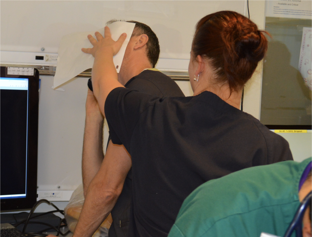
  • Agitated friends and family can be disruptive – allocate a rescuer to look after them
  • Having blood products to give is essential
  • Don’t rely on the memory of individuals, who are fallible, to pack your equipment. “I was sure I put them in” didn’t cut it when the team needed forceps to clamp the pulmonary hilum and stop the bleeding. Checklists are the in thing, for good reason.
  • Luckily, you don’t need to clamp the hilum (which is tricky) in massive unilateral thoracic haemorrhage. You can just twist the lung 180 degrees on the hilum so it’s upside down. This can prevent further haemorrhage and air embolism.

What’s a hilar twist then?

The hilar twist manoeuvre, as it’s called, is worth learning if you’re a clinician who is prepared to do resuscitative clamshell thoracotomy for penetrating traumatic cardiac arrest. The clamshell is quick and provides excellent exposure(1) and is preferred to lateral thoracotomy(2).

The primary purpose of clamshell thoracotomy in penetrating traumatic arrest is to relieve cardiac tamponade and control a cardiac wound(3). It is well described and continues to save lives in the prehospital setting(4).

However, sometimes you’ll open the chest and the pericardium will be empty (other than containing the heart of course), and there will be massive haemorrhage on one side of the chest. Although most of these patients will be unsalvageable outside a trauma centre’s operating room, it’s worth trying something once you’ve gone to all the trouble of opening the chest. The hilar twist(5) is probably the best option for the non-surgeon, especially when some muppet’s forgotten to pack a clamp.

In order to make the lung mobile enough to twist, it’s first necessary to cut through the inferior pulmonary ligament. This is also known as simply the pulmonary ligament (because there’s no superior equivalent) and sometimes the inferior hilar ligament. It’s not actually a ligament, but an extension of the parietal pleura extending downwards in a fold from the hilum. Some describe it as hanging down from the hilum like a ‘wizard’s sleeve’, which invariably gets a giggle from some of our trainees from the United Kingdom for some reason.


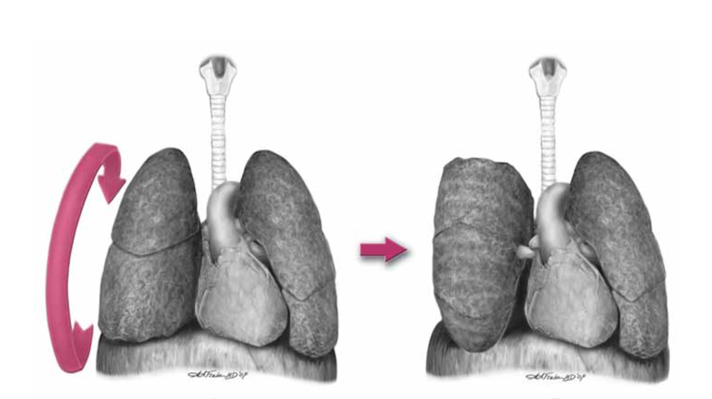
After cutting the ligament completely to the level of the inferior pulmonary vein, the lung is then twisted ‘lower lobe towards you’, ie. lower lobe is rotated anteriorly over the upper lobe until the lung is oriented ‘upside down’. The twisted vessels around the hilum become occluded and further haemorrhage from that side should be limited. Other priorities in the arrested patient will be aortic occlusion, internal cardiac massage, and blood products. Packs may be required to keep the lung from untwisting, and if return of spontaneous circulation is achieved, there is a risk of dysrhythmia, right heart failure, and refractory hypoxaemia.

I’ve only done this on pigs and human cadavers so am not speaking from any reassuring level of experience or competence. The literature is out there to read, and it’s up to you to decide how you want to expand or limit your options when you’ve cracked that chest in an arrested patient.

References

1. Flaris AN, Simms ER, Prat N, Reynard F, Caillot J-L, Voiglio EJ. Clamshell incision versus left anterolateral thoracotomy. Which one is faster when performing a resuscitative thoracotomy? The tortoise and the hare revisited. World J Surg. 2015 May;39(5):1306–11.

2. Simms ER, Flaris AN, Franchino X, Thomas MS, Caillot J-L, Voiglio EJ. Bilateral Anterior Thoracotomy (Clamshell Incision) Is the Ideal Emergency Thoracotomy Incision: An Anatomic Study. World J Surg. 2013 Feb 23;37(6):1277–85.

3. Wise D. Emergency thoracotomy: “how to do it.” Emerg Med J. 2005 Jan 1;22(1):22–4.

4. Davies GE, Lockey DJ. Thirteen Survivors of Prehospital Thoracotomy for Penetrating Trauma: A Prehospital Physician-Performed Resuscitation Procedure That Can Yield Good Results. The Journal of Trauma: Injury, Infection, and Critical Care. 2011 May;70(5):E75–8.

5. Wilson A, Wall MJ Jr., Maxson R, Mattox K. The pulmonary hilum twist as a thoracic damage control procedure. The American Journal of Surgery. 2003 Jul;186(1):49–52.
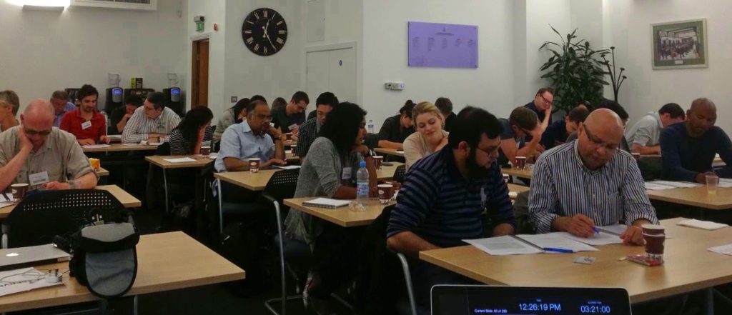