Tag Archives: CNS

Endovascular stroke treatment

Two randomised controlled trials have been published which compare endovascular stroke treatments with intravenous tPA. Both the American Interventional Management of Stroke (IMS) III trial (1) and the Italian SYNTHESIS Expansion trial (2) had Modified Rankin Scores as their primary endpoint. No significant differences in this outcome or in mortality or intracranial haemorrhage rates were found in either trial, and IMS III was terminated early due to futility.
A third trial, from North America, called MR RESCUE, randomised patients within 8 hours after the onset of large vessel, anterior-circulation strokes to undergo mechanical embolectomy or receive standard care(3). No clinical outcome differences were demonstrated.
An accompanying editorial (4) draws the following conclusion:


“The IMS III and SYNTHESIS Expansion studies show that intravenous thrombolysis should continue to be the first-line treatment for patients with acute ischemic stroke within 4.5 hours after stroke onset, even if imaging shows an occluded major intracranial artery. Beyond 4.5 hours, the MR RESCUE trial does not provide data supporting the use of endovascular treatment in patients with an ischemic penumbra of any size.”

Many might argue that showing endovascular treatment is equivalent to thrombolysis just means endovascular treatment doesn’t work, because a significant proportion of the emergency medicine community views this as the correct interpretation of a thorough analysis of the stroke thrombolysis literature.
1. Endovascular Therapy after Intravenous t-PA versus t-PA Alone for Stroke
NEJM Feb 8, 2013 Full Text Link
2. Endovascular Treatment for Acute Ischemic Stroke
NEJM Feb 8, 2013 Full Text Link
3. A Trial of Imaging Selection and Endovascular Treatment for Ischemic Stroke
NEJM Feb 8, 2013 Full Text Link
4.Endovascular Treatment for Acute Ischemic Stroke — Still Unproven
NEJM Feb 8, 2013 Full Text Link

Offensive medicine: CT before LP

I’m getting worn down by clinicians – often other specialists – who insist that CT imaging of the brain is mandatory prior to lumbar puncture in all patients. There is surely a subgroup of patients (especially young ones) in whom the benefit:harm balance of CT comes out in favour of NOT doing the imaging. In these cases, getting the scan is not ‘defensive medicine’ but ‘offensive medicine’ – offending the principle of primum non nocere. During ED shifts I have recently had to perform online searches in order to furnish colleagues and patients’ medically qualified relatives with printouts of the literature on this. This page is here to save me having to repeat those searches. Regarding the practice of performing a routine head CT prior to lumbar puncture to rule out risk of herniation:

  • Mass effect on CT does not predict herniation
  • Lack of mass effect on CT does not rule out raised ICP or herniation
  • Herniation has occurred in patients who did not undergoing lumbar puncture because of CT findings
  • Clinical predictors of raised ICP are more reliable than CT findings
  • CT may delay diagnosis and treatment of meningitis
  • Even in patients in whom LP may be considered contraindicated (cerebral abscess, mass effect on CT), complications from LP were rare in several studies

Best practice, it would seem, is the following

  • If you think CT will show a cause for the headache, do a CT
  • If a CT is indicated for other reasons (depressed conscious level, focal neurology), do a CT
  • If a GCS 15 patient is to undergo LP for suspected (or to rule out) meningitis, and they have a normal neurological exam (including fundi), and are not elderly or immunosuppressed, there is no need to do a CT first.
  • If you’re seriously worried about meningitis and are intent on getting a CT prior to LP, don’t let the imaging delay antimicrobial therapy.

Here are some useful references:

1. The CT doesn’t help

CT head before lumbar puncture in suspected meningitis BestBET evidence summary: In cases of suspected meningitis it is very unlikely that patients without clinical risk factors (immunocompromise/ history of CNS disease/seizures) or positive neurological findings will have a contraindication to lumbar puncture on their CT scan If CT scan is deemed to be necessary, administration of antibiotics should not be delayed. BestBETS website

Computed Tomography of the Head before Lumbar Puncture in Adults with Suspected Meningitis Much cited NEJM paper from 2001 which concludes: “In adults with suspected meningitis, clinical features can be used to identify those who are unlikely to have abnormal findings on CT of the headN Engl J Med. 2001 Dec 13;345(24):1727-33 Full Text

Cranial CT before Lumbar Puncture in Suspected Meningitis Correspondence in 2002 NEJM including study of 75 patients with pneumococcal meningitis: CT cannot rule out risk of herniation Cranial CT before Lumbar Puncture in Suspected Meningitis N Engl J Med. 2002 Apr 18;346(16):1248-51 Full Text

2. The CT may harm

Cancer risk from CT Paucis verbis card, from the wonderful Academic Life in EM

3. Guidelines say CT is not always needed

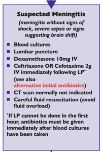
National (UK) guidelines on meningitis (community acquired meningitis in the immunocompetent host) available from meningitis.org. , including this box:
Practice Guidelines for the Management of Bacterial Meningitis These 2004 guidelines from the Infectious Diseases Society of America provide the following table listing the recommended criteria for adult patients with suspected bacterial meningitis who should undergo CT prior to lumbar puncture:
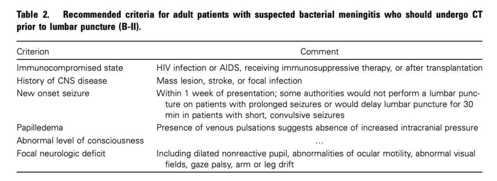
Clin Infect Dis. (2004) 39 (9): 1267-1284 Full text

4. This is potentially even more of an issue with paediatric patients

Fatal Lumbar Puncture: Fact Versus Fiction—An Approach to a Clinical Dilemma

An excellent summary of the above mentioned issues presented in a paediatric context, including the following:

On initial consideration a cranial CT would seem to be an appropriate and potentially useful diagnostic study for confirming the diagnosis of cerebral herniataion. The fallacy in this assessment has been emphasized by the finding that no clinically significant CT abnormalities are found that are not suspected on clinical assessments. Further, as previously noted, a normal CT examination may be found at about the time of a fatal herniation. Thus, the practical usefulness of a cranial CT in the majority of pediatric patients is limited to those rare patients whose increased ICP is secondary to mass lesions, not in the initial approach to acute meningitis.

Pediatrics. 2003 Sep;112(3 Pt 1):e174-6 Full Text

The last words should go to Dr Brad Spellberg, who in response to the IDSA’s guidelines wrote an excellent letter summarising much of the evidence at the time, confessed:

Why do we persist in using the CT scan for this purpose, despite the lack of supportive data? I am as guilty of this practice as anyone else, and the reason is simple: I am a chicken.

Clin Infect Dis. (2005) 40 (7): 1061 Full Text

Burr holes by emergency physicians

Emergency physicians at Hennepin County Medical Centre (HCMC) are trained in skull trephination (drilling a burr hole) for patients with coma, anisocoria and epidural (extradural) haematoma (EDH) who have not responded to osmotic agents and hyperventilation. This may be particularly applicable in centres remote from neurosurgical centres where delays caused by interfacility transfer are associated with increased morbidity and mortality.
Dr Smith and colleagues from HCMC describe a series of five talk-and-deteriorate patients with EDH who underwent skull trephination. 3 had complete recovery without disability, and 2 others had mild to moderate disability but with good to excellent cognitive function. None had complications from the procedure other than external bleeding from the already lacerated middle meningeal artery. In 4 of 5 cases, the times were recorded. Mean time from ED presentation to trephination was 55 min, and mean time from ED to craniotomy was 173 min. The mean time saved was 118 min, or approximately 2 h.
All trephinations were done by emergency physicians, who had received training in skull trephination as part of the HCMC Emergency Medicine Residency or as part of the Comprehensive Advanced Life Support (CALS) course. Training was very brief and involved discussion of the treatment of EDH, review of a CT scan of EDH, and hands-on practice on the skull of a dead sheep, using the Galt trephinator.

An excellent point made by the authors reminds us that patients with EDH who talk-and-deteriorate (those with the traditionally described “lucid interval”) have minimal primary brain injury and frequently have no brain parenchymal injury. Thus, if the EDH is rapidly decompressed, the outcome is significantly better than for deterioration due to other aetiologies. The authors recommend in EDH that the procedure should be done within 60–90 min of onset of anisocoria. A review of other studies on the procedure would suggest that case selection is critical in defining the appropriateness of the procedure: talk-and-deteriorate, coma, anisocoria, and a delay to neurosurgical decompression.
Emergency Department Skull Trephination for Epidural Hematoma in Patients Who Are Awake But Deteriorate Rapidly
J Emerg Med. 2010 Sep;39(3):377-83

New meningococcal guideline

The UK’s National Institute for Health and Clinical Excellence has produced a guideline on the management of bacterial meningitis and meningococcal septicaemia in children.
The guidelines cover when to treat a petechial rash, when to give steroids, when to do an LP (and what to test), how much fluid to give, and a number of other areas that otherwise can cause confusion.
The management of bacterial meningitis and meningococcal septicaemia in children and young people younger than 16 years in primary and secondary care
NICE guidance

Delays to neurosurgery

Further evidence from the UK shows that patients with acute traumatic brain injury suffer delays in the neurosurgical evacuation of intracranial haematomas which are increased from an average of 3.7 hours to 5.4 hours if they have to undergo interhospital transfer. Coordinated regional trauma systems please!
A prospective study of the time to evacuate acute subdural and extradural haematomas.
Anaesthesia. 2009 Mar;64(3):277-81