Tag Archives: neuroprotection

Fever in head injury might not be bad

Thanks to Michael McGonigal MD for highlighting this in his excellent Trauma Professional’s Blog:

  • Body temperature does not necessarily reflect brain temperature
  • Low brain temperature was independently associated with a worse outcome in a recent study
  • Brain temperature within the range of 36.5°C to 38°C was associated with a lower probability of death in this study
  • There are no randomised studies on which to base the practice of aggressive cooling of febrile patients with traumatic brain injury

There are few prospective studies reporting the effect of spontaneous temperature changes on outcome after severe traumatic brain injury (TBI). Where studies have been conducted, results are based on systemic rather than brain temperature per se. However, body temperature is not a reliable surrogate for brain temperature. Consequently, the effect of brain temperature changes on outcome in the acute phase after TBI is not clear. Continuous intraparenchymal brain temperature was measured in consecutive admissions of severe TBI patients during the course of the first 5 days of admission to the intensive care unit (ICU). Patients received minimal temperature altering therapy during their ICU stay. Logistic regression was used to explore the relationship between the initial, the 24-h mean, and the 48-h mean brain temperature with outcome for mortality at 30 days and outcome at 3 months. Multifactorial analysis was performed to account for potential confounders. At the 24-h time point, brain temperature within the range of 36.5°C to 38°C was associated with a lower probability of death (10-20%). Brain temperature outside of this range was associated with a higher probability of death and poor 3-month neurological outcome. After adjusting for other predictors of outcome, low brain temperature was independently associated with a worse outcome. Lower brain temperatures (below 37°C) are independently associated with a higher mortality rate after severe TBI. The results suggest that, contrary to current opinion, temperatures within the normal to moderate fever range during the acute post-TBI period do not impose an additional risk for a poor outcome after severe TBI.

The effect of spontaneous alterations in brain temperature on outcome: a prospective observational cohort study in patients with severe traumatic brain injury.
J Neurotrauma. 2010 Dec;27(12):2157-64

Nasal cooling method

More data on the RhinoChill device from an in-hospital study of post-cardiac arrest patients in Germany. The RhinoChill device (BeneChill Inc., San Diego, USA) allows evaporative cooling by spraying an inert liquid coolant (a perfluorochemical) into the nasal cavity. The liquid evaporates instantaneously, thereby removing heat. It can make your nose discoloured, and in one patient with cardiogenic shock, tissue damage of nose and cheeks due to freezing occurred. Several of the authors are linked with the company that manufactures the device.

AIM: Mild therapeutic hypothermia improves survival and neurologic recovery in primary comatose survivors of cardiac arrest. Cooling effectivity, safety and feasibility of nasopharyngeal cooling with the RhinoChill device (BeneChill Inc., San Diego, USA) were determined for induction of therapeutic hypothermia.
METHODS: Eleven emergency departments and intensive care units participated in this multi-centre, single-arm descriptive study. Eighty-four patients after successful resuscitation from cardiac arrest were cooled with nasopharyngeal delivery of an evaporative coolant for 1h. Subsequently, temperature was controlled with systemic cooling at 33 degrees C. Cooling rates, adverse events and neurologic outcome at hospital discharge using cerebral performance categories (CPC; CPC 1=normal to CPC 5=dead) were documented. Temperatures are presented as median and the range from the first to the third quartile.
RESULTS: Nasopharyngeal cooling for 1h reduced tympanic temperature by median 2.3 (1.6; 3.0) degrees C, core temperature by 1.1 (0.7; 1.5) degrees C. Nasal discoloration occurred during the procedure in 10 (12%) patients, resolved in 9, and was persistent in 1 (1%). Epistaxis was observed in 2 (2%) patients. Periorbital gas emphysema occurred in 1 (1%) patient and resolved spontaneously. Thirty-four of 84 patients (40%) patients survived, 26/34 with favorable neurological outcome (CPC of 1-2) at discharge.
CONCLUSIONS: Nasopharyngeal evaporative cooling used for 1h in primary cardiac arrest survivors is feasible and safe at flow rates of 40-50L/min in a hospital setting.
Safety and feasibility of nasopharyngeal evaporative cooling in the emergency department setting in survivors of cardiac arrest
Resuscitation. 2010 Aug;81(8):943-9

ILCOR neonatal cooling guideline

On the basis of the published data to date the Neonatal Task Force of the International Liaison Committee on Resuscitation (ILCOR) made the following recommendation on February 2010 with regard to therapeutic hypothermia:

  • Newly born infants born at term or near-term with evolving moderate to severe hypoxic-ischemic encephalopathy should be offered therapeutic hypothermia.
  • Whole-body cooling and selective head cooling are both appropriate strategies.
  • Cooling should be initiated and conducted in neonatal intensive care facilities using protocols consistent with those used in the randomized clinical trials i.e. commence within 6 h, continue for 72 h and rewarm over at least 4 h.
  • Carefully monitor for known adverse effects of cooling – thrombocytopenia and hypotension.
  • All treated infants should be followed longitudinally.

Therapeutic hypothermia following intrapartum hypoxia-ischemia. An advisory statement from the Neonatal Task Force of the International Liaison Committee on Resuscitation
Resuscitation 2010;81(11):1459-1461

Therapeutic hypothermia for newborns

More evidence that cooling the hypoxic neonatal brain improves outcomes….
OBJECTIVE Mild hypothermia after perinatal hypoxic-ischemic encephalopathy (HIE) reduces neurologic sequelae without significant adverse effects, but studies are needed to determine the most-efficacious methods.
METHODS In the neo.nEURO.network trial, term neonates with clinical and electrophysiological evidence of HIE were assigned randomly to either a control group, with a rectal temperature of 37°C (range: 36.5–37.5°C), or a hypothermia group, cooled and maintained at a rectal temperature of 33.5°C (range: 33–34°C) with a cooling blanket for 72 hours, followed by slow rewarming. All infants received morphine (0.1 mg/kg) every 4 hours or an equivalent dose of fentanyl. Neurodevelopmental outcomes were assessed at the age of 18 to 21 months. The primary outcome was death or severe disability.
RESULTS A total of 129 newborn infants were enrolled, and 111 infants were evaluated at 18 to 21 months (53 in the hypothermia group and 58 in the normothermia group). The rates of death or severe disability were 51% in the hypothermia group and 83% in the normothermia group (P = .001; odds ratio: 0.21 [95% confidence interval [CI]: 0.09–0.54]; number needed to treat: 4 [95% CI: 3–9]). Hypothermia also had a statistically significant protective effect in the group with severe HIE (n = 77; P = .005; odds ratio: 0.17 [95% CI: 0.05–0.57]). Rates of adverse events during the intervention were similar in the 2 groups except for fewer clinical seizures in the hypothermia group.
CONCLUSION Systemic hypothermia in the neo.nEURO.network trial showed a strong neuroprotective effect and was effective in the severe HIE group.
Systemic Hypothermia After Neonatal Encephalopathy: Outcomes of neo.nEURO.network RCT
Pediatrics. 2010 Oct;126(4):e771-8
Update Dec 2014:
An RCT to determine if longer duration cooling (120 hours), deeper cooling (32.0°C), or both are superior to cooling at 33.5°C for 72 hours in neonates who are full-term with moderate or severe hypoxic ischemic encephalopathy.
Longer cooling, deeper cooling, or both compared with hypothermia at 33.5°C for 72 hours did not reduce NICU death. Small study.
Effect of depth and duration of cooling on deaths in the NICU among neonates with hypoxic ischemic encephalopathy: a randomized clinical trial
JAMA. 2014 Dec 24;312(24):2629-39

GCS in intubated patients

We use the Glasgow Coma Score to describe conscious level, derived from eye opening, verbal response, and motor response.
One problem is that if your patient is intubated, there can’t be a verbal response. There are some ways round this. Imagine your intubated patient opens eys to a painful stimulus and withdraws his limb from one:

  • Just give him the lowest score (1) for the verbal component – E2M4V1
  • Write ‘V’ (ventilated) or ‘T’ (tube), eg. E2M4VT
  • Make it up, based on what you would expect the V score to be based on the E and M scores.

Weird as it sounds, there is a model for this, demonstrated in the paper abstracted below. The Derived Verbal Score = -0.3756 + Motor Score * (0.5713) + Eye Score * (0.4233).

Don’t worry…if you really want to use this, you don’t have to memorise that equation; there is an online calculator for it here and if you try it you’ll see this patient gets a derived verbal score of 2.3, and therefore a GCS of 7.3! Your decision now whether to round up or down. (In the meantime, I’ve given the patient a V of 1 and called it GCS E2M4VT=7.)
Alternatively, of course, you could try a better validated score that gives more information, the FOUR score, as validated here. The problem is, most people won’t know what you’re talking about.
The conundrum of the Glasgow Coma Scale in intubated patients: a linear regression prediction of the Glasgow verbal score from the Glasgow eye and motor scores.
Meredith W, Rutledge R, Fakhry SM, Emery S, Kromhout-Schiro S.
BACKGROUND: The Glasgow Coma Scale (GCS), which is the foundation of the Trauma Score, Trauma and Injury Severity Score, and the Acute Physiology and Chronic Health Evaluation scoring systems, requires a verbal response. In some series, up to 50% of injured patients must be excluded from analysis because of lack of a verbal component for the GCS. The present study extends previous work evaluating derivation of the verbal score from the eye and motor components of the GCS.
METHODS: Data were obtained from a state trauma registry for 24,565 unintubated patients. The eye and motor scores were used in a previously published regression model to predict the verbal score: Derived Verbal Score = -0.3756 + Motor Score * (0.5713) + Eye Score * (0.4233). The correlation of the actual and derived verbal and GCS scales were assessed. In addition the ability of the actual and derived GCS to predict patient survival in a logistic regression model were analyzed using the PC SAS system for statistical analysis. The predictive power of the actual and the predicted GCS were compared using the area under the receiver operator characteristic curve and Hosmer-Lemeshow goodness-of-fit testing.
RESULTS: A total of 24,085 patients were available for analysis. The mean actual verbal score was 4.4 +/- 1.3 versus a predicted verbal score of 4.3 +/- 1.2 (r = 0.90, p = 0.0001). The actual GCS was 13.6 + 3.5 versus a predicted GCS of 13.7 +/- 3.4 (r = 0.97, p = 0.0001). The results of the comparison of the prediction of survival in patients based on the actual GCS and the derived GCS show that the mean actual GCS was 13.5 + 3.5 versus 13.7 + 3.4 in the regression predicted model. The area under the receiver operator characteristic curve for predicting survival of the two values was similar at 0.868 for the actual GCS compared with 0.850 for the predicted GCS.
CONCLUSIONS: The previously derived method of calculating the verbal score from the eye and motor scores is an excellent predictor of the actual verbal score. Furthermore, the derived GCS performed better than the actual GCS by several measures. The present study confirms previous work that a very accurate GCS can be derived in the absence of the verbal component.
The conundrum of the Glasgow Coma Scale in intubated patients: a linear regression prediction of the Glasgow verbal score from the Glasgow eye and motor scores.
J Trauma. 1998 May;44(5):839-44 (if you have full text access to Journal of Trauma the best bit about this article is the discussion on pages 844-5 in which surgeons wrestle with the meaning of the word ‘conundrum’ and the spelling of ‘Glasgow’).

How to give cold saline in the field

Pre-hospital therapeutic hypothermia might be a good thing, but there may be difficulties in achieving it if the 4 degrees C saline warms up during the infusion. What’s the optimal way of administering it? Czech investigators attempt to answer the question:
Background The cooling efficacy of intravenous administration of cold crystalloids can be enhanced by optimisation of the procedure. This study assessed the temperature stability of different application regimens of cold normal saline (NS) in simulated prehospital conditions.

Methods Twelve different application regimens of 4°C cold NS (volumes of 250, 500 and 1000 ml applied at infusion rates of 1000, 2000, 4000 and 6000 ml/h) were investigated for infusion temperature changes during administration to an artificial detention reservoir in simulated prehospital conditions.
Results An increase in infusion temperature was observed in all regimens, with an average of 8.163.38C (p<0.001). This was most intense during application of the residual 20% of the initial volume. The lowest rewarming was exhibited in regimens with 250 and 500 ml bags applied at an infusion rate of 6000 ml/h and 250 ml applied at 4000 ml/h. More intense, but clinically acceptable, rewarming presented in regimens with 500 and 1000 ml bags administered at 4000 ml/h, 1000 ml at 6000 ml/h and 250 ml applied at 2000 ml/h. Other regimens were burdened by excessive rewarming.
Conclusion Rewarming of cold NS during application in prehospital conditions is a typical occurrence. Considering that the use of 250 ml bags means the infusion must be exchanged too frequently during cooling, the use of 500 or 1000 ml NS bags applied at an infusion rate of $4000 ml/h and termination of the infusion when 80% of the infusion volume has been administered is regarded as optimal.
Prehospital cooling by cold infusion: searching for the optimal infusion regimen
Emerg Med J. 2010 Aug 23. [Epub ahead of print]

Hypothermia and hypokalaemia

We all like to treat selected post cardiac arrest patients with hypothermia now, but isn’t hypothermia associated with a drop in potassium, which of course can precipitate pesky ventricular dysrhythmias in patients who would really rather not arrest again. Maybe the hypothermia itself is protective against the dysrhythmias?
A study from the Mayo Clinic updates our knowledge of this area:

METHODS: We retrospectively analyzed potassium variability with Therapeutic Hypothermia (TH) and performed correlative analysis of QT intervals and the incidence of ventricular arrhythmia.
RESULTS: We enrolled 94 sequential patients with OHCA, and serum potassium was followed intensively. The average initial potassium value was 3.9±0.7 mmol/l and decreased to a nadir of 3.2±0.7 mmol/l at 10 h after initiation of cooling (p<0.001). Eleven patients developed sustained polymorphic ventricular tachycardia (PVT) with eight of these occurring during the cooling phase. The corrected QT interval prolonged in relation to the development of hypothermia (p<0.001). Hypokalemia was significantly associated with the development of PVT (p=0.002), with this arrhythmia being most likely to develop in patients with serum potassium values of less than 2.5 mmol/l (p=0.002). Rebound hyperkalemia did not reach concerning levels (maximum 4.26±0.8 mmol/l at 40 h) and was not associated with the occurrence of ventricular arrhythmia. Furthermore, repletion of serum potassium did not correlate with the development of ventricular arrhythmia.
CONCLUSIONS: Therapeutic hypothermia is associated with a significant decline in serum potassium during cooling. Hypothermic core temperatures do not appear to protect against ventricular arrhythmia in the context of severe hypokalemia and cautious supplementation to maintain potassium at 3.0 mmol/l appears to be both safe and effective.
Hypokalemia during the cooling phase of therapeutic hypothermia and its impact on arrhythmogenesis
Resuscitation. 2010 Dec;81(12):1632-6

Ketamine update

Anaesthetist Dr Jan Persson from Stockholm has published an updated review of recent ketamine literature. The following interesting facts about our favourite drug are extracted from Dr Persson’s paper:

  • Action on multiple receptors earns it the nickname: ‘the nightmare of the pharmacologist’
  • Recently ketamine has also been shown to inhibit tumor necrosis factor-alpha (TNF- alpha) and interleukin 6 (IL-6) gene expressions in lipopolysaccharide (LPS)-activated macrophages. It has been speculated that these antiproinflammatory effects may be responsible for antihyperalgesic effects of ketamine
  • Ketamine can exist in two forms, or enantiomers; S-ketamine and R-ketamine. The physical properties of the enantiomers are identical, but their interactions with complex molecules, underlying PK/PD parameters, might differ. It has been well established that the elimination clearance of S-ketamine is larger than that of R-ketamine. The S-form has been commercially available for several years, probably based on the perception that it would have a better effect to side-effect ratio. The recent literature calls into question the proposed advantages of the S-enantiomer.

  • Ketamine has been shown to induce neuroapoptosis, or neuronal cell death, in newborn animals. This is obviously a concern in paediatrics, where ketamine plays an important role, both in anaesthesia and for sedation/analgesia during painful procedures. The relevance in humans of these effects, however, is unclear, and as pointed out by Green and Cote it does seem unlikely, for various reasons, that such an effect would be of major importance. It does not seem likely, though possible, that a clinically relevant effect would have passed unnoticed.
  • Another, somewhat unexpected, side effect that has emerged in recent years is bladder dysfunction. In some cases the bladder effects progress to ulcerative cystitis. Although the reported cases have mainly concerned recreational drug users, they are relevant for long-term analgesic use as well. The mechanisms involved are unknown. This side effect might turn out to be the most serious limitation to long-term analgesic treatment with ketamine.

Wherefore ketamine?
Curr Opin Anaesthesiol. 2010 Aug;23(4):455-60

Brain chain

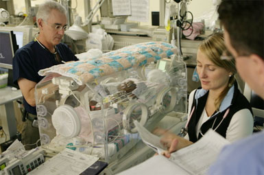
Therapeutic hypothermia (TH) has been associated with improved outcomes in term infants who present with moderate hypoxic-ischaemic encephalopathy (HIE). However, in the three major studies the time to initiate cooling was at approximately 4.5 postnatal hours. Many newborns are referred to specialist centres where cooling takes place from outlying hospitals (‘outborn’). It may be the case that earlier initiation of TH could improve outcomes, leading Takenouchi and colleagues to propose a ‘Chain of Brain Preservation’.
Given that most infants are outborn, a time sensitive education metaphor termed Chain of Brain Preservation may facilitate early recognition of high risk infants and thus earlier treatment.

Chain of Brain Preservation—A concept to facilitate early identification and initiation of hypothermia to infants at high risk for brain injury
Resuscitation. 2010 Dec;81(12):1637-41

Ultrasound measurement of optic nerve sheath diameter

Here’s the abstract from a new study contributing the literature on ED assessment of raised intracranial pressure using ocular ultrasound:
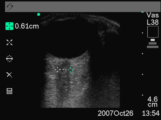
Background To assess if ultrasound measurement of the optic nerve sheath diameter (ONSD) can accurately predict the presence of raised intracranial pressure (ICP) and acute pathology in patients in the emergency department.
Methods This 3-month prospective observational study used ultrasound to measure the ONSD in adult patients who required CT from the emergency department. The mean ONSD from both eyes was measured using a 7.5 MHz ultrasound probe on closed eyelids. A mean ONSD value of >0.5 cm was taken as positive. Two radiologists independently assessed CT scans from patients in the study population for signs of raised ICP and signs of acute pathology (cerebrovascular accident, subarachnoid, subdural or extradural haemorrhage and tumour). Specificity, sensitivity and k values, for interobserver variability between reporting radiologists, were generated for the study data.

Results In all, 26 patients were enrolled into the study. The ONSD measurement was 100% specific (95% CI 79% to 100%) and 86% sensitive (95% CI 42% to 99%) for raised ICP. For any acute intracranial abnormality the value of ONSD was 100% specific (95% CI 76% to 100%) and 60% sensitive (95% CI 27% to 86%). k Values were 0.91 (95% CIs 0.73 to 1) for identification of raised ICP on CT and 0.84 (95% CIs 0.62 to 1) for any acute pathology on CT, between the radiologists.
Conclusions This study shows that ultrasound measurement of ONSD is sensitive and specific for raised ICP in the emergency department. Further observational studies are needed but this emerging technique could be used to focus treatment in unstable patients.
Ultrasound measurement of optic nerve sheath diameter in patients with a clinical suspicion of raised intracranial pressure
Emerg Med J. 2010 Aug 15. [Epub ahead of print]