Tag Archives: resuscitation

Beherrsche die Reanimation

TLsm-icon The whole purpose behind my career and this blog is to save life. Like most emergency physicians I don’t see a huge number of resuscitation patients myself in a given week, so my best hope in making a difference is to develop my teaching skills so that I can motivate and inspire others to improve their ability to manage resuscitation.
The highlight of my week therefore has been the receipt of some email feedback from a colleague in Germany. An intensivist, internist, and prehospital doctor (I like him already) who tells me he found my ‘Own the Resus‘ talk helpful:


Dear Dr. Reid,

Few days ago, too tired too sleep after a long shift on my ICU (18 beds internal medicine ICU, I am specialist in internal medicine specialized in intensive care and prehospital emergency medicine in a major German city) I watched your talk via emcrit podcast. I was immediately caught, I soaked in every word, I was fascinated, watched it twice in the middle of the night and next afternoon I listened to it in my car driving to work.

At this very day I did some overdue crap beyond the end of my shift when I heard the ominous shuffling of feet and rolling of the emergency cart from the other end of the ward… “I think we need your help….”

There it was, difficult airway situation. Patient crashing.

Then what followed was a kind of “out of body experience”. I did what was necessary, made things happen like calling anesthesia difficult airway code, calling the surgeons, organizing fiber optics and meanwhile trying to secure that airway myself until i could dispatch anesthesia to the head and surgeons to the neck. Within few minutes there were 6 doctors and 5 nurses shuffling on 9 square meters…

I found myself 1 meter behind the foot end of the pts bed and with your talk in my head I found me consciously controlling the crowd. There was suddenly the messages of your talk and there was me. I don’t know how to put it into words, I wouldn’t have done something else in medical terms but thanks to your talk I had the vocabulary, the tools to reflect myself as the leader to be in charge of the situation somehow with more distance, and after a successful resus the 10 people involved in this code went off with a good feeling that everybody contributed in what they could and all for the pts benefit.

Your talk was a kind of transition to the next level for me: from the colleague who asks how to get out of trouble in many situations because he was often deeply in trouble, to the one who leads out of trouble.

With your talk many things suddenly became clear and I am looking forward to be able to work harder on this role of leading.

Thank you very much.

D

Speeding things up through equipment design

Ever been at a cardiac arrest resuscitation where someone’s opening and closing drawers at great speed but failing to retrieve the drugs or equipment you’ve asked for urgently?
What if your resus trolley were designed by team of clinicians, engineers, and designers? Such a project was achieved through a collaboration between Imperial College London and the Helen Hamlyn Centre for Design, and the award-winning result was called the ‘Resus:Station’.
The trolley separates into three trolleys for airway, drugs and defibrillation, and circulation. The contents are visible from the outside.

Image from Pubmed Free Full Text Article

As well as improving access to equipment, the trolley can log the team’s actions during each resuscitation attempt. It can also provide an instant display of its readiness for use by recording the removal and replacement of each item.
In a randomised comparison with a standard resus trolley, a number of measures of efficiency and team performance were significantly better using the Resus:Station during simulated cardiac arrest resuscitations.
It appears to be specifically designed for cardiac arrest situations rather than ‘resus’ in its wider context. The most recent article (cited below) reports that a newer prototype is being developed prior to the manufacture of the final product.
For an in depth discussion of how resus room layout can optimise efficiency, check out Minh Le Cong’s PHARM blog and podcast with James French and Scott Weingart on Clinical Logistics

The “Resus:Station”: the use of clinical simulations in a randomised crossover study to evaluate a novel resuscitation trolley.
Resuscitation. 2012 Nov;83(11):1374-80 Free full text
[EXPAND Click for abstract]


BACKGROUND AND AIM: Inadequately designed equipment has been implicated in poor efficiency and critical incidents associated with resuscitation. A novel resuscitation trolley (Resus:Station) was designed and evaluated for impact on team efficiency, user opinion, and teamwork, compared with the standard trolley, in simulated cardiac arrest scenarios.

METHODS: Fifteen experienced cardiac arrest teams were recruited (45 participants). Teams performed recorded resuscitation simulations using new and conventional trolleys, with order of use randomised. After each simulation, efficiency (“time to drugs”, un-locatable equipment, unnecessary drawer opening) and team performance (OSCAR) were assessed from the video recordings and participants were asked to complete questionnaires scoring various aspects of the trolley on a Likert scale.

RESULTS: Time to locate the drugs was significantly faster (p=0.001) when using the Resus:Station (mean 5.19s (SD 3.34)) than when using the standard trolley (26.81s (SD16.05)). There were no reports of missing equipment when using the Resus:Station. However, during four of the fifteen study sessions using the standard trolley participants were unable to find equipment, with an average of 6.75 unnecessary drawer openings per simulation. User feedback results clearly indicated a highly significant preference for the newly designed Resus:Station for all aspects. Teams performed equally well for all dimensions of team performance using both trolleys, despite it being their first exposure to the Resus:Station.

CONCLUSION: We conclude that in this simulated environment, the new design of trolley is safe to use, and has the potential to improve efficiency at a resuscitation attempt.

[/EXPAND]

The Resus Room Life Guard

armstriconI was lucky to be accompanied through much of my emergency medicine training and specialist work in the UK by Bruce Armstrong. We shared many resuscitation cases together in hospitals and in prehospital care.
When preparing the team in resus, Armstrong used to appoint a ‘safety officer’. This could be a nurse or physician – it didn’t matter. Their role was to stay hands-off and be the eyes, ears, and mouth that would identify impending hazards and verbally intervene to thwart them.
This process seemed so natural that I rarely gave it a thought, but its glaring absence from every place I’ve worked since has only recently hit me.
Because my son goes swimming.
Photo on 29-12-12 at 10.31 AMMy three year old son attends a swimming class. There is usually one other child in the class. Recently a third child joined the class and I found myself getting uncomfortable. How could the instructor stay vigilant? What if while holding one child one of the others sank under water out of her field of view? My own obsessive reading about the limitations of human perception and cognition has convinced me that no-one can really focus on more than one thing at a time.
A friend of mine has coached kids at swimming so I asked him how they solve this. The answer was obvious – you rely on the life guards whose sole role is look out for everyone’s safety. Duh.
And then it came to me. Armstrong knew this all along. He got this idea from his prehospital experience working with fire & rescue crews and brought it into the ED. It didn’t occur to me that no-one else did this. It was just him.
Keen to explore whether anyone else had embraced this idea, I decided to go to the top when it comes to patient safety, and contacted Martin Bromiley. He told me he hadn’t come across the role in this specific setting, although did point out a great example from the BBC Documentary ‘Operation Iceberg’, in which ‘a group of scientists boarded an iceberg with someone watching over the big picture of polar bears and the berg cracking as well as fog etc’. Martin directed me to the Clinical Human Factors Group on LinkedIn, where interest was shown in the concept although it was apparent others haven’t come across it.
I went back to Armstrong to push him on further thoughts:


Yes a thought….in every other high risk environment they have a specific safety officer, whether it be nuclear industry, airline etc.

The role is specific not an add on to another role.

In healthcare we are seen as successful the more we do by one person. Think lean… think ‘efficiencies’ in the health service. Other industries focus on safety. Get safety right, your brand is safe and the public go with you. If you don’t put safety first it is only a matter of time before disaster strikes. In healthcare we have too many serious incidents. The time has come to believe in and practice safety in health care rather than ticking boxes and not applying CRM and human factors.

So here’s a proposed checklist for a Resuscitation Room Safety Officer. It’s a first draft to get the idea out there and start the conversation – just click the image below to enlarge. I’ve written (and used) checklists in resus before, but none specifically for a safety officer.

Safety Officer Checklistsm

I would like to hear if anyone’s already doing this anywhere, and how it’s been working.

Cliff

Perimortem Caesarean Delivery: Late is Better than Not


“To date, approximately one-third of the women who die during pregnancy remain undelivered at the time of death”

Guidelines recommend cardiac arrest in pregnant women beyond 20 weeks gestation should be treated with perimortem caesarean delivery (PMCD) commenced within 4 minutes of arrest and completed within 5. These time intervals come from two papers, neither of which is current or used robust review methodology.
To address this, an up-to-date fairly comprehensive review was undertaken of published cases of maternal cardiac arrests occurring prior to delivery. The primary outcome measures were maternal and neonatal survival to hospital discharge and the relationship between PMCD and this outcome.

The Arrests

94 cases were included in the final analysis.Most pregnancies were singleton (90.4%, n = 85) with an average gestational age at the time of the arrest of 33 ± 7 weeks (median 35, range 10–42).
The most common causes of arrest were trauma, maternal cardiac problems, severe pre-eclampsia and amniotic fluid embolism, together comprising about 70% of arrests; two thirds occurred in hospital.

The Outcomes

Overall, return of spontaneous circulation (ROSC) was achieved more often than not (60.6%) and overall survival to hospital discharge was 54.3%
Only 57 cases (75%) reported the time from arrest to delivery; the average time was 16.6 ± 12.5 min (median 10, range 1–60), with only 4 cases making it under the advocated 4-min time limit.

Timing of PMCD and Maternal Survival

In cases undergoing PMCD the average time elapsing from arrest to PMCD was significantly different between surviving (27/57) and non-surviving (30/57) mothers [10.0 ± 7.2 min (median 9, range 1–37) and 22.6 ± 13.3 min (median 20, range 4–60) respectively (p < 0.001, 95%CI 6.9–18.2)].

Timing of PMCD and Neonatal Survival

Mean times to PMCD were 14±11min (median=10, range=1–47) and 22 ± 13 min (median = 20, range = 4–60) in neonatal survivors and non-survivors respectively (p=0.016)
In cases with PMCD which reported outcome, the overall neonatal survival rate was 63.6% (42/66).


“The 4-min time frame advocated for PMCD usually remains unmet yet neonatal survival is still likely if delivery occurs within 10 or even 15 min of arrest”

Both maternal & neonatal mortality were higher with prehospital arrest location.

Summary

The study may be limited by recall bias, under-reporting and publication bias, but provides a more comprehensive evidence base on which to base resuscitation recommendations. The authors provide a useful warning against becoming fixated with the recommended four minute window, which may lead teams to fail to attempt a potentially life-saving intervention:


“Fixation on specific time frames for PMCD may not be ideal. It may be more important to focus on event recognition and good overall performance…. It may be wise to advocate a short time frame for performance of PMCD in order to achieve better outcomes; however, blanket endorsement of an unrealistic time frame may well create a defeatist attitude when that time frame cannot be met.”

Maternal cardiac arrest and perimortem caesarean delivery: Evidence or expert-based?
Resuscitation. 2012 Oct;83(10):1191-200
[EXPAND Click for abstract]


AIM: To examine the outcomes of maternal cardiac arrest and the evidence for the 4-min time frame from arrest to perimortem caesarean delivery (PMCD) recommended in current resuscitation and obstetric guidelines.

DATA SOURCES AND METHODS: Review and data extraction from all reported maternal cardiac arrests occurring prior to delivery (1980-2010). Cases were included if they provided details regarding both the event and outcomes. Outcomes of arrest were assessed using survival, Cerebral Performance Category (CPC) and maternal/neonatal harm/benefit from PMCD. Outcome measures were maternal and neonatal survival.

RESULTS: Of 1594 manuscripts screened, 156 underwent full review. Data extracted from 80 relevant papers yielded 94 included cases. Maternal outcome: 54.3% (51/94) of mothers survived to hospital discharge, 78.4% (40/51) with a CPC of 1/2. PMCD was determined to have been beneficial to the mother in 31.7% of cases and was not harmful in any case. In-hospital arrest and PMCD within 10 min of arrest were associated with better maternal outcomes (ORs 5.17 and 7.42 respectively, p<0.05 both). Neonatal outcome: mean times from arrest to delivery were 14±11 min and 22±13 min in survivors and non-survivors respectively (receiver operating area under the curve 0.729). Neonatal survival was only associated with in-hospital maternal arrest (OR 13.0, p<0.001).

CONCLUSIONS: Treatment recommendations should include a low admission threshold to a highly monitored area for pregnant women with cardiorespiratory decompensation, good overall performance of resuscitation and delivery within 10 min of arrest. Cognitive dissonance may delay both situation recognition and the response to maternal collapse.

[/EXPAND]

London Calling – part 2

Notes from Days 2 & 3 of the London Trauma Conference
Day 2 of the LTC was really good. There were some cracking speakers who clearly had the ‘gift’ when it comes to entertaining the audience. No death by PowerPoint here (although it seems Keynote is now the presentation software of choice!). The theme of the day was prehospital care and major incidents.
The golden nuggets to take away include: (too many to list all of course)

  • ‘Pull’ is the key to rapid extrication from cars if time critical from the Norweigan perspective. Dr Lars Wik of the Norweigen air ambulance presented their method of rapid extrication. Essentially they drag the car back on the road or away from what ever it has crashed into to control the environment and make space (360 style). They put a paramedic in the car whilst this is happening. They then make a cut in the A post near the roof, secure the rear of the car to a fire truck or fixed object with a chain and put another chain around the lower A post and steering wheel that is then winched tight. This has the effect of ‘reversing’ the crash and a few videos showed really fast access to the patient. The car seems to peel open. As they train specifically for it, there doesn’t seem to be any safety problems so far and its much quicker than their old method. I guess it doesnt matter really how you organise a rapid extrication method as long as it is trained for and everyone is on the same page.
  • Dr Bob Winter presented his thoughts on hangings – to date no survivor of a non-judicial hanging has had a C-spine injury, so why do we collar them? Also there seems no point in cooling them. All imaging and concern for these patients should be based on the significant soft tissue injury that can be caused around the neck.
  • Drownings – if the patient is totally submerged probably reasonable to search for 30mins in water that is >6 degrees or 90mins if <6 degrees. After that it becomes a body recovery (unless there is an air pocket or some exceptional circumstance). Patients that have drowned should have early ventilatory support if they show any signs of resp distress.
  • Drs Julian Thompson and Mark Byers reassured us on a variety of safety issues at major incidents. It seems the risk to rescuers from secondary bombs at scene is low. Very few terrorist attacks world wide, ever, have had secondary devices so rescuers should be reassured (a bit). Greatest risk to the rescuer, like always, are the silly simple things that are a risk every day, like tripping over your own feet! With reference to chemical incidents, simple PPE seems to be sufficient for the vast majority of incidents, even fairly significant chemical ones, all this mucking about in full air tight suits is probably pointless and means patients cant be treated (at all). This led to the debate of how much risk should we, as rescue staff, accept? Clearly there are no absolute answers but minimising all risk to the rescuer is often at conflict with your ability to rescue. Where the balance should lie is a matter for organisations and individuals I guess.
  • Sir Prof Keith Porter also gave us an update on the future of Prehospital emergency medicine as a recognised medical specialty. As those in the know, know, the specialty has been recognised by the GMC and the first draft of trainees are currently in post. More deaneries will be following suit soon to begin training but it is likely to take some time to build up large numbers of trained specialists. Importantly for those of us who already have completed our training there will be an option to sub specialise in PHEM but it will involve undertaking the FIMC exam. Great, more exams – see you there.

 
Day 3 – Major trauma
The focus of day 3 was that of damage control. Damage control surgery and damage control resucitation. We had indepth discussions about how to manage pelvic trauma and some of the finer points of trauma resuscitation.
Specific points raised were:

  • Pelvic binders are great and can replace an ex fix if the abdomen needs opening to fix a spleen for example.
  • You can catheterise patients with pelvic fractures (one gentle try).
  • Most pelvic bleeds are venous which is why surgeons who can pack a pelvis is better than a radiologist who can mainly only treat arterial bleeds.
  • Coagulopathy in trauma is not DIC and is probably caused by peripheral hypoperfusion.
  • All the standard clotting tests that we use (INR etc) are useless and take too long to do. ROTEM or TEG is much better but still not perfect.

Also, as I am sure will please many – pressure isn’t flow so dont use pressors in trauma!


Chris Hill is an emergency and prehospital care physician based in the United Kingdom

Transtracheal airways in kids. Well, pigs' kids anyway

Ever had to do a surgical airway in a child? Thought not. They’re pretty rare. Bill Heegaard MD from Henepin County Medical Center taught me a few approaches (with the help of an anaesthetised rabbit) which really got me thinking. It’s something I’d often trained for in my internal simulator, and I even keep the equipment for it in my house (listen out for an upcoming podcast on that). Research and experience has demonstrated that open surgical airway techniques are more reliable than transtracheal needle techniques in adults, but what about kids, in whom traditional teaching cautions against open techniques?

Australian investigators who were experienced airway proceduralists evaluated transtracheal needle techniques using a rabbit model (an excellent model for the infant airway). Their success rate was only 60% and they perforated the posterior tracheal wall in 42% of attempts. Of 13 attempts to insert a dedicated paediatric tracheotomy device, the Quicktrach Child, none were successful(1) (they did not use the Quicktrach Infant model as it is not available in Australia).

Danish investigators used fresh piglet cadavers weighing around 8 kg to assess two transtracheal cannulas, in which they achieved success rates of 65.6% and 68.8%(2). There was also a very high rate of posterior tracheal wall perforation. Using an open surgical tracheostomy technique, they were successful in 97% of attempts. These were also experienced operators, with a median anaesthetic experience of 12.5 years.
Their tracheotomy technique was nice and simple, and used just a scalpel, scissors, and surgical towel clips. Here’s their technique:

Simple tracheotomy procedure described by Holm-Knudsen et al

  1. Identify larynx and proximal trachea by palpation
  2. Vertical incision through the skin and subcutaneous tissue from the upper part of larynx to the sternal notch
  3. Grasp strap muscles with two towel forceps and separate in the midline
  4. Palpate and identify the trachea (palpate rather than look for tracheal rings, as in a live patient one would expect bleeding to obscure the view)
  5. Stabilise the trachea by grasping it with a towel forceps
  6. Insert sharp tip of the scissors between two tracheal rings and lift the trachea anteriorly to avoid damage to the posterior wall
  7. Cut vertically in the midline of the trachea with the scissors – they chose to use the scissors to cut the tracheal rings to facilitate tube insertion
  8. Insert the tracheal tube

Using ultrasound and CT to evaluate comparative airway dimensions, the authors concluded that the pig model is most useful for training emergency airway management in older children aged 5–10 years.
Why were they doing a tracheotomy rather than a cricothyroidotomy? Reasons given by the authors include:

  • The infant cricothyroid membrane is very small
  • Palpation of the thyroid notch may be hindered by the overlying hyoid bone
  • The mandible may obstruct needle access to the cricothyroid membrane given the cephalad position in the neck of the infant larynx.

From an emergency medicine point of view, there are a couple of other reasons why we need to be able to access the trachea lower than the cricothyroid membrane. One is fractured larynx or other blunt or penetrating airway injury where there may be anatomical disruption at the cricothyroid level. The other situation is foreign body airway obstruction, when objects may lodge at the level of the cricoid ring which is functionally the narrowest part of the pediatric upper airway. Of course, alternative methods might be considered to remove the foreign body prior to tracheotomy, such as employing basic choking algorithms, and other techniques depending on whether you do or don’t have equipment.

Take home messages

  • Transtracheal airways in kids are so rare, we can’t avoid extrapolating animal data
  • Whichever infant or paediatric model is used, transtracheal needle techniques have a high rate of failure even by ‘experienced’ operators
  • The small size and easy compressibility of the airway probably contributes to this failure rate, including the high rate of posterior wall puncture
  • In keeping with adult audit data, open surgical techniques may have a higher success rate
  • Tracheotomy may be necessary rather than cricothyroidotomy in infants and children depending on clinical scenario and accessibility of anatomy
  • The stress and blood that is not simulated in cadaveric animal models will make open tracheotomy harder in a live patient, and so these success rates may not translate. However these factors do mean that whatever technique is used must be kept simple and should employ readily available and familiar equipment
  • Something to maintain control and anterior position of the anterior trachea wall should be used during incision and intubation of the trachea. The study reported here used towel clips; sutures around the tracheal rings may also be used (see image below)

I recommend you add ‘paediatric tracheotomy’ to the list of procedures you might need to do (if it’s not already there). Identify what equipment you would use and run the simulation in your head and in your work environment.
Have fun.

1. The ‘Can’t Intubate Can’t Oxygenate’ scenario in Pediatric Anesthesia: a comparison of different devices for needle cricothyroidotomy
Paediatr Anaesth. 2012 Dec;22(12):1155-8

BACKGROUND: Little evidence exists to guide the management of the ‘Can’t Intubate, Can’t Oxygenate’ (CICO) scenario in pediatric anesthesia.

OBJECTIVES: To compare two intravenous cannulae for ease of use, success rate and complication rate in needle tracheotomy in a postmortem animal model of the infant airway, and trial a commercially available device using the same model.

METHODS: Two experienced proceduralists repeatedly attempted cannula tracheotomy in five postmortem rabbits, alternately using 18-gauge (18G) and 14-gauge (14G) BD Insyte(™) cannulae (BD, Franklin Lakes, NJ, USA). Attempts began at the first tracheal cartilage, with subsequent attempts progressively more caudad. Success was defined as intratracheal cannula placement. In each rabbit, an attempt was then made by each proceduralist to perform a cannula tracheotomy using the Quicktrach Child(™) device (VBM Medizintechnik GmbH, Sulz am Neckar, Germany).

RESULTS: The rabbit tracheas were of similar dimensions to a human infant. 60 attempts were made at cannula tracheotomy, yielding a 60% success rate. There was no significant difference in success rate, ease of use, or complication rate between cannulae of different gauge. Successful aspiration was highly predictive (positive predictive value 97%) and both sensitive (89%) and specific (96%) for tracheal cannulation. The posterior tracheal wall was perforated in 42% of tracheal punctures. None of 13 attempts using the Quicktrach Child(™) were successful.

CONCLUSION: Cannula tracheotomy in a model comparable to the infant airway is difficult and not without complication. Cannulae of 14- and 18-gauge appear to offer similar performance. Successful aspiration is the key predictor of appropriate cannula placement. The Quicktrach Child was not used successfully in this model. Further work is required to compare possible management strategies for the CICO scenario

2. Emergency airway access in children – transtracheal cannulas and tracheotomy assessed in a porcine model
Paediatr Anaesth. 2012 Dec;22(12):1159-65

OBJECTIVES: In the rare scenario when it is impossible to oxygenate or intubate a child, no evidence exists on what strategy to follow.

AIM: The aim of this study was to compare the time and success rate when using two different transtracheal needle techniques and also to measure the success rate and time when performing an emergency tracheotomy in a piglet cadaver model.

METHODS: In this randomized cross-over study, we included 32 anesthesiologists who each inserted two transtracheal cannulas (TTC) using a jet ventilation catheter and an intravenous catheter in a piglet model. Second, they performed an emergency tracheotomy. A maximum of 2 and 4 min were allowed for the procedures, respectively. The TTC procedures were recorded using a video scope.

RESULTS: Placement of a transtracheal cannula was successful in 65.6% and 68.8% of the attempts (P = 0.76), and the median duration of the attempts was 69 and 42 s (P = 0.32), using the jet ventilation catheter and the intravenous catheter, respectively. Complications were frequent in both groups, especially perforation of the posterior tracheal wall. Performing an emergency tracheotomy was successful in 97%, in a median of 88 s.

CONCLUSIONS: In a piglet model, we found no significant difference in success rates or time to insert a jet ventilation cannula or an intravenous catheter transtracheally, but the incidence of complications was high. In the same model, we found a 97% success rate for performing an emergency tracheotomy within 4 min with a low rate of complications.

Echo for cardiac arrest outcome prediction

A meta-analysis of studies evaluation transthoracic echo as a means of predicting return of spontaneous circulation in cardiac arrest (ROSC) provides some likelihood ratios to what we already know: absence of sonographic cardiac activity means a very low chance of ROSC.

The authors report a pooled negative LR of 0.18 (95% CI = 0.10 to 0.31), and a positive likelihood ratio of 4.26 (95% CI = 2.63 to 6.92).

They conclude that focused transthoracic echo is a fairly effective (although not definitive) test for predicting death if no cardiac activity is noted during resuscitation, and recommend interpreting the echo in the light of the test characteristics and the clinical pre-test probability, as one should do for all imaging investigations:


“An elderly patient with an unwitnessed cardiac arrest already has very poor odds for survival. Confirmation of asystole on echo lowers those pretest odds by a factor of 5.6 and therefore might lead to termination of resuscitation. However, in the case of a 50-year-old rescued from drowning, detection of cardiac contractility on echo would increase his already fair odds of survival by a factor of 4.3, prompting continued aggressive resuscitation.”

Only five relatively small studies contributed to the findings. A more definitive answer to this question should be provided in the future by the multi-centre REASON 1 trial.

Objectives:  The objective was to determine if focused transthoracic echocardiography (echo) can be used during resuscitation to predict the outcome of cardiac arrest.

Methods:  A literature search of diagnostic accuracy studies was conducted using MEDLINE via PubMed, EMBASE, CINAHL, and Cochrane Library databases. A hand search of references was performed and experts in the field were contacted. Studies were included for further appraisal and analysis only if the selection criteria and reference standards were met. The eligible studies were appraised and scored by two independent reviewers using a modified quality assessment tool for diagnostic accuracy studies (QUADAS) to select the papers included in the meta-analysis.

Results:  The initial search returned 2,538 unique papers, 11 of which were determined to be relevant after screening criteria were applied by two independent researchers. One additional study was identified after the initial search, totaling 12 studies to be included in our final analysis. The total number of patients in these studies was 568, all of whom had echo during resuscitation efforts to determine the presence or absence of kinetic cardiac activity and were followed up to determine return of spontaneous circulation (ROSC). Meta-analysis of the data showed that as a predictor of ROSC during cardiac arrest, echo had a pooled sensitivity of 91.6% (95% confidence interval [CI] = 84.6% to 96.1%), and specificity was 80.0% (95% CI = 76.1% to 83.6%). The positive likelihood ratio for ROSC was 4.26 (95% CI = 2.63 to 6.92), and negative likelihood ratio was 0.18 (95% CI = 0.10 to 0.31). Heterogeneity of the results (sensitivity) was nonsignificant (Cochran’s Q: χ(2) = 10.63, p = 0.16, and I(2) = 34.1%).

Conclusions:  Echocardiography performed during cardiac arrest that demonstrates an absence of cardiac activity harbors a significantly lower (but not zero) likelihood that a patient will experience ROSC. In selected patients with a higher likelihood of survival from cardiac arrest at presentation, based on established predictors of survival, echo should not be the sole basis for the decision to cease resuscitative efforts. Echo should continue to be used only as an adjunct to clinical assessment in predicting the outcome of resuscitation for cardiac arrest.

Bedside Focused Echocardiography as Predictor of Survival in Cardiac Arrest Patients: A Systematic Review
Acad Emerg Med. 2012 Oct;19(10):1119-1126

Surgical treatment for acute massive pulmonary embolism

A recent paper reminds us that surgery is an option in the management of massive pulmonary embolism(1), to be considered in the patient for whom thrombolysis has failed or is contraindicated. Good outcomes were produced when surgery was performed in a centre capable of cardiopulmonary bypass (6% 30-day postoperative mortality), but is surgery an option when these facilities are unavailable?

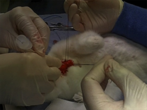
The “venous inflow occlusion” technique involves clamping the venae cavae prior to removing clot directly from the pulmonary artery and its branches after median sternotomy, and can be performed in any hospital with surgical facilities. Under normothermic conditions, speed is of the essence once cardiac arrest occurs, since the irreversible anoxic cerebral injury will occur after just a few minutes.
Clarke and Abrams wrote in the Lancet in 1972(2):

Our use of venous inflow-occlusion has given results which compare well with those obtained with extracorporeal circulation. 50% of our patients survived. All patients who had emboli removed without an episode of ventricular asystole survived surgery. Late deaths in 3 patients were from causes unrelated to pulmonary embolism, and from a further massive pulmonary embolus a week later. The technique has been applied with equal success in a major hospital fully equipped for cardiac surgery and in hospitals where resident and nursing staff had no experience of either thoracic or cardiac surgery. The simplicity and speed of the method has enabled the obstructed right ventricle to be relieved within thirty minutes of the onset of symptoms. The interval between induction of anaesthesia and the skin incision should be kept as short as possible, and drugs to maintain the blood pressure should be given. The period between skin incision and the restoration of the circulation has, with practice, been reduced to ten minutes.

But this clearly still requires surgical expertise and facilities. Emergency physicians can open the chest to deal with penetrating trauma. Could an ED thoracotomy facilitate clot removal from the pulmonary artery?

In 1969, a lady in her 50s arrested on the ward after an operation to remove a mass via a left lateral thoractomy. Pulmonary embolism was suspected and her thoracotomy wound was re-opened and the pulmonary artery incised, resulting in the removal of large amounts of clot. Return of spontaneous circulation resulted after a brief period of internal cardiac massage. Her case was written up decades later, in 1998(3):

The patient recovered rapidly and left the hospital on the 21st day without signs of cerebral damage. This patient is now 86 years old, mentally normal, living alone, and doing her own housekeeping. She remembers the hospital stay and the past years as worth living

Some patients may be considered too high risk for surgery and in some centres Extracorporeal Membrane Oxygenation (ECMO) is an option. It has been used both as life support pending surgery(4), or as an alternative to surgery to allow heparinisation to be used(5,6).

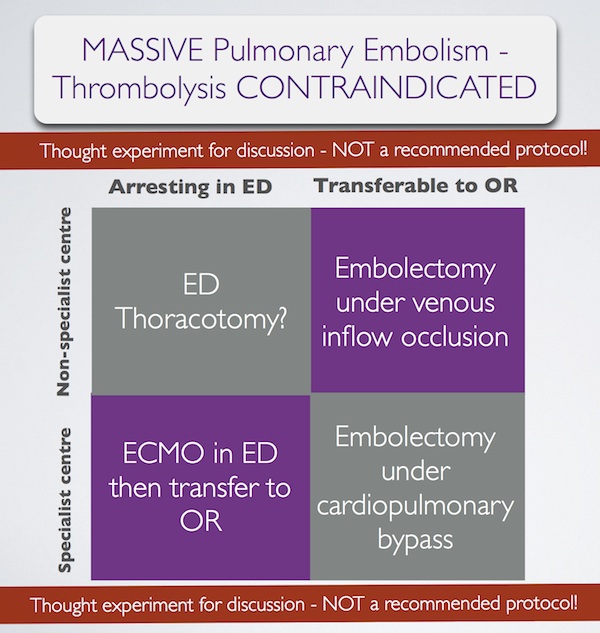
In summary, some patients with massive pulmonary embolism may benefit from surgery (contraindication to ‘lysis or failed ‘lysis). Getting them to surgery alive, or operating on them during cardiac arrest, is a challenge. Ideally they would undergo embolectomy under cardopulmonary bypass in the operating room, or could be placed on ECMO in the ED prior to going to the OR. If they present to a centre without these facilities, then the venous inflow occlusion technique could be used in the OR without bypass. Just rarely a patient may present in extremis with PE to an ED without these options. If that patient has major contraindications to thrombolysis, would an ED thoracotomy be something you would entertain?

I have done several thoracotomies for penetrating trauma but never for PE. I do not pretend to know how, and cannot find a case report of ED thoracotomy for pulmonary embolism in the literature. I’m therefore NOT recommending it. However, I would love to know people’s views on its feasibility. A possible approach could be summarised as:

Massive pulmonary embolism fascinates me, because it’s seen in the ‘talk and die’ patient. It is a single, treatable pathology that if diagnosed and treated appropriately truly makes the difference between life and death. When medicine presents us with an opportunity ‘on a plate’ like that to save a life, we need to be prepared. I have had great saves with this diagnosis and sadly have seen disastrous failures to act. When the time comes, we need to ask: ‘have we explored all options?’.

1. Surgical treatment of acute pulmonary embolism–a 12-year retrospective analysis.
Scand Cardiovasc J. 2012 Jun;46(3):172-6. Epub 2012 Mar 27.

OBJECTIVES: Surgical embolectomy for acute pulmonary embolism (PE) is considered to be a high risk procedure and therefore a last treatment option. We wanted to evaluate the procedures role in modern treatment of acute PE.
DESIGN: All data on patients treated with surgical embolectomy for acute PE were retrieved from our clinical database. The mortality was extracted from the Danish mortality register.
RESULTS: From October 1998 to July 2010, 33 patients underwent surgical embolectomy. All procedures were done through a median sternotomy and extracorporeal circulation. Twenty-six patients were diagnosed with a high risk PE and 7 with an intermediate risk PE and intracardial pathology. Six patients had been insufficiently treated with thrombolysis. Thirteen patients had contraindication for thrombolysis. Six patients were brought to the operating theatre in cardiogenic shock, 8 needed ventilator support, and 1 was in cardiac arrest. The postoperative 30-day mortality was 6% and during the 12-year follow-up the cumulative survival was 80% with 4 late deaths.
CONCLUSION: Surgical pulmonary embolectomy can be performed with low mortality although the treated patients belong to the most compromised part of the PE population. The results support surgical embolectomy as a vital part of the treatment algorithm for acute PE.

2. Pulmonary embolectomy with venous inflow-occlusion.
The Lancet 1972;1(7754):767–769

Massive pulmonary emboli have been removed surgically from 26 patients. The technique of normothermic circulatory arrest by venous inflow-occlusion was used in 25 patients. 13 patients survived. There were 10 operative deaths and 3 hospital deaths. Diagnosis was based upon clinical findings supplemented by electrocardiography and a plain radiograph of the chest. Surgery was offered to patients having a pulmonary embolus sufficiently massive to produce sustained hypotension. All patients whose hearts stopped beating before the embolectomy died. 6 successful operations were performed in hospitals without facilities for cardiac surgery. The method is recommended for its simplicity.

3. Left Anterior Thoracotomy for Pulmonary Embolectomy With 29-Year Follow-up
The Annals of Thoracic Surgery 1998, 66(4):1420-1421

Pulmonary embolectomy is usually performed in cardiopulmonary bypass. In acute situations too much time can be lost in setting up and connecting the pump oxygenator; this delay can cause cerebral damage in a patient with circulatory arrest. In such a situation left anterior thoracotomy can provide an ideal approach. An emergency thoracotomy can be performed in a few seconds. The lung automatically retracts. The phrenic nerve, pulmonary artery, and pericardium are clearly seen, and they outline the area for embolectomy. A case in which such an approach was successfully used is described.

4.ECMO treatment saved life of a young woman with acute pulmonary embolism
Lakartidningen 2004, 101(44):3420-3421

A 42-year old obese female using contraceptive medication was admitted to the emergency room because of sudden onset of dyspnoea and hypoxia. Computed tomography showed massive pulmonary emboli. Despite initial treatment with thrombolysis her condition deteriorated further and she was referred for acute surgery to our clinic. Before putting the patient to sleep extracorporeal circulation was instituted with access from the groin. After anaesthesia a median sternotomy was performed. With the heart beating, the main pulmonary artery was incised and a 9 cm long thrombus was removed. Immediate weaning from the heart-lung machine was not possible, mainly because of bleeding to the airways. The right atrium and the aorta was therefore cannulated and an extracorporeal circulation membrane oxygenator (ECMO) was used for three days. The patient required several re-entries for bleeding and a tracheotomy during the postoperative course. She was fully recovered three months after the operation.

5. Extracorporeal membrane oxygenator for pulmonary embolism.
The Annals of Thoracic Surgery 1997, 64(3):883-884 Free full text

6. Peripheral Extracorporeal Membrane Oxygenation: Comprehensive Therapy for High-Risk Massive Pulmonary Embolism
Ann Thorac Surg 2012;94:104–8

Background: Although commonly reserved as a last line of defense, experienced centers have reported excellent results with pulmonary embolectomy for massive and submassive pulmonary embolism (PE). We present a contemporary surgical series for PE that demonstrates the utility of peripheral extracorporeal membrane oxygenation (pECMO) for high-risk surgical candidates.
Methods: Between June 2005 and April 2011, 29 patients were treated for massive or submassive pulmonary embolism, with surgical embolectomy performed in 26. Four high-risk patients were placed on pECMO, established by percutaneously cannulating the right atrium through a femoral vein and perfusing by a Dacron graft anastomosed to the axillary artery. A small, extracorporeal, rotary assist device was used, interposing a compact oxygenator in the circuit, and maintaining anticoagulation with heparin.
Results: Extracorporeal membrane oxygenation was weaned in 3 of 4 patients after 5.3 days (5, 5, and 6), with normalization of right ventricular dysfunction and pulmonary artery pressure (44.0 ± 2.0 to 24.5 ± 5.5 mm Hg) by ECHO. Follow-up computed tomographies showed several peripheral, nearly resorbed emboli in 1 case and complete resolution in 2 others. The fourth patient, not improving after 10 days, underwent surgery where an embolic liposarcoma was extracted. For all 29 cases, hospital and 30-day mortality was 0% and all patients were discharged, with average postoperative length of stay of 15 days for embolectomy and 17 days for pECMO.
Conclusions: Heparin therapy with pECMO support is a rapid, effective option for patients who might benefit from pulmonary embolectomy but are at high risk for surgery.

More on the meaning of lactate values

A newly published study(1) reminds us that we need to do better than just identify a raised lactate in patients with sepsis; we need to make sure it’s not increasing when they leave the ED (if we can). An incremental rise is associated with mortality.

The authors comment:


We found that the prognostic value of lactate continues to rise across a wide range of values, from 0 to 20 mmol/L…. These data suggest that grouping patients into less granular and larger groups, such as low, intermediate, and high, potentially underutilizes the prognostic value of the test. Furthermore, we did not find any value of lactate, up to a maximum value of 20 mmol/L, where mortality failed to increase with an increase in lactate concentration.

The paper does not state whether the lactate was arterial or venous, although either can be used. The Surviving Sepsis Campaign provides this comment:


In the course of the Campaign the question has been raised many times as to whether an arterial or venous lactate sample is appropriate. While there is no consensus of settled literature on this question, an elevated lactate of any variety is typically abnormal, although this may be influenced by other conditions..

This relationship between lactate trend and mortality has also been demonstrated in a study of all patients admitted to hospital (with or without sepsis), which also showed good correlation between arterial and venous lactate(2).

Lactate clearance has been shown to be an acceptable alternative to central venous oxygen saturation as a goal for therapy in ED severe sepsis patients(3), which is good because it provides one less reason for a central line.

Always remember the good emergency physician / critical care practitioner will consider other causes of a raised lactate, particularly when things don’t add up. I invented the ‘LACTATES’ acronym to help me remember them(4), and it’s come in handy several times.

Craving more info on lactate? Check out the EMCrit site with its great lactate reference sheet.

1. Prognostic Value of Incremental Lactate Elevations in Emergency Department Patients With Suspected Infection
Acad Emerg Med. 2012 Aug;19(8):983-5
[EXPAND Click for abstract]


Objectives:  Previous studies have confirmed the prognostic significance of lactate concentrations categorized into groups (low, intermediate, high) among emergency department (ED) patients with suspected infection. Although the relationship between lactate concentrations categorized into groups and mortality appears to be linear, the relationship between lactate as a continuous measurement and mortality is uncertain. This study sought to evaluate the association between blood lactate concentrations along an incremental continuum up to a maximum value of 20 mmol/L and mortality.

Methods:  This was a retrospective cohort analysis of adult ED patients with suspected infection from a large urban ED during 2007–2010. Inclusion criteria were suspected infection evidenced by administration of antibiotics in the ED and measurement of whole blood lactate in the ED. The primary outcome was in-hospital mortality. Logistic and polynomial regression were used to model the relationship between lactate concentration and mortality.

Results:  A total of 2,596 patients met inclusion criteria and were analyzed. The initial median lactate concentration was 2.1 mmol/L (interquartile range [IQR] = 1.3 to 3.3 mmol/L) and the overall mortality rate was 14.4%. In the cohort, 459 patients (17.6%) had initial lactate levels >4 mmol/L. Mortality continued to rise across the continuum of incremental elevations, from 6% for lactate <1.0 mmol/L up to 39% for lactate 19–20 mmol/L. Polynomial regression analysis showed a strong curvilinear correlation between lactate and mortality (R = 0.72, p < 0.0001).
Conclusions:  In ED patients with suspected infection, we found a curvilinear relationship between incremental elevations in lactate concentration and mortality. These data support the use of lactate as a continuous variable rather than a categorical variable for prognostic purposes.

[/EXPAND]
2. Blood lactate as a predictor for in-hospital mortality in patients admitted acutely to hospital: a systematic review
Scand J Trauma Resusc Emerg Med. 2011 Dec 28;19:74 Free Full Text
[EXPAND Click for abstract]


BACKGROUND: Using blood lactate monitoring for risk assessment in the critically ill patient remains controversial. Some of the discrepancy is due to uncertainty regarding the appropriate reference interval, and whether to perform a single lactate measurement as a screening method at admission to the hospital, or serial lactate measurements. Furthermore there is no consensus whether the sample should be drawn from arterial, peripheral venous, or capillary blood. The aim of this review was: 1) To examine whether blood lactate levels are predictive for in-hospital mortality in patients in the acute setting, i.e. patients assessed pre-hospitally, in the trauma centre, emergency department, or intensive care unit. 2) To examine the agreement between arterial, peripheral venous, and capillary blood lactate levels in patients in the acute setting.

METHODS: We performed a systematic search using PubMed, Cochrane Central Register of Controlled Trials, Cochrane Database of Systematic Reviews, and CINAHL up to April 2011. 66 articles were considered potentially relevant and evaluated in full text, of these ultimately 33 articles were selected.

RESULTS AND CONCLUSION: The literature reviewed supported blood lactate monitoring as being useful for risk assessment in patients admitted acutely to hospital, and especially the trend, achieved by serial lactate sampling, is valuable in predicting in-hospital mortality. All patients with a lactate at admission above 2.5 mM should be closely monitored for signs of deterioration, but patients with even lower lactate levels should be considered for serial lactate monitoring. The correlation between lactate levels in arterial and venous blood was found to be acceptable, and venous sampling should therefore be encouraged, as the risk and inconvenience for this procedure is minimal for the patient. The relevance of lactate guided therapy has to be supported by more studies.

[/EXPAND]
3. Lactate clearance vs central venous oxygen saturation as goals of early sepsis therapy: a randomized clinical trial
JAMA. 2010 Feb 24;303(8):739-46
[EXPAND Click for abstract]


CONTEXT: Goal-directed resuscitation for severe sepsis and septic shock has been reported to reduce mortality when applied in the emergency department.

OBJECTIVE: To test the hypothesis of noninferiority between lactate clearance and central venous oxygen saturation (ScvO2) as goals of early sepsis resuscitation.

DESIGN, SETTING, AND PATIENTS: Multicenter randomized, noninferiority trial involving patients with severe sepsis and evidence of hypoperfusion or septic shock who were admitted to the emergency department from January 2007 to January 2009 at 1 of 3 participating US urban hospitals.

INTERVENTIONS: We randomly assigned patients to 1 of 2 resuscitation protocols. The ScvO2 group was resuscitated to normalize central venous pressure, mean arterial pressure, and ScvO2 of at least 70%; and the lactate clearance group was resuscitated to normalize central venous pressure, mean arterial pressure, and lactate clearance of at least 10%. The study protocol was continued until all goals were achieved or for up to 6 hours. Clinicians who subsequently assumed the care of the patients were blinded to the treatment assignment.

MAIN OUTCOME MEASURE: The primary outcome was absolute in-hospital mortality rate; the noninferiority threshold was set at Delta equal to -10%.

RESULTS: Of the 300 patients enrolled, 150 were assigned to each group and patients were well matched by demographic, comorbidities, and physiological features. There were no differences in treatments administered during the initial 72 hours of hospitalization. Thirty-four patients (23%) in the ScvO2 group died while in the hospital (95% confidence interval [CI], 17%-30%) compared with 25 (17%; 95% CI, 11%-24%) in the lactate clearance group. This observed difference between mortality rates did not reach the predefined -10% threshold (intent-to-treat analysis: 95% CI for the 6% difference, -3% to 15%). There were no differences in treatment-related adverse events between the groups.

CONCLUSION: Among patients with septic shock who were treated to normalize central venous and mean arterial pressure, additional management to normalize lactate clearance compared with management to normalize ScvO2 did not result in significantly different in-hospital mortality.

[/EXPAND]
4. Non-septic hyperlactataemia in the emergency department
Emerg Med J. 2010 May;27(5):411-2