Tag Archives: TBI

CRASH-2 and head injury

The overall effect of the antifibrinolytic drug tranexamic acid on outcome from major trauma was assessed in the CRASH-2 trial, reported here and here. Its effect on a nested cohort of 270 patients from the trial who had traumatic brain injury has now been published1.
Previous evaluation in nontraumatic subarachnoid haemorrhage patients showed tranexamic acid to be associated with cerebral ischaemia, whereas in CRASH-2 (in which a lower dose of tranexamic acid was used) there was a trend to fewer ischaemic lesions as well as smaller haematoma growth and decreased mortality. None of these outcomes were statistically significant so further research is warranted.
An accompanying editorial2 states:

…the CRASH-2 study also justifies a re-evaluation of the possible benefit of low dose short term TXA in patients with other types of intracranial haemorrhage. Many patients with aneurysmal subarachnoid haemorrhage still have to wait for one or two days before the aneurysm is occluded. In addition, at least 30% of patients with spontaneous intracerebral haemorrhage experience substantial haematoma growth in the first 24 hours after the onset of the haemorrhage. As well as the CRASH-2 trial we therefore need new trials investigating short course low dose TXA in patients with aneurysmal subarachnoid haemorrhage and intracerebral haemorrhage.

It looks like considerable enthusiasm for this drug will be around for a while. I look forward to more outcome data, particularly in regard to this challenging group of patients with traumatic and non-traumatic intracranial bleeding.


OBJECTIVE: To assess the effect of tranexamic acid (which reduces bleeding in surgical patients and reduces mortality due to bleeding in trauma patients) on intracranial haemorrhage in patients with traumatic brain injury.

METHODS: A nested, randomised, placebo controlled trial. All investigators were masked to treatment allocation. All analyses were by intention to treat. Patients 270 adult trauma patients with, or at risk of, significant extracranial bleeding within 8 hours of injury, who also had traumatic brain injury.

INTERVENTIONS: Patients randomly allocated to tranexamic acid (loading dose 1 g over 10 minutes, then infusion of 1 g over 8 hours) or matching placebo.

MAIN OUTCOME MEASURES: Intracranial haemorrhage growth (measured by computed tomography) between hospital admission and then 24-48 hours later, with adjustment for Glasgow coma score, age, time from injury to the scans, and initial haemorrhage volume.

RESULTS: Of the 133 patients allocated to tranexamic acid and 137 allocated to placebo, 123 (92%) and 126 (92%) respectively provided information on the primary outcome. All patients provided information on clinical outcomes. The mean total haemorrhage growth was 5.9 ml (SD 26.8) and 8.1 mL (SD 29.2) in the tranexamic acid and placebo groups respectively (adjusted difference -3.8 mL (95% confidence interval -11.5 to 3.9)). New focal cerebral ischaemic lesions occurred in 6 (5%) patients in the tranexamic acid group versus 12 (9%) in the placebo group (adjusted odds ratio 0.51 (95% confidence interval 0.18 to 1.44)). There were 14 (11%) deaths in the tranexamic acid group and 24 (18%) in the placebo group (adjusted odds ratio 0.47 (0.21 to 1.04)).

CONCLUSIONS: This trial shows that neither moderate benefits nor moderate harmful effects of tranexamic acid in patients with traumatic brain injury can be excluded. However, the analysis provides grounds for further clinical trials evaluating the effect of tranexamic acid in this population

1. Effect of tranexamic acid in traumatic brain injury: a nested randomised, placebo controlled trial (CRASH-2 Intracranial Bleeding Study)
BMJ. 2011 Jul 1;343:d379 (free text available)
2. Tranexamic acid for traumatic brain injury
BMJ. 2011 Jul 1;343:d3958

Decompressive craniectomy for high ICP head trauma

Bilateral decompressive craniectomy for severe diffuse traumatic brain injury and intracranial hypertension that was refractory to first line therapies did not improve neurological outcome. This was the Australasian DECRA study.

Emergency Medicine Ireland reviews the paper here.
Another study on decompressive craniectomy, the RESCUE-ICP study, is ongoing, with 306/400 patients now recruited. The RESCUE-ICP investigators make the following comment on the DECRA trial:
“The study showed a significant decrease in intracranial pressure in patients in the surgical group. However, although ICP was lowered by surgery, ICP was not excessively high in the medical group (mean ICP below 24 mmHg pre-randomisation).
RESCUE-ICP differs from DECRA in terms of ICP threshold (25 vs 20 mmHg), timing of surgery (any time after injury vs within 72 hours post-injury), acceptance of contusions and longer follow up (2 years).
The cohort profiles and criteria for entry and randomisation between the DECRA and RESCUE-ICP are therefore very different. Hence the results from the DECRA study should not deter recruitment into RESCUE-ICP. Randomising patients into the RESCUE-ICP study is now even more important!”

Background
It is unclear whether decompressive craniectomy improves the functional outcome in patients with severe traumatic brain injury and refractory raised intracranial pressure.
Methods
From December 2002 through April 2010, we randomly assigned 155 adults with severe diffuse traumatic brain injury and intracranial hypertension that was refractory to first-tier therapies to undergo either bifrontotemporoparietal decompressive craniectomy or standard care. The original primary outcome was an unfavorable outcome (a composite of death, vegetative state, or severe disability), as evaluated on the Extended Glasgow Outcome Scale 6 months after the injury. The final primary outcome was the score on the Extended Glasgow Outcome Scale at 6 months.
Results
Patients in the craniectomy group, as compared with those in the standard-care group, had less time with intracranial pressures above the treatment threshold (P<0.001), fewer interventions for increased intracranial pressure (P<0.02 for all comparisons), and fewer days in the intensive care unit (ICU) (P<0.001). However, patients undergoing craniectomy had worse scores on the Extended Glasgow Outcome Scale than those receiving standard care (odds ratio for a worse score in the craniectomy group, 1.84; 95% confidence interval [CI], 1.05 to 3.24; P=0.03) and a greater risk of an unfavorable outcome (odds ratio, 2.21; 95% CI, 1.14 to 4.26; P=0.02). Rates of death at 6 months were similar in the craniectomy group (19%) and the standard-care group (18%).
Conclusions
In adults with severe diffuse traumatic brain injury and refractory intracranial hypertension, early bifrontotemporoparietal decompressive craniectomy decreased intracranial pressure and the length of stay in the ICU but was associated with more unfavorable outcomes

Decompressive Craniectomy in Diffuse Traumatic Brain Injury
N Engl J Med. 2011 Apr 21;364(16):1493-502

Salt or sugar on the brain

A meta-analysis suggests hypertonic saline may be more effective at lowering intracranial pressure than mannitol. An accompanying editorial cleverly entitled ‘Salt or sugar on the brain: Does it matter except for taste?’ suggests one reason hypertonic saline (HTS) has not replaced mannitol in clinical practice is that too many different regimens of HTS, in terms of concentration, dose, bolus vs. continuous infusions, and plus or minus supplementation of colloids, have been utilised. Because only 112 patients with 184 episodes of increased ICP were treated with each medication in this meta-analysis, the editorialist agrees with the authors in suggesting a larger randomised study is needed.

OBJECTIVES: Randomized trials have suggested that hypertonic saline solutions may be superior to mannitol for the treatment of elevated intracranial pressure, but their impact on clinical practice has been limited, partly by their small size. We therefore combined their findings in a meta-analysis.
DATA SOURCES: We searched for relevant studies in MEDLINE, EMBASE, the Cochrane Central Register of Controlled Trials (CENTRAL), Scopus, and ISI Web of Knowledge.
STUDY SELECTION: Randomized trials were included if they directly compared equiosmolar doses of hypertonic sodium solutions to mannitol for the treatment of elevated intracranial pressure in human subjects undergoing quantitative intracranial pressure measurement.
DATA EXTRACTION: Two investigators independently reviewed potentially eligible trials and extracted data using a preformed data collection sheet. Disagreements were resolved by consensus or by a third investigator if needed. We collected data on patient demographics, type of intracranial pathology, baseline intracranial pressure, osms per treatment dose, quantitative change in intracranial pressure, and prespecified adverse events. Our primary outcome was the proportion of successfully treated episodes of elevated intracranial pressure.
DATA SYNTHESIS: Five trials comprising 112 patients with 184 episodes of elevated intracranial pressure met our inclusion criteria. In random-effects models, the relative risk of intracranial pressure control was 1.16 (95% confidence interval, 1.00-1.33), and the difference in mean intracranial pressure reduction was 2.0 mm Hg (95% confidence interval, -1.6 to 5.7), with both favoring hypertonic saline over mannitol. A mild degree of heterogeneity was present among the included trials. There were no significant adverse events reported.
CONCLUSIONS: We found that hypertonic saline is more effective than mannitol for the treatment of elevated intracranial pressure. Our meta-analysis is limited by the small number and size of eligible trials, but our findings suggest that hypertonic saline may be superior to the current standard of care and argue for a large, multicenter, randomized trial to definitively establish the first-line medical therapy for intracranial hypertension.

Hypertonic saline versus mannitol for the treatment of elevated intracranial pressure: A meta-analysis of randomized clinical trials
Crit Care Med. 2011 Mar;39(3):554-9

Pre-hospital transcranial Doppler

The SAMU (Service d’aide médicale urgente) guys have had a run of interesting pre-hospital publications lately. In this study, one of their ultrasound-wielding physicians travelled in a car to meet comatose head injured patients in a large semi-rural territory area with up to a 120–160-min transport time to a hospital with emergency neurosurgical capability. Pre-hospital transcranial Doppler was done, the results of which appear to have influenced treatment decisions, including the pre-hospital administration of noradrenaline (norepinephrine). I think this study has answered the ‘can it be done?’ question, but further work is needed to determine whether it really makes a difference to outcome.

Background: Investigation of the feasibility and usefulness of pre-hospital transcranial Doppler (TCD) to guide early goal-directed therapy following severe traumatic brain injury (TBI).
Methods: Prospective, observational study of 18 severe TBI patients during pre-hospital medical care. TCD was performed to estimate cerebral perfusion in the field and upon arrival at the Level 1 trauma centre. Specific therapy (mannitol, noradrenaline) aimed at improving cerebral perfusion was initiated if the initial TCD was abnormal (defined by a pulsatility index >1.4 and low diastolic velocity).
Results: Nine patients had a normal initial TCD and nine an abnormal one, without a significant difference between groups in terms of the Glasgow Coma Scale or the mean arterial pressure. Among patients with an abnormal TCD, four presented with an initial areactive bilateral mydriasis. Therapy normalized TCD in five patients, with reversal of the initial mydriasis in two cases. Among these five patients for whom TCD was corrected, only two died within the first 48 h. All four patients for whom the TCD could not be corrected during transport died within 48 h. Only patients with an initial abnormal TCD required emergent neurosurgery (3/9). Mortality at 48 h was significantly higher for patients with an initial abnormal TCD.
Conclusions: Our preliminary study suggests that TCD could be used in pre-hospital care to detect patients whose cerebral perfusion may be impaired.

Pre-hospital transcranial Doppler in severe traumatic brain injury: a pilot study
Acta Anaesthesiol Scand. 2011 Apr;55(4):422-8

Fever in head injury might not be bad

Thanks to Michael McGonigal MD for highlighting this in his excellent Trauma Professional’s Blog:

  • Body temperature does not necessarily reflect brain temperature
  • Low brain temperature was independently associated with a worse outcome in a recent study
  • Brain temperature within the range of 36.5°C to 38°C was associated with a lower probability of death in this study
  • There are no randomised studies on which to base the practice of aggressive cooling of febrile patients with traumatic brain injury

There are few prospective studies reporting the effect of spontaneous temperature changes on outcome after severe traumatic brain injury (TBI). Where studies have been conducted, results are based on systemic rather than brain temperature per se. However, body temperature is not a reliable surrogate for brain temperature. Consequently, the effect of brain temperature changes on outcome in the acute phase after TBI is not clear. Continuous intraparenchymal brain temperature was measured in consecutive admissions of severe TBI patients during the course of the first 5 days of admission to the intensive care unit (ICU). Patients received minimal temperature altering therapy during their ICU stay. Logistic regression was used to explore the relationship between the initial, the 24-h mean, and the 48-h mean brain temperature with outcome for mortality at 30 days and outcome at 3 months. Multifactorial analysis was performed to account for potential confounders. At the 24-h time point, brain temperature within the range of 36.5°C to 38°C was associated with a lower probability of death (10-20%). Brain temperature outside of this range was associated with a higher probability of death and poor 3-month neurological outcome. After adjusting for other predictors of outcome, low brain temperature was independently associated with a worse outcome. Lower brain temperatures (below 37°C) are independently associated with a higher mortality rate after severe TBI. The results suggest that, contrary to current opinion, temperatures within the normal to moderate fever range during the acute post-TBI period do not impose an additional risk for a poor outcome after severe TBI.

The effect of spontaneous alterations in brain temperature on outcome: a prospective observational cohort study in patients with severe traumatic brain injury.
J Neurotrauma. 2010 Dec;27(12):2157-64

Paramedic RSI in Australia

A prospective, randomized, controlled trial compared paramedic rapid sequence intubation with hospital intubation in adults with severe traumatic brain injury in four cities in Victoria, Australia. The primary outcome was neurologic outcome at 6 months postinjury.
Training
Paramedics already experienced in ‘cold’ intubation (without drugs) undertook an additional 16-hour training program in the theory and practice of RSI, including class time (4 hours), practical intubating experience in the operating room under the supervision of an anesthesiologist (8 hours), and completion of a simulation-based examination (4 hours).
Methods
Patients included in the study were those assessed by paramedics on road ambulances as having all the following: evidence of head trauma, Glasgow Coma Score ≤9, age ≥15 years, and ‘intact airway reflexes’, although this is not defined or explained. Patients were excluded if any of the following applied: within 10 minutes of a designated trauma hospital, no intravenous access, allergy to any of the RSI drugs (as stated by relatives or a medical alert bracelet), or transport planned by medical helicopter. Drug therapy for intubation consisted of fentanyl (100μg), midazolam (0.1 mg/kg), and succinylcholine (1.5 mg/kg) administered in rapid succession. Atropine (1.2 mg) was administered for a heart rate <60/min. A minimum 500 mL fluid bolus (lactated Ringers Solution) was administered. A half dose of the sedative drugs was used in patients with hypotension (systolic blood pressure <100 mm Hg) or older age (>60 years).

Cricoid pressure was applied in all patients. After intubation and confirmation of the position of the endotracheal tube using the presence of the characteristic waveform on a capnograph, patients received a single dose of pancuronium (0.1 mg/kg), and an intravenous infusion of morphine and midazolam at 5 to 10 mg/h each. If intubation was not achieved at the first attempt, or the larynx was not visible, one further attempt at placement of the endotracheal tube over a plastic airway bougie was permitted. If this was unsuccessful, ventilation with oxygen using a bag/mask and an oral airway was commenced and continued until spontaneous respirations returned. Insertion of a laryngeal mask airway was indicated if bag/mask ventilation using an oral airway appeared to provide inadequate ventilation. Cricothyroidotomy was indicated if adequate ventilation could not be achieved with the above interventions. In all patients, a cervical collar was fitted, and hypotension (systolic blood pressure <100 mm Hg) was treated with a 20 mL/kg bolus of lactated Ringers Solution that could be repeated as indicated. Other injuries such as fractures were treated as required. In the hospital emergency department, patients who were not intubated underwent immediate RSI by a physician prior to chest x-ray and computed tomography head scan.
Follow up
At 6 months following injury, surviving patients or their next-of-kin were interviewed by telephone using a structured questionnaire and allocated a score from 1 (deceased) to 8 (normal) using the extended Glasgow Outcome Scale (GOSe). The interviewer was blinded to the treatment allocation.
Statistical power
A sample size of 312 patients was calculated to achieve 80% power at an alpha error of 0.05. Three hundred twenty-eight patients met the enrollment criteria. Three hundred twelve patients were randomly allocated to either paramedic intubation (160 patients) or hospital intubation (152 patients). A mean Injury Severity Score of 25 indicated that many patients had multiple injuries.
Success of intubation
Of the 157 patients administered RSI drugs, intubation was successful in 152 (97%) patients. The remaining 5 patients had esophageal placement of the endotracheal tube recognized immediately on capnography. The endotracheal tube was removed and the patients were managed with an oropharyngeal airway and bag/mask ventilation with oxygen and transported to hospital. There were no cases of unrecognised esophageal intubation on arrival at the emergency department during this study and no patient underwent cricothyroidotomy.
Outcome
After admission to hospital, both groups appeared to receive similar rates of neurosurgical interventions, including initial CT scan, urgent craniotomy (if indicated), and monitoring of intracranial pressure in the intensive care unit.
Favorable neurologic outcome was increased in the paramedic intubation patients (51%) compared with the hospital intubation patients (39%), just reaching statistical significance with P = 0.046. A limitation is that 13 of 312 patients were lost to follow-up and the majority of these were in the hospital intubation group. The authors do point out that the difference in outcomes would no longer be statistically significant whether one more patient had a positive outcome in the treatment group (P = 0.059) or one less in the control group (P = 0.061). The median GOSe was higher in the paramedic intubation group compared with hospital intubation (5 vs. 3), however, this did not reach statistical significance (P = 0.28).
More patients in the paramedic intubation group suffered prehospital cardiac arrest. There were 10 cardiac arrests prior to hospital arrival in the paramedic RSI group and 2 in the patients allocated to hospital intubation. Further detail on these patients is provided in the paper. The authors state that it is likely that the administration of sedative drugs followed by positive pressure ventilation had adverse hemodynamic consequences in patients with uncontrolled bleeding, and that it is possible that the doses of sedative drugs administered in this study to hemodynamically unstable patients were excessive and consideration should be given to a decreasing the dose of sedation.
Authors’ conclusions
The authors overall conclusion is that patients with severe TBI should undergo prehospital intubation using a rapid sequence approach to increase the proportion of patients with favorable neurologic outcome at 6 months postinjury. Further studies to determine the optimal protocol for paramedic rapid sequence intubation that minimize the risk of cardiac arrest should be undertaken.
Prehospital rapid sequence intubation improves functional outcome for patients with severe traumatic brain injury: a randomized controlled trial.
Ann Surg. 2010 Dec;252(6):959-65.
Victorian Ambulance Service protocols are available here, which include their current paramedic RSI protocol

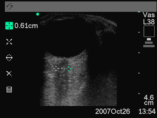
Ultrasound measurement of optic nerve sheath diameter

Here’s the abstract from a new study contributing the literature on ED assessment of raised intracranial pressure using ocular ultrasound:
Background To assess if ultrasound measurement of the optic nerve sheath diameter (ONSD) can accurately predict the presence of raised intracranial pressure (ICP) and acute pathology in patients in the emergency department.
Methods This 3-month prospective observational study used ultrasound to measure the ONSD in adult patients who required CT from the emergency department. The mean ONSD from both eyes was measured using a 7.5 MHz ultrasound probe on closed eyelids. A mean ONSD value of >0.5 cm was taken as positive. Two radiologists independently assessed CT scans from patients in the study population for signs of raised ICP and signs of acute pathology (cerebrovascular accident, subarachnoid, subdural or extradural haemorrhage and tumour). Specificity, sensitivity and k values, for interobserver variability between reporting radiologists, were generated for the study data.

Results In all, 26 patients were enrolled into the study. The ONSD measurement was 100% specific (95% CI 79% to 100%) and 86% sensitive (95% CI 42% to 99%) for raised ICP. For any acute intracranial abnormality the value of ONSD was 100% specific (95% CI 76% to 100%) and 60% sensitive (95% CI 27% to 86%). k Values were 0.91 (95% CIs 0.73 to 1) for identification of raised ICP on CT and 0.84 (95% CIs 0.62 to 1) for any acute pathology on CT, between the radiologists.
Conclusions This study shows that ultrasound measurement of ONSD is sensitive and specific for raised ICP in the emergency department. Further observational studies are needed but this emerging technique could be used to focus treatment in unstable patients.
Ultrasound measurement of optic nerve sheath diameter in patients with a clinical suspicion of raised intracranial pressure
Emerg Med J. 2010 Aug 15. [Epub ahead of print]

Burr holes by emergency physicians

Emergency physicians at Hennepin County Medical Centre (HCMC) are trained in skull trephination (drilling a burr hole) for patients with coma, anisocoria and epidural (extradural) haematoma (EDH) who have not responded to osmotic agents and hyperventilation. This may be particularly applicable in centres remote from neurosurgical centres where delays caused by interfacility transfer are associated with increased morbidity and mortality.
Dr Smith and colleagues from HCMC describe a series of five talk-and-deteriorate patients with EDH who underwent skull trephination. 3 had complete recovery without disability, and 2 others had mild to moderate disability but with good to excellent cognitive function. None had complications from the procedure other than external bleeding from the already lacerated middle meningeal artery. In 4 of 5 cases, the times were recorded. Mean time from ED presentation to trephination was 55 min, and mean time from ED to craniotomy was 173 min. The mean time saved was 118 min, or approximately 2 h.
All trephinations were done by emergency physicians, who had received training in skull trephination as part of the HCMC Emergency Medicine Residency or as part of the Comprehensive Advanced Life Support (CALS) course. Training was very brief and involved discussion of the treatment of EDH, review of a CT scan of EDH, and hands-on practice on the skull of a dead sheep, using the Galt trephinator.

An excellent point made by the authors reminds us that patients with EDH who talk-and-deteriorate (those with the traditionally described “lucid interval”) have minimal primary brain injury and frequently have no brain parenchymal injury. Thus, if the EDH is rapidly decompressed, the outcome is significantly better than for deterioration due to other aetiologies. The authors recommend in EDH that the procedure should be done within 60–90 min of onset of anisocoria. A review of other studies on the procedure would suggest that case selection is critical in defining the appropriateness of the procedure: talk-and-deteriorate, coma, anisocoria, and a delay to neurosurgical decompression.
Emergency Department Skull Trephination for Epidural Hematoma in Patients Who Are Awake But Deteriorate Rapidly
J Emerg Med. 2010 Sep;39(3):377-83

RCT of 7.5% saline in head injury

Over a thousand patients in North America with blunt traumatic head injury and coma who did not have hypovolaemic shock were randomised to different fluids pre-hospital. 250 ml Hypertonic (7.5%) saline was compared with normal (0.9%) saline and hypertonic saline dextran (7.5% saline/6% dextran 70). There was no difference in 6-month neurologic outcome or survival.

Out-of-Hospital Hypertonic Resuscitation Following Severe Traumatic Brain Injury
JAMA. 2010;304(13):1455-1464.

No benefit from pre-hospital trauma doctor in Holland

Being human, I suffer from confirmation bias: I’ve become aware that I’m always on the look out for studies that show benefit from physician-provided pre-hospital care and therefore it’s possible I miss the ones that show no benefit. Of course, no ‘level 1’ evidence is out there yet. This study isn’t hugely impressive, but worth adding to the list. After adjusting for injury severity, trauma patients treated on scene by Dutch physicians had no difference in mortality compared with those that received standard care. In the subgroup analysis for patients with severe traumatic brain injury, the mortality rate with physician involvement was lower than that without, but was not statistically significant. On scene times averaged 2.7 minutes longer in the physician group although factors that might have contributed to this, such as entrapment or on scene interventions, were not recorded.

A Dutch Doctor

A major limitation in study design is that patients who died while under care at the scene or during transport were excluded from the analysis. The on scene time in these patients could have been prolonged by medical interventions in the field possibly contributing to the adverse outcome.
Take home message? More evidence needed.
The Association of Mobile Medical Team Involvement on On-Scene Times and Mortality in Trauma Patients
J Trauma. 2010 Sep;69(3):589-94