Tag Archives: anaesthesia

Transtracheal airways in kids. Well, pigs' kids anyway

Ever had to do a surgical airway in a child? Thought not. They’re pretty rare. Bill Heegaard MD from Henepin County Medical Center taught me a few approaches (with the help of an anaesthetised rabbit) which really got me thinking. It’s something I’d often trained for in my internal simulator, and I even keep the equipment for it in my house (listen out for an upcoming podcast on that). Research and experience has demonstrated that open surgical airway techniques are more reliable than transtracheal needle techniques in adults, but what about kids, in whom traditional teaching cautions against open techniques?

Australian investigators who were experienced airway proceduralists evaluated transtracheal needle techniques using a rabbit model (an excellent model for the infant airway). Their success rate was only 60% and they perforated the posterior tracheal wall in 42% of attempts. Of 13 attempts to insert a dedicated paediatric tracheotomy device, the Quicktrach Child, none were successful(1) (they did not use the Quicktrach Infant model as it is not available in Australia).

Danish investigators used fresh piglet cadavers weighing around 8 kg to assess two transtracheal cannulas, in which they achieved success rates of 65.6% and 68.8%(2). There was also a very high rate of posterior tracheal wall perforation. Using an open surgical tracheostomy technique, they were successful in 97% of attempts. These were also experienced operators, with a median anaesthetic experience of 12.5 years.
Their tracheotomy technique was nice and simple, and used just a scalpel, scissors, and surgical towel clips. Here’s their technique:

Simple tracheotomy procedure described by Holm-Knudsen et al

  1. Identify larynx and proximal trachea by palpation
  2. Vertical incision through the skin and subcutaneous tissue from the upper part of larynx to the sternal notch
  3. Grasp strap muscles with two towel forceps and separate in the midline
  4. Palpate and identify the trachea (palpate rather than look for tracheal rings, as in a live patient one would expect bleeding to obscure the view)
  5. Stabilise the trachea by grasping it with a towel forceps
  6. Insert sharp tip of the scissors between two tracheal rings and lift the trachea anteriorly to avoid damage to the posterior wall
  7. Cut vertically in the midline of the trachea with the scissors – they chose to use the scissors to cut the tracheal rings to facilitate tube insertion
  8. Insert the tracheal tube

Using ultrasound and CT to evaluate comparative airway dimensions, the authors concluded that the pig model is most useful for training emergency airway management in older children aged 5–10 years.
Why were they doing a tracheotomy rather than a cricothyroidotomy? Reasons given by the authors include:

  • The infant cricothyroid membrane is very small
  • Palpation of the thyroid notch may be hindered by the overlying hyoid bone
  • The mandible may obstruct needle access to the cricothyroid membrane given the cephalad position in the neck of the infant larynx.

From an emergency medicine point of view, there are a couple of other reasons why we need to be able to access the trachea lower than the cricothyroid membrane. One is fractured larynx or other blunt or penetrating airway injury where there may be anatomical disruption at the cricothyroid level. The other situation is foreign body airway obstruction, when objects may lodge at the level of the cricoid ring which is functionally the narrowest part of the pediatric upper airway. Of course, alternative methods might be considered to remove the foreign body prior to tracheotomy, such as employing basic choking algorithms, and other techniques depending on whether you do or don’t have equipment.

Take home messages

  • Transtracheal airways in kids are so rare, we can’t avoid extrapolating animal data
  • Whichever infant or paediatric model is used, transtracheal needle techniques have a high rate of failure even by ‘experienced’ operators
  • The small size and easy compressibility of the airway probably contributes to this failure rate, including the high rate of posterior wall puncture
  • In keeping with adult audit data, open surgical techniques may have a higher success rate
  • Tracheotomy may be necessary rather than cricothyroidotomy in infants and children depending on clinical scenario and accessibility of anatomy
  • The stress and blood that is not simulated in cadaveric animal models will make open tracheotomy harder in a live patient, and so these success rates may not translate. However these factors do mean that whatever technique is used must be kept simple and should employ readily available and familiar equipment
  • Something to maintain control and anterior position of the anterior trachea wall should be used during incision and intubation of the trachea. The study reported here used towel clips; sutures around the tracheal rings may also be used (see image below)
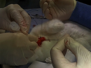
I recommend you add ‘paediatric tracheotomy’ to the list of procedures you might need to do (if it’s not already there). Identify what equipment you would use and run the simulation in your head and in your work environment.
Have fun.

1. The ‘Can’t Intubate Can’t Oxygenate’ scenario in Pediatric Anesthesia: a comparison of different devices for needle cricothyroidotomy
Paediatr Anaesth. 2012 Dec;22(12):1155-8

BACKGROUND: Little evidence exists to guide the management of the ‘Can’t Intubate, Can’t Oxygenate’ (CICO) scenario in pediatric anesthesia.

OBJECTIVES: To compare two intravenous cannulae for ease of use, success rate and complication rate in needle tracheotomy in a postmortem animal model of the infant airway, and trial a commercially available device using the same model.

METHODS: Two experienced proceduralists repeatedly attempted cannula tracheotomy in five postmortem rabbits, alternately using 18-gauge (18G) and 14-gauge (14G) BD Insyte(™) cannulae (BD, Franklin Lakes, NJ, USA). Attempts began at the first tracheal cartilage, with subsequent attempts progressively more caudad. Success was defined as intratracheal cannula placement. In each rabbit, an attempt was then made by each proceduralist to perform a cannula tracheotomy using the Quicktrach Child(™) device (VBM Medizintechnik GmbH, Sulz am Neckar, Germany).

RESULTS: The rabbit tracheas were of similar dimensions to a human infant. 60 attempts were made at cannula tracheotomy, yielding a 60% success rate. There was no significant difference in success rate, ease of use, or complication rate between cannulae of different gauge. Successful aspiration was highly predictive (positive predictive value 97%) and both sensitive (89%) and specific (96%) for tracheal cannulation. The posterior tracheal wall was perforated in 42% of tracheal punctures. None of 13 attempts using the Quicktrach Child(™) were successful.

CONCLUSION: Cannula tracheotomy in a model comparable to the infant airway is difficult and not without complication. Cannulae of 14- and 18-gauge appear to offer similar performance. Successful aspiration is the key predictor of appropriate cannula placement. The Quicktrach Child was not used successfully in this model. Further work is required to compare possible management strategies for the CICO scenario

2. Emergency airway access in children – transtracheal cannulas and tracheotomy assessed in a porcine model
Paediatr Anaesth. 2012 Dec;22(12):1159-65

OBJECTIVES: In the rare scenario when it is impossible to oxygenate or intubate a child, no evidence exists on what strategy to follow.

AIM: The aim of this study was to compare the time and success rate when using two different transtracheal needle techniques and also to measure the success rate and time when performing an emergency tracheotomy in a piglet cadaver model.

METHODS: In this randomized cross-over study, we included 32 anesthesiologists who each inserted two transtracheal cannulas (TTC) using a jet ventilation catheter and an intravenous catheter in a piglet model. Second, they performed an emergency tracheotomy. A maximum of 2 and 4 min were allowed for the procedures, respectively. The TTC procedures were recorded using a video scope.

RESULTS: Placement of a transtracheal cannula was successful in 65.6% and 68.8% of the attempts (P = 0.76), and the median duration of the attempts was 69 and 42 s (P = 0.32), using the jet ventilation catheter and the intravenous catheter, respectively. Complications were frequent in both groups, especially perforation of the posterior tracheal wall. Performing an emergency tracheotomy was successful in 97%, in a median of 88 s.

CONCLUSIONS: In a piglet model, we found no significant difference in success rates or time to insert a jet ventilation cannula or an intravenous catheter transtracheally, but the incidence of complications was high. In the same model, we found a 97% success rate for performing an emergency tracheotomy within 4 min with a low rate of complications.

From BURP to BILP: backwards internal laryngeal pressure

A burns patient whose tracheal tube was accidentally dislodged and ended up placed in the oesophagus on day 2 of his ICU stay continued to spontaneously ventilate and maintain saturations on a midazolam infusion. The oesophageal tube was left in during laryngoscopy (after propofol but no muscle relaxant due to anticipated difficult airway) which revealed a cormack-lehane grade 3 view. The operator’s hand which was holding a bougie rested on the oesophageal tube, which displaced it backwards. This resulted in backwards displacement of the larynx and improved the glottic view to 2b, facilitating intubation.
The discovery of this ‘backwards internal laryngeal pressure’ manoeuvre led the authors to make the recommendation that during difficult intubation an inadvertently placed oesophageal tube should be left in place to allow a BILP manouevre, but removed if it impedes the passage of the tracheal tube.
I love anything that might improve success rates of critical procedures and this one could conceivably come in handy. I can just see Minh Le Cong inventing a transoesophageal posterior laryngal retractor for under 50 bucks…
The use of “Internal Laryngeal Pressure” to improve the laryngeal view following inadvertent oesophageal intubation in a patient with difficult airway
Anaesth Intensive Care. 2012 Jul;40(4):736-7

Size matters when you're sick

A nice example of a difference between elective anaesthesia and critical care practice when it comes to airway management is the selection of appropriate tracheal tube size when intubating, which is highlighted in a recent Anaesthesia article.
In recent years progressively smaller tubes have been used in anaesthesia in pursuit of decreased tracheal injury, sore throat, and hoarseness and increased ease of placement.
Patients likely to remain intubated for some time due to critical illness, however, may benefit from larger diameter tubes for the following reasons:

  • Accumulation of biofilm debris, which increases with duration of intubation – this can significantly decrease the luminal internal diameter, but is less likely to be significant with larger tubes.
  • Work of breathing during weaning: spontaneous breathing trials prior to extubation require patients to breathe through tracheal tubes. Volunteer studies have demonstrated that work of breathing increases as tube diameter decreases.
  • Bronchoscopes and suction catheters: the standard adult ICU fibreoptic bronchoscope has a diameter of 5.7 mm with a 2-mm suction channel to enable adequate suction, which limits the tracheal tube to those larger than 7.5–8.0 mm, and even with an 8.0-mm tube, the bronchoscope occupies more than 50% of the tube diameter, which can lead to ventilation issues during bronchoscopy.

The authors conclude by recommending:


‘If admission to ICU is contemplated then the time-honoured ‘8.0 for females, 9.0 for males’ is a reasonable rule of thumb, unless circumstances dictate otherwise, e.g. in difficult airways or particularly small patients.’

Size matters: choosing the right tracheal tube
Anaesthesia. 2012 Aug;67(8):815-9

Airway lessons relearned


A UK study examined all out-of-operating room intubations over a one month period in nine hospitals1.
Patients whose indication for tracheal intubation was cardiac arrest and who were intubated without the use of drugs were excluded from analysis, as were neonatal intubations.
Disappointing – but not surprising – findings were the lack of universally applied capnography and the use of propofol as the most commonly used induction agent. However more senior intubators were less likely to use propofol than more junior ones (who used it in 93% of intubations!), and the seniors were also more likely to use non-depolarising neuromuscular blocking drugs (NMBDs) than juniors.
The authors report that in seven (4%) patients, pre-oxygenation “was felt to be impossible“. I find it hard to imagine this situation unless RSI is being done on combative patients without prior sedation, which if this is the case makes me shudder.
The authors express their understandable concern over the absence of an alternative airway such as a laryngeal mask in 12% of cases.
Although the adverse event rate seems high, they point out that they used the term ‘adverse events’ rather than ‘complications’ as the events may not be directly attributable to the intubation. In other words, some patients may have been hypoxaemic or hypotensive to start with due to their underlying clinical problem.
I find this study interesting because the results are similar to those reported in a study I and my colleagues conducted a decade ago2, in which ICU intubations were shown to be more hazardous that ED intubations. This can be explained by the higher proportion of patients on ICU with shock and/or respiratory failure. On the other hand, ED patients more commonly required intubation for neurological presentations, with relatively stable cardiorespiratory physiology.
Take a look at the breakdown of cases in the recent study:

and compare this with our findings:

…this is why I have to argue when I hear it occasionally stated that ‘all ED airways are difficult airways’ – some are actually easy, in patients with long stable apnoea times who make great teaching cases.
The authors “speculate that the low rate of hypoxaemia and airway complications may be related to the high proportion of intubations undertaken by those with anaesthesia as a base speciality, and to the almost universal use of NMBDs.” They do not provide strong data to support the first half of their statement. The supplementary data available online indeed show that the majority of intubators were anaesthesia-based, but how their adverse event rates compare with those of the emergency physicians and paediatricians who also undertook intubations is not available.
I don’t want to detract from the important message Dr Bowles and colleagues are conveying: that the lessons from the 4th National Audit Project on major complications of airway management in the UK still need to be applied.
This paper is one aspect of the potentially life-saving work done by this team, which includes the intubation checklist they created.


BACKGROUND: Tracheal intubation is commonly performed outside the operating theatre and is associated with higher risk than intubation in theatre. Recent guidelines and publications including the 4th National Audit Project of the Royal College of Anaesthetists have sought to improve the safety of out-of-theatre intubations.

METHODS: We performed a prospective observational study examining all tracheal intubations occurring outside the operating theatre in nine hospitals over a 1 month period. Data were collected on speciality and grade of intubator, presence of essential safety equipment and monitoring, and adverse events.

RESULTS: One hundred and sixty-four out-of-theatre intubations were identified (excluding those where intubation occurred as part of the management of cardiac arrest). The most common indication for intubation was respiratory failure [74 cases (45%)]. Doctors with at least 6 month’s experience in anaesthesia performed 136 intubations (83%); consultants were present for 68 cases (41%), and overall a second intubator was present for 94 procedures (57%). Propofol was the most common induction agent [124 cases (76%)] and 157 patients (96%) received neuromuscular blocking agents. An airway rescue device was available in 139 cases (87%). Capnography was not used in 52 cases (32%). Sixty-four patients suffered at least one adverse event (39%) around the time of tracheal intubation.

CONCLUSIONS: Out-of-theatre intubation frequently occurs in the absence of essential safety equipment, despite the existing guidelines. The associated adverse event rate is high.

1. Out-of-theatre tracheal intubation: prospective multicentre study of clinical practice and adverse events
Br J Anaesth. 2011 Nov;107(5):687-92


BACKGROUND: Emergency rapid sequence intubation (RSI) performed outside the operating room on emergency patients is the cornerstone of emergency airway management. Complication rates are unknown for this procedure in the United Kingdom and the factors contributing to immediate complications have not been identified.

AIMS: To quantify the immediate complications of RSI and to assess the contribution made by environmental, patient, and physician factors to overall complication rates.

METHODS: Prospective observational study of 208 consecutive adult and paediatric patients undergoing RSI over a six month period.

RESULTS: Patients were successfully intubated by RSI in all cases. There were no deaths during the procedure and no patient required a surgical airway. Patient diagnostic groups requiring RSI are described. Immediate complications were hypoxaemia 19.2%, hypotension 17.8%, and arrhythmia 3.4%. Hypoxaemia was more common in patients with pre-existing respiratory or cardiovascular conditions than in patients with other diagnoses (p<0.01). Emergency department intubations were associated with a significantly lower complication rate than other locations (16.9%; p = 0.004). This can be explained by the difference in diagnostic case mix. Intubating teams comprised anaesthetists, non-anaesthetists, or both. There were no significant differences in complication rates between these groups.
CONCLUSIONS: RSI has a significant immediate complication rate, although the clinical significance of transient events is unknown. The likelihood of immediate complications depends on the patient’s underlying condition, and relevant diagnoses should be emphasised in airway management training. Complication rates are comparable between anaesthetists and non-anaesthetists. The significantly lower complication rates in emergency department RSI can be explained by a larger proportion of patients with comparatively stable cardiorespiratory function.

2. The who, where, and what of rapid sequence intubation: prospective observational study of emergency RSI outside the operating theatre
Emerg Med J. 2004 May;21(3):296-301 Free Full Text

Steroid replacement after etomidate: no benefit

More fuel for the etomidate debate…
In essence:

  • Etomidate has been a useful induction agent for RSI for many years due to its greater haemodynamic stability compared with thiopentone or propofol
  • It is widely used in the USA
  • It inhibits the 11β-hydroxylase enzyme that converts 11β-deoxycortisol into cortisol in the adrenal gland
  • A single dose of etomidate has been demonstrated to inhibit cortisol production for up to 48 hrs
  • This has led to concerns about its use in the critically ill, particular in patients with severe sepsis / septic shock
  • This small study randomised patients receiving etomidate to hydrocortisone or placebo, with no significant difference in these patient-oriented outcomes: duration of mechanical ventilation, intensive care unit length of stay, or 28-day mortality
  • This study suggests that replacement doses of hydrocortisone are not required after a single dose of etomidate
  • No randomised study has conclusively demonstrated increased mortality due to etomidate; however while controversy and the possibility of harm remain, I personally see no reason not to use ketamine for RSI in haemodynamically compromised patients.
  • Ketamine was compared with etomidate in a previous controlled trial


OBJECTIVE: To investigate the effects of moderate-dose hydrocortisone on hemodynamic status in critically ill patients throughout the period of etomidate-related adrenal insufficiency.

DESIGN: Randomized, controlled, double-blind trial (NCT00862381).

SETTING: University hospital emergency department and three intensive care units.

INTERVENTIONS: After single-dose etomidate (H0) for facilitating endotracheal intubation, patients without septic shock were randomly allocated at H6 to receive a 42-hr continuous infusion of either hydrocortisone at 200 mg/day (HC group; n = 49) or saline serum (control group; n = 50).

MEASUREMENTS AND MAIN RESULTS: After completion of a corticotrophin stimulation test, serum cortisol and 11β-deoxycortisol concentrations were subsequently assayed at H6, H12, H24, and H48. Forty-eight patients were analyzed in the HC group and 49 patients in the control group. Before treatment, the diagnostic criteria for etomidate-related adrenal insufficiency were fulfilled in 41 of 45 (91%) and 38 of 45 (84%) patients in the HC and control groups, respectively. The proportion of patients with a cardiovascular Sequential Organ Failure Assessment score of 3 or 4 declined comparably over time in both HC and control groups: 65% vs. 67% at H6, 65% vs. 69% at H12, 44% vs. 54% at H24, and 34% vs. 45% at H48, respectively. Required doses of norepinephrine decreased at a significantly higher rate in the HC group compared with the control group in patients treated with norepinephrine at H6. No intergroup differences were found regarding the duration of mechanical ventilation, intensive care unit length of stay, or 28-day mortality.

CONCLUSION: These findings suggest that critically ill patients without septic shock do not benefit from moderate-dose hydrocortisone administered to overcome etomidate-related adrenal insufficiency.

Corticosteroid after etomidate in critically ill patients: A randomized controlled trial
Crit Care Med. 2012 Jan;40(1):29-35

Preoxygenation and Prevention of Desaturation

This paper is an excellent review article citing the cogent relevant evidence for optimal preoxygenation prior to RSI in the critically ill patient. The evidence has been interpreted with pertinent recommendations by two of the world’s heavy hitters in emergency medicine – Scott Weingart and Rich Levitan. If you can get a full text copy of the paper, laminate Figure 3 (‘Sequence of Preoxygenation and Prevention of Desaturation‘) and stick it to the wall in your resus bay!
The points covered include:

  • Why preoxygenate? Preoxygenation extends the duration of safe apnoea and should be considered mandatory, even in the crashing patient.
  • Standard non-rebreather facemasks set to the highest flow rate of oxygen possible should be used.
  • Allow 8 vital capacity breaths for co-operative patients or 3 minutes for everyone else.
  • Increasing mean airway pressure by CPAP/NIV or PEEP valves improves preoxygenation. However caution should be used in hypovolaemic shocked patients (decreased venous return) and should be reserved for patients who cannot preoxygenate >93-95% with high FiO2.
  • 20-degree head up or reverse Trendelenburg (in suspected trauma) improves pre oxygenation.
  • Apnoeic diffusion oxygenation can extend safe duration of apnoea after the RSI. Set nasal cannulae at 15L/min and leave on during intubation attempts. Ensure upper airway patency (ear to sternal notch and jaw thrust).
  • Active ventilation during onset of muscle relaxation should be assessed on a case by case basis and reserved for patients at high risk of desaturation (6-8 breaths per minute slowly, TV 6-7ml/kg).
  • If there is a high risk of desaturation rocuronium (1.2 mg/kg) may provide a longer duration of safe apnoea than suxamethonium with similar onset time.

Preoxygenation and Prevention of Desaturation During Emergency Airway Management
Ann Emerg Med. 2011 Nov 1. [Epub ahead of print]
[EXPAND Abstract]

Patients requiring emergency airway management are at great risk of hypoxemic hypoxia because of primary lung pathology, high metabolic demands, anemia, insufficient respiratory drive, and inability to protect their airway against aspiration. Tracheal intubation is often required before the complete information needed to assess the risk of periprocedural hypoxia is acquired, such as an arterial blood gas level, hemoglobin value, or even a chest radiograph. This article reviews preoxygenation and peri-intubation oxygenation techniques to minimize the risk of critical hypoxia and introduces a risk-stratification approach to emergency tracheal intubation. Techniques reviewed include positioning, preoxygenation and denitrogenation, positive end expiratory pressure devices, and passive apneic oxygenation.

[/EXPAND]

Oxygen prevented hypoxia. Yep.


Why wouldn’t you give oxygen prophylactically to someone undergoing procedural sedation? One argument is that this will delay the detection of respiratory depression since a pre-oxygenated patient can be hyponoeic/apnoeic for longer prior to desaturation. This is not an issue for those of us who use non-invasive capnography during sedation.
In this randomized trial of oxygen vs air during ED propofol procedural sedation there was less hypoxia when high-flow supplemental oxygen was added. The authors made the following observations:

  • There was no difference between groups in the incidence of respiratory depression, confirming previous research that supplemental oxygen does not exacerbate respiratory depression
  • 5 patients in the compressed air group developed hypoxia without preceding respiratory depression, so capnography cannot be completely relied on in this setting.

They summarise:
“…assuming that capnography is in place to monitor ventilatory function, our results strongly support the routine use of high-flow oxygen during ED propofol sedation”


STUDY OBJECTIVE: We determine whether high-flow oxygen reduces the incidence of hypoxia by 20% in adults receiving propofol for emergency department (ED) sedation compared with room air.

METHODS: We randomized adults to receive 100% oxygen or compressed air at 15 L/minute by nonrebreather mask for 5 minutes before and during propofol procedural sedation. We administered 1.0 mg/kg of propofol, followed by 0.5 mg/kg boluses until the patient was adequately sedated. Physicians and patients were blinded to the gas used. Hypoxia was defined a priori as an oxygen saturation less than 93%; respiratory depression was defined as an end tidal CO(2) greater than 50 mm Hg, a 10% absolute change from baseline, or loss of waveform.

RESULTS: We noted significantly less hypoxia in the 59 patients receiving high-flow oxygen compared with the 58 receiving compressed air (19% versus 41%; P=.007; difference 23%; 95% confidence interval 6% to 38%). Respiratory depression was similar between groups (51% versus 48%; difference 2%; 95% confidence interval -15% to 22%). We observed 2 adverse events in the high-flow group (1 hypotension, 1 bradycardia) and 2 in the compressed air group (1 assisted ventilation, 1 hypotension).

CONCLUSION: High-flow oxygen reduces the frequency of hypoxia during ED propofol sedation in adults.

The Utility of High-Flow Oxygen During Emergency Department Procedural Sedation and Analgesia With Propofol: A Randomized, Controlled Trial
Ann Emerg Med. 2011 Oct;58(4):360-364

Is there nothing ketamine can't do?

As well as the benefits of cardiovascular stability, maintenance of cerebral perfusion pressure, possibly lowering ICP and providing other neuroprotective benefits, ketamine may have other advantages. These are reviewed in a British Journal of Anaesthesia article from which I’ve selected those benefits of interest to practitioners of emergency medicine and critical care.


Additional Beneficial Effects of Ketamine

  • the dysphoric, or ’emergence’ reactions associated with ketamine may be reduced by pre-administration or co-administration of sedatives, such as benzodiazepines, propofol, dexmedetomidine, or droperidol.
  • ketamine potentiates opioid analgesia in multiple settings, reducing opioid total dose and in some groups of patients reducing postoperative desaturation
  • ketamine has possible anti-inflammatory effects demonstrated in some types of surgical patients
  • ketamine may prevent awareness, recall, or both during general anaesthesia

Ketamine: new uses for an old drug?
Br J Anaesth. 2011 Aug;107(2):123-6

Ultrasound to detect difficult laryngoscopy

A pilot study suggests sonographic measurements of neck soft tissue thickness may predict difficult laryngoscopy. Laryngoscopy was difficult in patients with increased thickness of the anterior neck soft tissue at the level of the hyoid bone and thyrohyoid membrane. The authors suggest that anterior neck soft tissue thickness cutoff value of 2.8 cm at the thyrohyoid membrane level can potentially be used to detect difficult laryngoscopy, but that this would require further validation since in this pilot study there were only six subjects in the difficult laryngoscopy group.


Objectives:  Prediction of difficult laryngoscopy in emergency care settings is challenging. The preintubation clinical screening tests may not be applied in a large number of emergency intubations due to the patient’s clinical condition. The objectives of this study were 1) to determine the utility of sonographic measurements of thickness of the tongue, anterior neck soft tissue at the level of the hyoid bone, and thyrohyoid membrane in distinguishing difficult and easy laryngoscopies and 2) to examine the association between sonographic measurements (thickness of tongue and anterior neck soft tissue) and difficult airway clinical screening tests (modified Mallampati score, thyromental distance, and interincisor gap).

Methods:  This was a prospective observational study at an academic medical center. Adult patients undergoing endotracheal intubation for an elective surgical procedure were included. The investigators involved in data collection were blinded to each other’s assessments. Demographic variables were collected preoperatively. The clinical screening tests to predict a difficult airway were performed. The ultrasound (US) measurements of tongue and anterior neck soft tissue were obtained. The laryngoscopic view was graded using Cormack and Lehane classification by anesthesia providers on the day of surgery. To allow for comparisons between difficult airway and easy airway groups, a two-sided Student’s t-test and Fisher’s exact test were employed as appropriate. Spearman’s rank correlation coefficients were used to examine the association between screening tests and sonographic measurements.

Results:  The mean (±standard deviation [SD]) age of 51 eligible patients (32 female, 19 male) was 53.1 (±13.2) years. Six of the 51 patients (12%, 95% confidence interval [CI] = 3% to 20%) were classified as having difficult laryngoscopy by anesthesia providers. The distribution of laryngoscopy grades for all subjects was 63, 25, 4, and 8% for grades 1, 2, 3, and 4, respectively. In this study, 83% of subjects with difficult airways were males. No other significant differences were noted in the demographic variables and difficult airway clinical screening tests between the two groups. The sonographic measurements of anterior neck soft tissue were greater in the difficult laryngoscopy group compared to the easy laryngoscopy group at the level of the hyoid bone (1.69, 95% CI = 1.19 to 2.19 vs. 1.37, 95% CI = 1.27 to 1.46) and thyrohyoid membrane (3.47, 95% CI = 2.88 to 4.07 vs. 2.37, 95% CI = 2.29 to 2.44). No significant correlation was found between sonographic measurements and clinical screening tests.

Conclusions:  This pilot study demonstrated that sonographic measurements of anterior neck soft tissue thickness at the level of hyoid bone and thyrohyoid membrane can be used to distinguish difficult and easy laryngoscopies. Clinical screening tests did not correlate with US measurements, and US was able to detect difficult laryngoscopy, indicating the limitations of the conventional screening tests for predicting difficult laryngoscopy.

Pilot Study to Determine the Utility of Point-of-care Ultrasound in the Assessment of Difficult Laryngoscopy
Acad Emerg Med. 2011 Jul;18(7):754-8

Effect of physician specialty on pre-hospital intubation success

Researchers from the London Helicopter Emergency Medical Service describe the success of pre-hospital laryngoscopy according to the grade and specialty of the HEMS physician…

There is conflicting evidence concerning the role and safety of prehospital intubation, and which providers should deliver it. Success rates for physician-performed rapid sequence induction are reported to be 97-100%, with limited evidence of improved survival in some patient groups. However, there is also evidence that prehospital intubation and ventilation can do harm. Prospective data were recorded on the success of intubation, the quality of the laryngeal view obtained and the number of attempts at intubation. These data were then analysed by the grade of intubating doctor and whether the intubating doctor had a background in anaesthesia or emergency medicine. All groups had a similar success rate after two attempts at intubation. Doctors with a background in anaesthesia and consultant emergency physicians had a significantly better first-pass intubation rate than emergency medicine trainees. The quality of laryngeal view was significantly better in doctors with an anaesthetics background.

Success in physician prehospital rapid sequence intubation: what is the effect of base speciality and length of anaesthetic training?
Emerg Med J. 2011 Mar;28(3):225-9