Tag Archives: airway

Head Rotation for Mask Ventilation

This is a guest post from Dr Per Bredmose, anaesthetist and retrieval medicine physician in Norway, also known as Viking One

I struggle to ventilate the patient in the resus room, airway pressures are high, the bag doesn’t empty properly. In my mind I plan ahead for the next step. Through my mind goes the thought – is this the one, the one that I cannot ventilate? Statistically it is not likely to be, but I am prepared to add two-person technique, airway adjuncts like nasopharyngeal or oropharyngeal, or supraglottic devices that I use frequently in theatre. I feel confident in the use of these methods, and (in the worst case) in cricothyroidotomy. I have practiced that numerous times on our live-tissue course on anaesthetised pigs. However – before I start any of these actions.. I routinely, almost as a reflex from theatre turn the patient’s head 45 degrees to the left, and then the bag suddenly empties easily – and I can ventilate the patient.

Some people think that time with TIVA in theatre has little value for emergency medicine and advanced prehospital care. I strongly disagree. This is some of the most relevant and valuable time I have for keeping and optimising my practical skills. Bag-valve-mask (BVM) ventilation is an essential core skill for any prehospital provider. In theatre this manoeuvre is well known and frequently practiced. It is my impression that this head rotation is less used, and even maybe less well known outside theatre, and especially in the prehospital field. Therefore this is a reminder of an old technique.

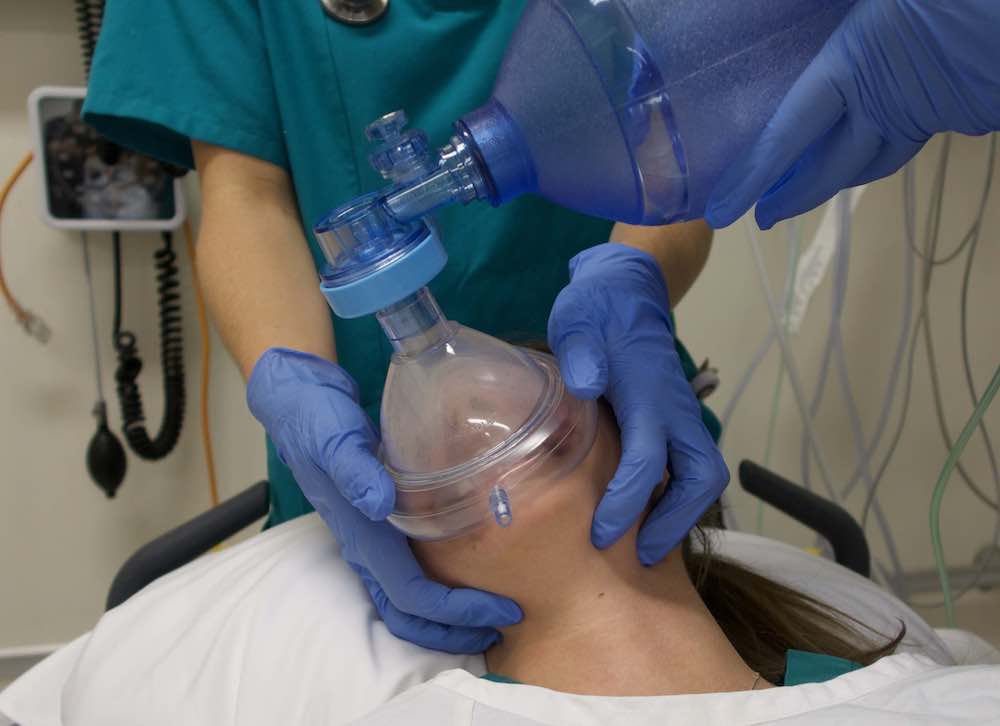
When to do it: When encountering difficulties in conventional BVM ventilation, either when you cannot ventilate or when it’s just difficult to ventilate.

How to do it: Keep a firm one hand grip and gently rotate the head 45 degrees towards the side of the hand with the jaw grip. At the same time, one can try to optimise the one-hand-jaw thrust that goes along with BVM ventilation. Occasionally one needs to extend (dorsiflex) the neck a bit further to fully open the airway. The technique can also be used as a two-person technique, although this is rarely needed.

Opposition: Frequently I hear that I cannot transfer practice from theatre to the prehospital field. Well, this seems to work well in theatre, in ICU and in the field – airways are airways!!


Recently an article in European Journal of Anaesthesiology by Itagaki et al(1) with a cross over design showed an increase in tidal volume when the patients were ventilated in a head rotated position compared to neutral position with the same airway pressure. Their conclusion was as follows: Head rotation of 45° in anaesthetised apnoeic adults significantly increases the efficiency of mask ventilation compared with the neutral head position. Head rotation is an effective alternative to improve mask ventilation if airway obstruction is encountered. Therefore – this is a useful tool that one always should have in the “practical toolbox”. It is not always the solution, but occasionally it saves you (and the patient) a lot of trouble.

Thoughts from Dr Cliff Reid
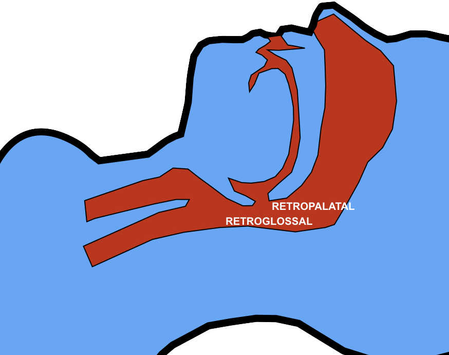
I haven’t used this approach and wasn’t aware of previous research showing an increase in the retroglossal (but not retropalatine) spaces in (awake) patients with head rotation(2).


The mechanism is thought to be gravitational. It is also possible that neck rotation increases upper airway wall tension that reduces collapsibility of the lumen.

In this elegantly designed new study, a two handed BMV technique was used, similar to that advocated in my prehospital & emergency medicine environments. The rotation was always to the right, although the authors comment that they would expect the same results on the left. The increased tidal volume effect with head rotation occurred mostly in younger patients and patients with Mallampati classification I. Such patients are unlikely to be difficult to mask-ventilate, limiting the applicability of these findings to patients who are difficult to ventilate. However having one more option to employ to improve BMV efficacy (after two person technique, optimising ear-to-sternal-notch positioning, and inserting oro- and/or nasopharyngeal airways) may be useful, and the experience and perspective of my anaesthetic colleague Viking One is definitely food for thought. Obviously one should avoid this if there is potential neck injury so I won’t be trying it my trauma patients.


1. Itagaki T, Oto J, Burns SM, Jiang Y, Kacmarek RM, Mountjoy JR. The effect of head rotation on efficiency of face mask ventilation in anaesthetised apnoeic adults. Eur J Anaesthesiol. 2017 Jul;34(7):432–40.

2. Ono T, Otsuka R, Kuroda T, Honda E, Sasaki T. Effects of head and body position on two- and three-dimensional configurations of the upper airway. J Dent Res. 2000 Nov;79(11):1879–84.

Difficult Airway? What Kind Exactly?

crazed-nutter-sm
Mention the term ‘difficult airway’ and many of us will conjure mental images of some kind of distorted anatomy. However, airway management may be ‘difficult’ for a number of reasons, and no internationally agreed definition of the term exists. Given the wrong staff and circumstances, an ‘easy’ airway in your or my hands may indeed become very difficult. In their editorial “The myth of the difficult airway: airway management revisited” (1) Huitink & Bouwman state:

“In our opinion, the ‘difficult airway’ does not exist. It is a complex situational interplay of patient, practitioner, equipment, expertise and circumstances.”

Airways that are anatomically difficult (eg. limited mouth opening, short thyromental distance, large tongue, neck immobility, etc.) and physiologically difficult (hypoxaemia, hypotension, acidosis) are well described among FOAM resources (2-4). In addition to these, a third category of difficulty is well worth considering.

At the smaccDUB conference, intensivist and human factors legend Peter Brindley described three types of difficult airway:

  1. Anatomically difficult
  2. Physiologically difficult
  3. Situationally difficult
    brindleysm
    Brindley = Legend

This last category probably surfaces more commonly than realised, particularly outside the operating room.

Imagine attending a cardiac arrest call on a medical ward. The patient is a 70 year old 120 kg male. The nurses have flattened the bed and discarded the pillow to optimise supine position for CPR. Gobs of vomitus splash from the patient’s pharynx with each compression. The wall suction system is disconnected. There is no bougie in the crash cart’s airway drawer. The nearest capnograph is on another floor of the hospital. In this scenario, no matter how excellent the critical care practitioner’s airway skills, this is a damned difficult airway.

I think Brindley’s third category is a term that should catch on, as a way of helping analyse cases that progress suboptimally and to identify factors during pre-intubation checks that can be addressed. It is terminology that I have added to my own Resuscitese Lexicon, particularly for case discussions during morbidity & mortality and airway audit meetings.

I would like to hear the ‘Situationally Difficult Airway‘ become more widely used, as it fills a gap in how we describe this important area of resuscitation practice.


1. Huitink JM, Bouwman RA. The myth of the difficult airway: airway management revisited. Anaesthesia. 2015 Mar;70(3):244–9. (Full text)

2. LITFL: Airway Assessment

3. EMCrit: HOP Killers

4. PulmCCM: The Physiologically Difficult Airway

Difficult airways can't be reliably predicted

This paper1 proves what Rich Levitan has been saying (and writing) for years – that there is no method of prediction of difficult intubation that is both highly sensitive (the test wouldn’t miss many difficult airways) and highly specific (meaning those predicted to be difficult would indeed turn out to be difficult). Most importantly, this means one should always have a plan for failure to intubate and failure to mask-ventilate regardless of how ‘easy’ the airway may appear.
This study of a large prospectively collected database captured anaesthetists’ clinical assessment of likelihood of difficult intubation and difficult mask-ventilation, and compared them with actual findings. These studies are always difficult, due in part to the lack of standard definitions of difficult airways, but the take home was clear – the large majority of difficulties were unanticipated and not suspected from pre-operative clinical assessment.
This issue was brilliantly summed up by Yentis in a 2002 Editorial2:
I dare to suggest that attempting to predict difficult intubation is unlikely to be useful – does that mean one shouldn’t do it at all? To this I say no, for there is another important benefit of this ritual: it forces the anaesthetist at least to think about the airway, and for this reason we should encourage our trainees (and ourselves) to continue doing it.”
1. Diagnostic accuracy of anaesthesiologists’ prediction of difficult airway management in daily clinical practice: a cohort study of 188 064 patients registered in the Danish Anaesthesia Database
Anaesthesia. 2014 Dec 16. doi: 10.1111/anae.12955. [Epub ahead of print]
[EXPAND Abstract]

Both the American Society of Anesthesiologists and the UK NAP4 project recommend that an unspecified pre-operative airway assessment be made. However, the choice of assessment is ultimately at the discretion of the individual anaesthesiologist. We retrieved a cohort of 188 064 cases from the Danish Anaesthesia Database, and investigated the diagnostic accuracy of the anaesthesiologists’ predictions of difficult tracheal intubation and difficult mask ventilation. Of 3391 difficult intubations, 3154 (93%) were unanticipated. When difficult intubation was anticipated, 229 of 929 (25%) had an actual difficult intubation. Likewise, difficult mask ventilation was unanticipated in 808 of 857 (94%) cases, and when anticipated (218 cases), difficult mask ventilation actually occurred in 49 (22%) cases. We present a previously unpublished estimate of the accuracy of anaesthesiologists’ prediction of airway management difficulties in daily routine practice. Prediction of airway difficulties remains a challenging task, and our results underline the importance of being constantly prepared for unexpected difficulties.

[/EXPAND]
2. Predicting difficult intubation–worthwhile exercise or pointless ritual?
Anaesthesia. 2002 Feb;57(2):105-9

High flow systems for apnoeic oxygenation

nascaniconApnoeic oxygenation during laryngoscopy via nasal prongs has really taken off in the last couple of years in emergency department RSI, and is associated with decreased desaturation rates in out-of-hospital RSI.
More effective oxygenation and a small amount of PEEP can be provided by high flow nasal cannulae with humidified oxygen (HFNC)
A logical step in the progression of this topic is to consider HFNC for apnoeic oxygenation, and Reuben Strayer wrote about this nearly three years ago.
In a Twitter conversation today, Dr Pete Sherren highlighted a new article describing its use in anaesthesia for patients with difficult airways. This is labelled Transnasal Humidified Rapid-Insufflation Ventilatory Exchange (THRIVE). A reply from Dr Neil Brain points out that when used in kids, the bulkiness of the apparatus may get in the way of bag-mask ventilation (if that becomes necessary).
But does HFNC apnoeic oxygenation confer any advantages over standard nasal cannulae?
In an apnoeic patient, 15l/min via standard cannulae should fill the pharyngeal space with 100% oxygen, and you can’t improve on 100%.
HFNC provide some continuous positive pressure, but this may be cancelled by the necessary mouth opening for laryngoscopy.
One issue with apnoea is of course a rise in carbon dioxide with consequent acidosis. The authors of the THRIVE paper (abstract below) point out that in previous apnoeic oxygenation studies, the rate of rise of carbon dioxide levels was between 0.35 and 0.45 kPa/min (2.7-3.4 mmHg/min), whereas with THRIVE the rise was 0.15 kPa/min (1.1 mmHg/min). They suggest that continuous insufflation with high flow oxygen facilitates oxygenation AND carbon dioxide clearance through gaseous mixing and flushing of the deadspace.
So should we switch from standard nasal cannula to high flow cannulae for apnoeic oxygenation? I think not routinely, but perhaps consider it in patients:
(1) with pressure-dependent oxygenation (eg. ARDS) although I’m not sure any CPAP effect would be sustained during laryngoscopy
and
(2) in patients with significant acidosis in whom a significant rise in carbon dioxide could be detrimental (eg. diabetic ketoacidosis).
I look forward to reading more studies on this, and to hearing from anyone with experience of this technique in the comments section.
Transnasal Humidified Rapid-Insufflation Ventilatory Exchange (THRIVE): a physiological method of increasing apnoea time in patients with difficult airways.
Anaesthesia. 2014 Nov 10. doi: 10.1111/anae.12923. [Epub ahead of print]
[EXPAND Abstract]

Emergency and difficult tracheal intubations are hazardous undertakings where successive laryngoscopy-hypoxaemia-re-oxygenation cycles can escalate to airway loss and the ‘can’t intubate, can’t ventilate’ scenario.
Between 2013 and 2014, we extended the apnoea times of 25 patients with difficult airways who were undergoing general anaesthesia for hypopharyngeal or laryngotracheal surgery. This was achieved through continuous delivery of transnasal high-flow humidified oxygen, initially to provide pre-oxygenation, and continuing as post-oxygenation during intravenous induction of anaesthesia and neuromuscular blockade until a definitive airway was secured. Apnoea time commenced at administration of neuromuscular blockade and ended with commencement of jet ventilation, positive-pressure ventilation or recommencement of spontaneous ventilation. During this time, upper airway patency was maintained with jaw-thrust.
Transnasal Humidified Rapid-Insufflation Ventilatory Exchange (THRIVE) was used in 15 males and 10 females. Mean (SD [range]) age at treatment was 49 (15 [25-81]) years. The median (IQR [range]) Mallampati grade was 3 (2-3 [2-4]) and direct laryngoscopy grade was 3 (3-3 [2-4]). There were 12 obese patients and nine patients were stridulous. The median (IQR [range]) apnoea time was 14 (9-19 [5-65]) min. No patient experienced arterial desaturation < 90%. Mean (SD [range]) post-apnoea end-tidal (and in four patients, arterial) carbon dioxide level was 7.8 (2.4 [4.9-15.3]) kPa. The rate of increase in end-tidal carbon dioxide was 0.15 kPa.min(-1) .
We conclude that THRIVE combines the benefits of ‘classical’ apnoeic oxygenation with continuous positive airway pressure and gaseous exchange through flow-dependent deadspace flushing. It has the potential to transform the practice of anaesthesia by changing the nature of securing a definitive airway in emergency and difficult intubations from a pressured stop-start process to a smooth and unhurried undertaking.

[/EXPAND]

Time to change thinking on 'cricoid pressure'

I don’t like cricoid pressure. Some people do. There is insufficient evidence that it is of any benefit. There is some consistent evidence that it worsens laryngoscopic view.
In my clinical practice of critical care in and out of hospital, I can’t afford to risk delaying the securing of my patients’ airways with a procedure in which in my view the risks of harm outweigh any unproven chance of benefit.
I had erroneously thought after many online ‘debates’ that the critical care community had settled on a compromise – if you want to use it great, just take it off if it’s causing a problem. If you don’t want to use it, then that’s equally fine.
However a Google Plus conversation last week ignited a storm! There was a suggestion that cricoid pressure represented a ‘standard of care’, and that not to use it in a critical care intubation would potentially invite legal proceedings, catalysed by colleagues prepared to testify against those of us who have carefully weighed the balance of evidence and selected what we feel is the best approach for our patients.
I wrote a post to challenge the very thinking that what might be considered a ‘standard of care’ in elective anaesthesia in some guidelines should ever be applied to a critical care airway. I proposed a tongue in cheek change of terminology to emphasis what we know about cricoid pressure in the critically ill: that it can delay intubation, distort and compress the airway, and move rather than compress the oesophagus (although I concede the latter point may be irrelevant in terms of CP’s proposed mechanism).
Some people got upset. I reworded the post and added a big fat disclaimer to avoid any perception of ad hominen attack. I wanted to attack and ridicule the procedure, not its proponents. I still got attacked using some bizarrely offensive comparisons by people you would expect to know better. It got ugly.
The combination of support by some people I hold in very high regard and a currently crazy schedule (I’ve been in the UK for three hours having just travelled from Australia) meant the post stayed up for a while until I could consider the feedback. I still haven’t read it all. But I’ve read enough.
I respect the people I disagree with. I respect absolutely their right to hold different views from my own. But I don’t respect all their views, and I don’t necessarily think people have a right not to be offended by my views. However if the WAY I EXPRESS those views causes UNNECESSARY offence I have to reconsider my message.
The science around cricoid pressure is there in the literature. The arguments that it can acceptably be discarded in critical care are powerful. If we need a new acronym it doesn’t need to be one that can be pronounced and construed in a way different to that which I’d envisaged. As Dr Brent May so insightfully put: ‘You can’t emphasise a syllable on Twitter‘.
I want to thank EVERYONE who provided constructive feedback on and off social media. I apologise unreservedly to anyone offended by the post. It’s gone. The battle against unthinking enforcement of a potentially harmful technique goes on, but the unwitting offence of innocent parties is not an acceptable consequence. I will try to be more intelligent in subsequent debate.
Cliff

Palpating neonatal tracheal tubes

infant-intubate-iconAfter neonatal intubation, the incidence of malposition of the tip of the tracheal tube is fairly high.
A technique was evaluated involving palpation of the tube tip in the suprasternal notch, which in this small study was superior to insertion length based on a weight-based nomogram.
The suprasternal notch was chosen because it anatomically corresponds to vertebral level T2, close to the optimal position at the mid-tracheal point. Correct position on the chest radiograph was defined as any position <0.5 cm above the interclavicular midpoint and more than 1 cm above the carina.
During tracheal tube placement, the tip was gently palpated in the suprasternal notch with the index or little finger of the left hand while holding the body of the tube with the fingers of the right hand. The tube tip was adjusted until the bevelled edge was just palpable in the the suprasternal notch.
Digital palpation of endotracheal tube tip as a method of confirming endotracheal tube position in neonates: an open-label, three-armed randomized controlled trial.
Paediatr Anaesth. 2013 Oct;23(10):934-9
[EXPAND Abstract]


OBJECTIVE: To compare the malposition rates of endotracheal tubes (ETTs) when the insertional length (IL) is determined by a weight-based nomogram versus when IL is determined by palpation of the ETT tip.

DESIGN: Open-label, randomized controlled trial (RCT).

SETTING: Level III neonatal intensive care unit (NICU).

SUBJECTS: All newborn babies admitted in NICU requiring intubation.

INTERVENTIONS: Subjects were randomly allocated to one of three groups, wherein IL was determined by (i) weight-based nomogram alone, (ii) weight-based nomogram combined with suprasternal palpation of ETT tip performed by specially trained neonatology fellows, or (iii) combination of weight-based and suprasternal methods by personnel not specially trained.

PRIMARY OUTCOME: Rate of malposition of ETT as judged on chest X-ray (CXR).

RESULTS: Fifty seven babies were randomized into group 1(n = 15), group 2 (n = 20), and group 3 (n = 22). The proportion of correct ETT placement was highest in group 2, being 66.7%, 83.3%, and 66.7% in groups 1 through 3, respectively (P value = 0.58). No complication was attributable to palpation technique.

CONCLUSION: Suprasternal palpation shows promise as a simple, safe, and teachable method of confirming ETT position in neonates.

[/EXPAND]

London Trauma Conference Day 3


Dr Louisa Chan reports on Day 3 of the London Trauma Conference
There was a jam-packed line up for the Pre-hospital and Air Ambulance Day which was Co-hosted by the Norwegian Air Ambulance Foundation.
 

My highlights were:

HEMS

Dr Rasmus Hesselfeldt works in Denmark where they have a pretty good EMS system with ambulances, RRV’s and PHC doctors. Road conditions are good with the longest travel distance of 114 miles. So would the introduction of a HEMS service improve outcomes? He did an observational study looking at year of data post-trial and compared this with 5 months pre-trial. Trauma patients with ISS > 15 and medical emergencies greater than 30 min by road to the Trauma Centre (TC). Primary endpoint was time to TC, secondary outcomes were number of secondary transfers and 30 day mortality.
Results: Increase in on scene time 20 min vs 28 min, time to hospital increased but time to TC was less – 218 min vs 90 min, reduced mortality, increased direct transfer to TC and fewer secondary transfers.
Full article here: A helicopter emergency medical service may allow faster access to highly specialised care. Dan Med J. 2013 Jul;60(7):A4647
 
Airway
Prof Dan Davis ran through pre-hospital intubation. It seems that this man has spent his life trying to perfect airway management. Peter Rosen was his mentor and imprinted on him that RSI is the cornerstone of airway management.
So surely pre-hospital intubation saves lives. The evidence however begs to differ, or does it? As with all evidence we need to consider the validity of the results and luckily Prof Davis has spent a lot of time thinking through the reasons why there no evidence.
During his research he opened a huge can of worms:
1. Hyperventilation was common – any EtCO2 <30mmHg lead to a doubling in mortality.
2. First pass intubation is great, but not if you let your patient become hypoxic or hypotension or worse still both!
3. Hospital practice had similar issues.
So really the RSI processes he was looking at weren’t great.
The good news is that things have improved and he can now boast higher first pass rates and lower complication rates for his EMS system. His puts this success down to training.
 
 
AIRPORT-LTCThe AIRPORT study was discussed at last years LTC. This year we have the results. 21 HEMS services in 6 countries were involved in the data collection including GSA HEMS. The headline findings are that intubation success rates are high (98%) with a complication rate of 10-12%. The more difficult airways were seen in the non-trauma group. 28.2% patients died (mainly cardiac arrest).
 
 
Matt Thomas reported on REVIVE – a pre-hospital feasibility study looking at airway management in OHCA (I-Gel vs LMA Supreme vs standard care). It was never powered to show a difference in these groups, the main aim was to see if research in this very challenging area was possible. And the answer is YES. The paramedics involved recruited more patients than expected and stuck to the protocol (prob better that docs would have!). A randomised controlled trial to look at the I-Gel vs ETT is planned.
 
(P)REBOA
ReboaLTCFinally, Pre-hospital Resuscitative Endovascular Balloon Occlusion of the Aorta (REBOA) seems eminently possible – Dr Nils Petter Oveland showed us the training manikin they developed for training. Through training on this manikin they achieved an average skin to balloon time of 3.3mins. Animal data supports this procedure as a bridge to definitive care in non compressible haemorrhage.
London HEMS will be starting (P)REBOA in the New Year.
So now it’s stand up science, I’m off for my glass of wine…………….
Check out what they’re saying about the London Trauma Conference on Twitter

Guidelines on prehospital drug-assisted LMA insertion

The UK’s Faculty of Prehospital Care has published a number of consensus guidelines in this month’s EMJ
Dr Minh Le Cong‘s PHARM blog has summaries of three of them:

The final one is the most contentious: Pharmacologically assisted laryngeal mask insertion: a consensus statement(1). Here is the summary:

  1. The PALM technique is an acceptable tool for managing the prehospital airway
  2. The PALM technique is indicated in a rare set of circumstances
  3. The PALM procedure is a rescue technique
  4. The PALM procedure should be checklist driven
  5. At least a second generation SAD should be used
  6. End-tidal CO2 monitoring is mandatory
  7. No preference is expressed for any particular drug
  8. No preference is expressed for any particular dosing regime
  9. Flumazenil is highly unlikely to have a role in managing the PALM patient
  10. The PALM procedure should only be carried out by practitioners of level 7 or above competences
  11. The availability of a trained assistant, familiar with the procedure would be advantageous
  12. The training required to achieve competency in performing the PALM procedure must include in-hospital insertion of SADs, simulation training and training in the transfer of critically ill patients
  13. Data should be collected and collated at a national level for all patients who receive the PALM procedure

They qualify the first point with the statement: The consensus group felt that, in the hands of a specific set of practitioners and in certain circumstances, patients would benefit from the technique. It was recognised that pre-hospital airway management can be very challenging, and deeming the technique unacceptable could deprive patients of a potentially life saving intervention. It was felt that having another tool available to clinicians which could potentially improve patient outcome was important. This was despite the lack of a robust evidence base. It was felt that the technique is indicated in, and should be limited to, a very specific set of circumstances as described below
The publication lists some ‘Organisations represented at the consensus meeting’, which include some commercial training and equipment companies.
It also states that ‘The Royal College of Anaesthetists, although represented at the initial meeting, was unable to support the outcomes agreed by the other represented organisations.
This is a very interesting development. I can see the pros and cons of this. Since practitioners are out there doing PALM anyway, it is in the interests of patients to produce a statement that encourages monitoring, checklists, training, and data collection. To meet all the requirements, one must undergo ‘training in the transfer of critically ill patients’, which would normally necessitate more advanced airway and anaesthesia skills anyway.
A tough one – what would you want if there was no RSI capability but you were hypoxic with trismus and basic airway maneouvres were failing? An all out ban on PALM, or PALM provided by someone trained in surgical airway if it fails (as per the consensus recommendations)?
This and some of the other statements can be downloaded in full at the Faculty of Pre-hospital Care site
1. Pharmacologically assisted laryngeal mask insertion: a consensus statement
Emerg Med J. 2013 Dec;30(12):1073-5

Even the dead exhale CO2

cadaverETCO2iconCardiac arrest patients sometimes have unrecognised oesophageal intubations because clinicians omit capnography, based on the assumption that circulatory arrest leads to an absence of exhaled CO2. This is wrong, and reassuringly the latest ILCOR cardiac arrest guidelines recommend waveform capnography during resuscitation.
Of interest is the fact that even corpses have CO2 in their lungs. While not clinically relevant, this may have value when fresh frozen cadavers are used for airway training, since we might be able to supplement the realism of airway instrumentation with the realism of connecting the capnography adaptor and circuit and seeing confirmation on the monitor.
This preliminary study, completed by my Sydney HEMS colleagues, needs further work, but it’s an interesting area.
Sustained life-like waveform capnography after human cadaveric tracheal intubation
Emerg Med J doi:10.1136/emermed-2013-203105
[EXPAND Abstract]


Introduction Fresh frozen cadavers are effective training models for airway management. We hypothesised that residual carbon dioxide (CO2) in cadaveric lung would be detectable using standard clinical monitoring systems, facilitating detection of tracheal tube placement and further enhancing the fidelity of clinical simulation using a cadaveric model.

Methods The tracheas of two fresh frozen unembalmed cadavers were intubated via direct laryngoscopy. Each tracheal tube was connected to a self-inflating bag and a sidestream CO2 detector. The capnograph display was observed and recorded in high-definition video. The cadavers were hand-ventilated with room air until the capnometer reached zero or the waveform approached baseline.

Results A clear capnographic waveform was produced in both cadavers on the first postintubation expiration, simulating the appearances found in the clinical setting. In cadaver one, a consistent capnographic waveform was produced lasting over 100 s. Maximal end-tidal CO2 was 8.5 kPa (65 mm Hg). In cadaver two, a consistent capnographic waveform was produced lasting over 50 s. Maximal end-tidal CO2 was 5.9 kPa (45 mm Hg).

Conclusions We believe this to be the first work to describe and quantify detectable end-tidal capnography in human cadavers. We have demonstrated that tracheal intubation of fresh frozen cadavers can be confirmed by life-like waveform capnography. This requires further validation in a larger sample size.

[/EXPAND]

The non-intubation checklist

no-sm

Scenario:
A 79 year old previously well female presents with loss of consciousness, having been found unresponsive by her daughter who saw her well one hour previously.
Examination reveals a GCS of E1V2M3 = 6 and reactive pupils with no lateralising signs. She is hypertensive. A VBG reveals a normal glucose and sodium and a pCO2 of 60 mmHg (7.9 kPa).
The emergency physician’s plan is to intubate and get a CT scan of her brain. This is explained to the daughter.
A no-brainer? You’d think so.

A consistent issue that recurs during discussions with UK emergency medicine colleagues is that of having to rely on anaesthesia and/or ICU colleagues for intubation of their patients in the ED. The pain comes not from disagreeing about who does the procedure or what drugs to use, but rather on the decision to intubate.
The refusal to intubate can stall or halt a resuscitation plan, delay care, result in risky transfers to the imaging suite, and even deny potential outcome-improving therapy (for example post-ROSC cooling). It can undermine team leadership and disrupt the team dynamic.
There are often different ways to ‘skin a cat’ and it is frequently helpful to invite the opinion of other critical care specialists. However, it is clear from multiple discussions with frustrated EM colleagues that the decision not to intubate is often influenced by non-clinical factors, most often ICU bed availability. Other times, it appears to be that the ‘gatekeeper’ to airway care (and to ICU beds) does not share the same appreciation of the clinical issues at stake. Examples here include the self-fulfilling pessimism post-ROSC based on inappropriate assignment of predictive value to neurological signs, and the assumption of non-treatable pathology in elderly patients presenting with coma.
The obvious solution to this is that the responsibility for managing the ‘A’ of ABC should not be delegated to non-emergency medicine personnel. Sadly, this is not achievable 24/7 in all UK departments right now for a number of awkward reasons.
So what’s a team leader to do when faced with a colleague’s refusal to intubate? The best approach would be to gently and politely persuade them to change their mind by stating some clinical facts that enable a shared mental model and agreed management plan, and to ensure the most senior available physicians are participating in the discussion.
Sometimes that fails. What next? Here’s a suggestion. This is slightly tongue-in-cheek but take away from it what you will.
It is imperative that the individual declining intubation appreciates the gravity of his or her decision. They must not be under the impression that they’ve done you (and the patient) a favour by giving their opinion after an ‘airway consult’. They have declined a resuscitative intervention requested by the emergency medicine team leader and should appreciate the consequences of this decision and the need to document it as such.
Perhaps say something along the lines of:

I see we haven’t managed to agree on this. We’ll just need you to complete the non-intubation form please for our quality improvement process. This will also help prevent your point being forgotten or misunderstood if we’re unlucky enough to face any complaints or litigation. I can fill it in on your behalf but I suspect you’d want to represent yourself as accurately as possible when documenting such a bold decision

And here’s the form. It is provocative, cheeky, and in no way should really be used in its current form:

nonintubationchecklistsm