Tag Archives: procedures

Hilar Twists & Human Error

An engaging scene from ‘Code Blue‘ demonstrated a Helicopter Emergency Medical Service team managing a patient with major thoracic haemorrhage. They did a right thoracotomy and wanted to clamp the hilum but there was some kit missing from the pack.

Unfortunately, the video is no longer available.

This scene had some great discussion points for prehospital professionals, even if the specific scenario is somewhat unlikely for most people’s practice:

  • Non-compressible haemorrhage is possibly the biggest single clinical challenge when you’re a long way from hospital
  • Agitated friends and family can be disruptive – allocate a rescuer to look after them
  • Having blood products to give is essential
  • Don’t rely on the memory of individuals, who are fallible, to pack your equipment. “I was sure I put them in” didn’t cut it when the team needed forceps to clamp the pulmonary hilum and stop the bleeding. Checklists are the in thing, for good reason.
  • Luckily, you don’t need to clamp the hilum (which is tricky) in massive unilateral thoracic haemorrhage. You can just twist the lung 180 degrees on the hilum so it’s upside down. This can prevent further haemorrhage and air embolism.

What’s a hilar twist then?

The hilar twist manoeuvre, as it’s called, is worth learning if you’re a clinician who is prepared to do resuscitative clamshell thoracotomy for penetrating traumatic cardiac arrest. The clamshell is quick and provides excellent exposure(1) and is preferred to lateral thoracotomy(2).

The primary purpose of clamshell thoracotomy in penetrating traumatic arrest is to relieve cardiac tamponade and control a cardiac wound(3). It is well described and continues to save lives in the prehospital setting(4).

However, sometimes you’ll open the chest and the pericardium will be empty (other than containing the heart of course), and there will be massive haemorrhage on one side of the chest. Although most of these patients will be unsalvageable outside a trauma centre’s operating room, it’s worth trying something once you’ve gone to all the trouble of opening the chest. The hilar twist(5) is probably the best option for the non-surgeon, especially when some muppet’s forgotten to pack a clamp.

In order to make the lung mobile enough to twist, it’s first necessary to cut through the inferior pulmonary ligament. This is also known as simply the pulmonary ligament (because there’s no superior equivalent) and sometimes the inferior hilar ligament. It’s not actually a ligament, but an extension of the parietal pleura extending downwards in a fold from the hilum. Some describe it as hanging down from the hilum like a ‘wizard’s sleeve’, which invariably gets a giggle from some of our trainees from the United Kingdom for some reason.
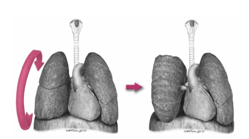
 

After cutting the ligament completely to the level of the inferior pulmonary vein, the lung is then twisted ‘lower lobe towards you’, ie. lower lobe is rotated anteriorly over the upper lobe until the lung is oriented ‘upside down’. The twisted vessels around the hilum become occluded and further haemorrhage from that side should be limited. Other priorities in the arrested patient will be aortic occlusion, internal cardiac massage, and blood products. Packs may be required to keep the lung from untwisting, and if return of spontaneous circulation is achieved, there is a risk of dysrhythmia, right heart failure, and refractory hypoxaemia.

I’ve only done this on pigs and human cadavers so am not speaking from any reassuring level of experience or competence. The literature is out there to read, and it’s up to you to decide how you want to expand or limit your options when you’ve cracked that chest in an arrested patient.

References

1. Flaris AN, Simms ER, Prat N, Reynard F, Caillot J-L, Voiglio EJ. Clamshell incision versus left anterolateral thoracotomy. Which one is faster when performing a resuscitative thoracotomy? The tortoise and the hare revisited. World J Surg. 2015 May;39(5):1306–11.

2. Simms ER, Flaris AN, Franchino X, Thomas MS, Caillot J-L, Voiglio EJ. Bilateral Anterior Thoracotomy (Clamshell Incision) Is the Ideal Emergency Thoracotomy Incision: An Anatomic Study. World J Surg. 2013 Feb 23;37(6):1277–85.

3. Wise D. Emergency thoracotomy: “how to do it.” Emerg Med J. 2005 Jan 1;22(1):22–4.

4. Davies GE, Lockey DJ. Thirteen Survivors of Prehospital Thoracotomy for Penetrating Trauma: A Prehospital Physician-Performed Resuscitation Procedure That Can Yield Good Results. The Journal of Trauma: Injury, Infection, and Critical Care. 2011 May;70(5):E75–8.

5. Wilson A, Wall MJ Jr., Maxson R, Mattox K. The pulmonary hilum twist as a thoracic damage control procedure. The American Journal of Surgery. 2003 Jul;186(1):49–52.

Open cardiac massage in asthmatic arrests?

This idea was provoked by a colleague some years ago who could not achieve a palpable pulse during CPR of an arrested asthmatic child. He wondered whether the severe hyperinflation was rendering external cardiac compressions ineffective and whether he should have done a (prehospital) thoracotomy.
The literature is not strong. The 2010 AHA Guidelines rightly focus on reducing hyperinflation by disconnecting the tracheal tube from the ventilator circuit, and they mention ECMO for refractory cases, but there is no mention of open chest CPR.
I can only find two papers discussing it, both pretty old. A case series in the British Medical Journal from 1968 describes three patients with asthma who had asystolic arrests but did not achieve femoral pulses with external compressions(1). In two, open cardiac massage was performed resulting in restoration of sinus rhythm and cardiac output, and one appeared to make a neurological recovery.
A case report in 1987 describes a 32 year old man in asystolic cardiac arrest due to asthma(2):

“Ventilation required very high inflation pressures and little air movement was heard within the chest despite the administration of Adrenaline 1 mg and Aminophylline 250mg intravenously, and Adrenaline 1mg via the endotracheal tube. This was followed by an intravenous infusion of 100 ml of 8.4% Sodium Bicarbonate solution. External cardiac massage failed to produce a palpable pulse in the carotid area. The chest was, therefore, opened through a left anterolateral thoracotomy. The lungs appeared hyperinflated, bulky and tense and did not collapse when the pleural cavity was opened. The pericardium was opened and asystole confirmed, following eight to ten compressions of the heart some intrinsic activity commenced, ventilation also became much easier.”

He achieved ROSC and became haemodynamically stable but failed to wake up and treatment was withdrawn some days later.
Neither reports include mention of disconnection strategies to reduce hyperinflation. The lack of neurological recovery is not surprising given the apparent prolonged state of arrest the patients were resuscitated from. However there does appear to be a survivor who may not have made it had standard resuscitation (at the time) been continued.
Does this mean I would open the chest in an arrested asthma patient?
Not straight away, no. I would treat dynamic hyperinflation with tube disconnection and external compressions. I would correct absolute and relative hypovolaemia with crystalloid. I would treat bronchospasm (and possible anaphylaxis) with intravenous adrenaline/epinephrine. And I would exclude pneumothorax, possibly with ultrasound or more likely with bilateral open thoracostomies. If however these measures resulted in no detectable carotid flow with external cardiac compressions, ECMO was not available, and the arrest was not prolonged, I would definitely consider doing internal cardiac massage via thoracotomy.
What about you?
1. Grant IW, Kennedy WP, Malone DN
Deaths from asthma
Br Med J. 1968 May 18;2(5602):429–30
2. Diament RH, Sloan JP
Failed resuscitation in acute severe asthma: a medical indication for emergency thoracotomy?
Arch Emerg Med. 1987 Dec;4(4):233–5

Profound hypothermia and no ECMO?

CPRsnow2sm
Patients in cardiac arrest due to severe hypothermia benefit from extracorporeal rewarming, and it is often recommended that they are treated at centres capable of providing cardiopulmonary bypass or extracorporeal membrane oxygenation (ECMO).
But what if they’re brought to a centre that doesn’t have those facilities?
If you work in such a centre do you have a plan, and are you familiar with what equipment you could use?
One option if you have an ICU is to provide extracorporeal warming using a haemofiltration machine used for renal replacement therapy(1). A double lumen haemofiltration catheter is inserted into a central vein and an ICU nurse can often do the rest, although some variables have to be set by the intensivist, often aided by a standard renal replacement therapy prescription chart. The machines are mobile and can be wheeled into the resus room (I have practiced this set up in resus). It might be worth discussing and practicing this option with your ICU.
Another extracorporeal option is to rig up a rapid infusion device such as a ‘Level 1’ to connect to arterial and venous catheters so that blood from the patient flows through and is warmed by the machine before being returned to the patient(2). Rapid rewarming has been achieved by this method but it requires some modification to the usual set up and so is much less likely to be a realistic option for most teams doing this on very rare occasions.
Less technical options are the traditionally taught warm saline lavage of body cavities such as the thorax and the peritoneal cavity. These can be achieved with readily available catheters and of course should be combined with ventilation with warmed gas and administration of warm intravenous fluid.
Thoracic lavage can be achieved with open thoracotomy or tube thoracostomy. One or two chest tubes can be placed on each side. One technique was described as:


Two 36 French chest tubes were placed in each hemithorax. One tube was placed in the fourth intercostal space in the mid-clavicular line. Another tube was placed into the sixth intercostal space in the mid-axillary line. Sterile saline at 39.0◦C was infused by gravity into each superior chest tube and allowed to drain passively through each inferior tube.(3)

Rapid rewarming at a rate of 6.8◦C per hour was achieved in an arrested hypothermic man using peritoneal lavage. It was done in the operating room with peritoneal lavage (saline 40◦C) with a rapid infusion system (Level 1) through two laparoscopic access sites. It was combined with external forced air rewarming and warm intravenous infusions(4).
Finally some devices manufactured for inducing hypothermia in post-cardiac arrest patients can also be used to rewarm patients, which might be endovascular devices, such as the Cool Line® catheter(5), or external, such as the Arctic Sun® Temperature Management System(6). It’s definitely worth finding out what your critical care services have as far as this equipment goes.
In summary, although the ‘exam answer’ for cardiac arrest due to profound hypothermia is often ECMO/cardiopulmonary bypass, in most centres that’s not an option. It’s helpful to remind ourselves that (1) other extracorporeal rewarming options exist and (2) non-extracorporeal techniques can provide rapid rewarming.
 
1. Spooner K, Hassani A. Extracorporeal rewarming in a severely hypothermic patient using venovenous haemofiltration in the accident and emergency department. J Accid Emerg Med. 2000 Nov;17(6):422–4. Full text
2. Gentilello LM, Cobean RA, Offner PJ, Soderberg RW, Jurkovich GJ. Continuous arteriovenous rewarming: rapid reversal of hypothermia in critically ill patients. The Journal of Trauma: Injury, Infection, and Critical Care. 1992 Mar;32(3):316–25 PubMed
3. Plaisier BR. Thoracic lavage in accidental hypothermia with cardiac arrest — report of a case and review of the literature. Resuscitation. 2005 Jul;66(1):99–104. PubMed
4. Gruber E, Beikircher W, Pizzinini R, Marsoner H, Pörnbacher M, Brugger H, et al. Non-extracorporeal rewarming at a rate of 6.8°C per hour in a deeply hypothermic arrested patient. Resuscitation. 2014 Aug;85(8):e119–20. PubMed
5. Kiridume K, Hifumi T, Kawakita K, Okazaki T, Hamaya H, Shinohara N, et al. Clinical experience with an active intravascular rewarming technique for near-severe hypothermia associated with traumatic injury. Journal of Intensive Care. BioMed Central Ltd; 2014;2(1):11. link to abstract
6. Cocchi MN, Giberson B, Donnino MW. Rapid rewarming of hypothermic patient using arctic sun device. Journal of Intensive Care Medicine. 2012 Mar;27(2):128–30. PubMed

Time to change thinking on 'cricoid pressure'

I don’t like cricoid pressure. Some people do. There is insufficient evidence that it is of any benefit. There is some consistent evidence that it worsens laryngoscopic view.
In my clinical practice of critical care in and out of hospital, I can’t afford to risk delaying the securing of my patients’ airways with a procedure in which in my view the risks of harm outweigh any unproven chance of benefit.
I had erroneously thought after many online ‘debates’ that the critical care community had settled on a compromise – if you want to use it great, just take it off if it’s causing a problem. If you don’t want to use it, then that’s equally fine.
However a Google Plus conversation last week ignited a storm! There was a suggestion that cricoid pressure represented a ‘standard of care’, and that not to use it in a critical care intubation would potentially invite legal proceedings, catalysed by colleagues prepared to testify against those of us who have carefully weighed the balance of evidence and selected what we feel is the best approach for our patients.
I wrote a post to challenge the very thinking that what might be considered a ‘standard of care’ in elective anaesthesia in some guidelines should ever be applied to a critical care airway. I proposed a tongue in cheek change of terminology to emphasis what we know about cricoid pressure in the critically ill: that it can delay intubation, distort and compress the airway, and move rather than compress the oesophagus (although I concede the latter point may be irrelevant in terms of CP’s proposed mechanism).
Some people got upset. I reworded the post and added a big fat disclaimer to avoid any perception of ad hominen attack. I wanted to attack and ridicule the procedure, not its proponents. I still got attacked using some bizarrely offensive comparisons by people you would expect to know better. It got ugly.
The combination of support by some people I hold in very high regard and a currently crazy schedule (I’ve been in the UK for three hours having just travelled from Australia) meant the post stayed up for a while until I could consider the feedback. I still haven’t read it all. But I’ve read enough.
I respect the people I disagree with. I respect absolutely their right to hold different views from my own. But I don’t respect all their views, and I don’t necessarily think people have a right not to be offended by my views. However if the WAY I EXPRESS those views causes UNNECESSARY offence I have to reconsider my message.
The science around cricoid pressure is there in the literature. The arguments that it can acceptably be discarded in critical care are powerful. If we need a new acronym it doesn’t need to be one that can be pronounced and construed in a way different to that which I’d envisaged. As Dr Brent May so insightfully put: ‘You can’t emphasise a syllable on Twitter‘.
I want to thank EVERYONE who provided constructive feedback on and off social media. I apologise unreservedly to anyone offended by the post. It’s gone. The battle against unthinking enforcement of a potentially harmful technique goes on, but the unwitting offence of innocent parties is not an acceptable consequence. I will try to be more intelligent in subsequent debate.
Cliff

Palpating neonatal tracheal tubes

infant-intubate-iconAfter neonatal intubation, the incidence of malposition of the tip of the tracheal tube is fairly high.
A technique was evaluated involving palpation of the tube tip in the suprasternal notch, which in this small study was superior to insertion length based on a weight-based nomogram.
The suprasternal notch was chosen because it anatomically corresponds to vertebral level T2, close to the optimal position at the mid-tracheal point. Correct position on the chest radiograph was defined as any position <0.5 cm above the interclavicular midpoint and more than 1 cm above the carina.
During tracheal tube placement, the tip was gently palpated in the suprasternal notch with the index or little finger of the left hand while holding the body of the tube with the fingers of the right hand. The tube tip was adjusted until the bevelled edge was just palpable in the the suprasternal notch.
Digital palpation of endotracheal tube tip as a method of confirming endotracheal tube position in neonates: an open-label, three-armed randomized controlled trial.
Paediatr Anaesth. 2013 Oct;23(10):934-9
[EXPAND Abstract]


OBJECTIVE: To compare the malposition rates of endotracheal tubes (ETTs) when the insertional length (IL) is determined by a weight-based nomogram versus when IL is determined by palpation of the ETT tip.

DESIGN: Open-label, randomized controlled trial (RCT).

SETTING: Level III neonatal intensive care unit (NICU).

SUBJECTS: All newborn babies admitted in NICU requiring intubation.

INTERVENTIONS: Subjects were randomly allocated to one of three groups, wherein IL was determined by (i) weight-based nomogram alone, (ii) weight-based nomogram combined with suprasternal palpation of ETT tip performed by specially trained neonatology fellows, or (iii) combination of weight-based and suprasternal methods by personnel not specially trained.

PRIMARY OUTCOME: Rate of malposition of ETT as judged on chest X-ray (CXR).

RESULTS: Fifty seven babies were randomized into group 1(n = 15), group 2 (n = 20), and group 3 (n = 22). The proportion of correct ETT placement was highest in group 2, being 66.7%, 83.3%, and 66.7% in groups 1 through 3, respectively (P value = 0.58). No complication was attributable to palpation technique.

CONCLUSION: Suprasternal palpation shows promise as a simple, safe, and teachable method of confirming ETT position in neonates.

[/EXPAND]

Guidelines on prehospital drug-assisted LMA insertion

The UK’s Faculty of Prehospital Care has published a number of consensus guidelines in this month’s EMJ
Dr Minh Le Cong‘s PHARM blog has summaries of three of them:

The final one is the most contentious: Pharmacologically assisted laryngeal mask insertion: a consensus statement(1). Here is the summary:

  1. The PALM technique is an acceptable tool for managing the prehospital airway
  2. The PALM technique is indicated in a rare set of circumstances
  3. The PALM procedure is a rescue technique
  4. The PALM procedure should be checklist driven
  5. At least a second generation SAD should be used
  6. End-tidal CO2 monitoring is mandatory
  7. No preference is expressed for any particular drug
  8. No preference is expressed for any particular dosing regime
  9. Flumazenil is highly unlikely to have a role in managing the PALM patient
  10. The PALM procedure should only be carried out by practitioners of level 7 or above competences
  11. The availability of a trained assistant, familiar with the procedure would be advantageous
  12. The training required to achieve competency in performing the PALM procedure must include in-hospital insertion of SADs, simulation training and training in the transfer of critically ill patients
  13. Data should be collected and collated at a national level for all patients who receive the PALM procedure

They qualify the first point with the statement: The consensus group felt that, in the hands of a specific set of practitioners and in certain circumstances, patients would benefit from the technique. It was recognised that pre-hospital airway management can be very challenging, and deeming the technique unacceptable could deprive patients of a potentially life saving intervention. It was felt that having another tool available to clinicians which could potentially improve patient outcome was important. This was despite the lack of a robust evidence base. It was felt that the technique is indicated in, and should be limited to, a very specific set of circumstances as described below
The publication lists some ‘Organisations represented at the consensus meeting’, which include some commercial training and equipment companies.
It also states that ‘The Royal College of Anaesthetists, although represented at the initial meeting, was unable to support the outcomes agreed by the other represented organisations.
This is a very interesting development. I can see the pros and cons of this. Since practitioners are out there doing PALM anyway, it is in the interests of patients to produce a statement that encourages monitoring, checklists, training, and data collection. To meet all the requirements, one must undergo ‘training in the transfer of critically ill patients’, which would normally necessitate more advanced airway and anaesthesia skills anyway.
A tough one – what would you want if there was no RSI capability but you were hypoxic with trismus and basic airway maneouvres were failing? An all out ban on PALM, or PALM provided by someone trained in surgical airway if it fails (as per the consensus recommendations)?
This and some of the other statements can be downloaded in full at the Faculty of Pre-hospital Care site
1. Pharmacologically assisted laryngeal mask insertion: a consensus statement
Emerg Med J. 2013 Dec;30(12):1073-5

Pleurodesis for spontaneous pneumothorax?

A Taiwanese study demonstrated a lower recurrence rate when primary spontaneous pneumothoraces requiring drainage received pleurodesis using minocycline(1). However, significantly more pleurodesed patients required opioid analgesia, and the success rates were less than are found with surgical methods, which are recommended in the West, where pleurodesis is reserved for patients unfit for surgery(2).
1. Simple aspiration and drainage and intrapleural minocycline pleurodesis versus simple aspiration and drainage for the initial treatment of primary spontaneous pneumothorax: an open-label, parallel-group, prospective, randomised, controlled trial
Lancet. 2013 Apr 13;381(9874):1277-82
[EXPAND Abstract]


BACKGROUND: Simple aspiration and drainage is a standard initial treatment for primary spontaneous pneumothorax, but the rate of pneumothorax recurrence is substantial. We investigated whether additional minocycline pleurodesis after simple aspiration and drainage reduces the rate of recurrence.

METHODS: In our open-label, parallel-group, prospective, randomised, controlled trial at two hospitals in Taiwan, patients were aged 15-40 years and had a first episode of primary spontaneous pneumothorax with a rim of air greater than 2 cm on chest radiographs, complete lung expansion without air leakage after pigtail catheter drainage, adequate haematological function, and normal renal and hepatic function. After simple aspiration and drainage via a pigtail catheter, patients were randomly assigned (1:1) to receive 300 mg of minocycline pleurodesis or no further treatment (control group). Randomisation was by computer-generated random numbers in sealed envelopes. Our primary endpoint was rate of pneumothorax recurrence at 1 year. This trial is registered with ClinicalTrials.gov (NCT00418392).

FINDINGS: Between Dec 31, 2006, and June 30, 2012, 214 patients were randomly assigned-106 to the minocycline group and 108 to the control group (intention-to-treat population). Treatment was unsuccessful within 7 days of randomisation in 14 patients in the minocycline group and 20 patients in the control group. At 1 year, pneumothoraces had recurred in 31 of 106 (29·2%) patients in the minocycline group compared with 53 of 108 (49·1%) in the control group (p=0·003). We noted no procedure-related complications in either group.

INTERPRETATION: Simple aspiration and drainage followed by minocycline pleurodesis is a safe and more effective treatment for primary spontaneous pneumothorax than is simple aspiration and drainage only. Minocycline pleurodesis should be an adjunct to standard treatment for primary spontaneous pneumothorax.

[/EXPAND]
2. Primary spontaneous pneumothorax: to pleurodese or not?
Lancet. 2013 Apr 13;381(9874):1252-4

Thenar eminence based medicine

A recent study showed superior effectiveness of one bag-mask ventilation style over another in novice providers. The technique recommended is the thenar eminence grip, in which downward pressure is applied with the thenar eminences while the four fingers of each hand pull the jaw upwards toward the mask.

Interestingly, in their crossover study in which the thenar emininence (TE) technique was compared with the traditionally taught ‘CE’ technique, they demonstrated a ‘sequence effect’. If subjects did TE first, they maintained good tidal volumes when doing CE. However if they did CE first, they achieved poor tidal volumes which were markedly improved when switching to TE.

The authors suggest: “A possible explanation for this sequence effect is that the TE grip is superior. When one used the TE grip first, he or she was more likely to learn how a good tidal volume “feels” and then more likely to apply good technique with the EC grip.“.

Some of us have been practicing and teaching this technique for a while. None have put it better than the brilliant Reuben Strayer of EM Updates in this excellent short video:

Emergency Ventilation in 11 Minutes from reuben strayer on Vimeo.

Efficacy of facemask ventilation techniques in novice providers
J Clin Anesth. 2013 May;25(3):193-7

STUDY OBJECTIVE: To determine which of two facemask grip techniques for two-person facemask ventilation was more effective in novice clinicians, the traditional E-C clamp (EC) grip or a thenar eminence (TE) technique.

DESIGN: Prospective, randomized, crossover comparison study.

SETTING: Operating room of a university hospital.

SUBJECTS: 60 novice clinicians (medical and paramedic students).

MEASUREMENTS: Subjects were assigned to perform, in a random order, each of the two mask-grip techniques on consenting ASA physical status 1, 2, and 3 patients undergoing elective general anesthesia while the ventilator delivered a fixed 500 mL tidal volume (VT). In a crossover manner, subjects performed each facemask ventilation technique (EC and TE) for one minute (12 breaths/min). The primary outcome was the mean expired VT compared between techniques. As a secondary outcome, we examined mean peak inspiratory pressure (PIP).

MAIN RESULTS: The TE grip provided greater expired VT (379 mL vs 269 mL), with a mean difference of 110 mL (P < 0.0001; 95% CI: 65, 157). Using the EC grip first had an average VT improvement of 200 mL after crossover to the TE grip (95% CI: 134, 267). When the TE grip was used first, mean VTs were greater than for EC by 24 mL (95% CI: -25, 74). When considering only the first 12 breaths delivered (prior to crossover), the TE grip resulted in mean VTs of 339 mL vs 221 mL for the EC grip (P = 0.0128; 95% CI: 26, 209). There was no significant difference in PIP values using the two grips: the TE mean (SD) was 14.2 (7.0) cm H2O, and the EC mean (SD) was 13.5 (9.0) cm H2O (P = 0.49).

CONCLUSIONS: The TE facemask ventilation grip results in improved ventilation over the EC grip in the hands of novice providers.

Awake intubation

I had some fun today getting intubated.
We used the Ambu aScope 2 and the Greater Sydney Area HEMS equipment and approach to airway management. I didn’t receive an antisialogogue or any analgesia or sedation.
The big learning point for me was how hard it was to anaesthetise the posterior part of my nasal cavity and nasopharynx. I thought the worst part would be any stimulation of my vocal cords or trachea with lidocaine or instrumentation, but this really was fine. Nebulised 2% lidocaine (the strongest concentration we have), atomised lidocaine (using a mucosal atomiser), and co-phenylcaine spray weren’t sufficient. I can see why people use pastes or gel to maintain mucosal contact while the lidocaine takes effect, but we don’t have those (yet). The best solution came from hooking up oxygen tubing to an iv cannula via a three way tap. Oxygen was run through at 2 l/min and lidocaine injected via the the three way tap. This enabled an atomised spray to be directed right onto the area concerned, and made the insertion of the nasotracheal tube more tolerable – although still unpleasant.
crazed-nutter-sm
The fact I could be intubated awake with reasonable topicalisation suggests most patients should tolerate it perhaps after even an analgesic dose of ketamine, eg. 30-40 mg in an adult. I suspect full dissocation would not be required, which is good for cooperation (“stick your tongue out sir”). I appreciate there are better agents, such as remifentanil or dexmedetomidine, but my area of interest is the retrieval setting – where I have neither the luxury of using these agents nor that of calling for anaesthetic back up.
Thanks to HEMS physicians Emily Stimson, Nirosha De Zoysa, Felicity Day, Chloe Tetlow, and Fergal McCourt for making it fun and safe.
Here’s the video:

Twitter has been helpful in gathering some advice, particularly from @DocJohnHinds:

Another argument for ED thoracotomy

ICM-iconA team from Los Angeles (including the great Kenji Inaba) has published a study on penetrating cardiac wounds in the pediatric population[1]. This is one of the largest studies on this thankfully rare event.
The outcome was poor which may be due to the high proportion of patients arriving at hospital without signs of life (SOL).
What I like about the paper is the discussion of their liberal policy for the use of resuscitative ED thoracotomy:


…we do not rely heavily on prehospital data regarding the precise timing of loss of SOL. Thus, at the discretion of the attending trauma surgeon, every penetrating injury to the chest with SOL lost during patient transport will be considered for ED thoracotomy.

In cases when a perfusing cardiac rhythm is regained, the patient will receive all operative and critical care support as standard of care. If the patient progresses to brain death, aggressive donor management will be implemented in accordance with consent obtained by the organ procurement organization.

In a recent publication, we observed two pediatric patients who underwent ED thoracotomy that subsequently became organ donors after brain death was declared [2]. A total of nine organs were recovered for transplantation. This contemporary outcome measure is of paramount importance in the current era of significant organ shortage.

When such aggressive resuscitative procedures are attempted on arrested trauma patients, there is a temptation to justify inaction on the grounds of futility or the risk of ‘creating a vegetable’. This paper reminds us that other outcome benefits may arise from attempted resuscitation even if the patient does not survive.

These benefits include the saving of other lives through organ donation. In addition to this, there is the opportunity for family members to be with their loved one on the ICU, to hold their warm hand for the last time, to hear the news broken by a team they have gotten to know and trust, to enact any spiritual or religious rites that may provide a source of comfort and closure, and to be there during withdrawal of life sustaining therapies after diagnosis of brain stem death. That will never be pleasant, but on the bleak spectrum of parental torture it may be better than being told the devastating news in the ED relatives’ room by a stranger they’ve never met but will remember forever.

The ED thoracotomy may at the very least remove any doubt that everything that could have been done, was done.
1. Penetrating cardiac trauma in adolescents: A rare injury with excessive mortality
Journal of Pediatric Surgery (2013) 48, 745–749
[EXPAND Abstract]


Background Penetrating cardiac injuries in pediatric patients are rarely encountered. Likewise, the in-hospital outcome measures following these injuries are poorly described.

Methods All pediatric patients (<18years) sustaining penetrating cardiac injuries between 1/2000 and 12/2010 were retrospectively identified using the trauma registry of an urban level I trauma center. Demographic and admission variables, operative findings, and hospital course were extracted. Outpatient follow-up data were obtained through chart reviews and cardiac-specific imaging studies.

Results During the 11-year study period, 32 of the 4569 pediatric trauma admissions (0.7%) sustained penetrating cardiac injuries. All patients were male and the majority suffered stab wounds (81.2%). The mean systolic blood pressure on admission was 28.8±52.9mmHg and the mean ISS was 46.9±27.7. Cardiac chambers involved were the right ventricle (46.9%), the left ventricle (43.8%), and the right atrium (18.8%). Overall, 9 patients (28.1%) survived to hospital discharge. Outpatient follow-up echocardiography was available for 4 patients (44.4%). An abnormal echocardiography result was found in 1 patient, demonstrating hypokinesia and tricuspid regurgitation.

Conclusions Penetrating cardiac trauma is a rare injury in the pediatric population. Cardiac chambers predominantly involved are the right and left ventricles. This injury is associated with a low in-hospital survival (<30%).

[/EXPAND]
2. Organ donation: an important outcome after resuscitative thoracotomy
J Am Coll Surg. 2010 Oct;211(4):450-5
[EXPAND Abstract]


BACKGROUND: The persistent shortage of transplantable organs remains a critical issue around the world. The purpose of this study was to investigate outcomes, including organ procurement, in trauma patients undergoing resuscitative emergency department thoracotomy (EDT). Our hypothesis was that potential organ donor rescue is one of the important outcomes after traumatic arrest and EDT.

STUDY DESIGN: Retrospective study at Los Angeles County and University of Southern California Medical Center. Patients undergoing resuscitative EDT from January 1, 2006 through June 30, 2009 were analyzed. Primary outcomes measures included survival. Secondary outcomes included organ donation and the brain-dead potential organ donor.

RESULTS: During the 42-month study period, a total of 263 patients underwent EDT. Return of a pulse was achieved in 85 patients (32.3%). Of those patients, 37 (43.5%) subsequently died in the operating room and 48 (56.5%) survived to the surgical intensive care unit. Overall, 5 patients (1.9%) survived to discharge and 11 patients (4.2%) became potential organ donors. Five of the 11 potential organ donors had sustained a blunt mechanism injury. Of the 11 potential organ donors, 8 did not donate: 4 families declined consent, 3 because of poor organ function, and 1 expired due to cardiopulmonary collapse. Eventually 11 organs (6 kidneys, 2 livers, 2 pancreases, and 1 small bowel) were harvested from 3 donors. Two of the 3 donors had sustained blunt injury and 1 penetrating mechanism of injury.

CONCLUSIONS: Procurement of organs is one of the tangible outcomes after EDT. These organs have the potential to alter the survival and quality of life of more recipients than the number of survivors of the procedure itself.

[/EXPAND]