Tag Archives: thoracic

Hilar Twists & Human Error

An engaging scene from ‘Code Blue‘ demonstrated a Helicopter Emergency Medical Service team managing a patient with major thoracic haemorrhage. They did a right thoracotomy and wanted to clamp the hilum but there was some kit missing from the pack.

Unfortunately, the video is no longer available.

This scene had some great discussion points for prehospital professionals, even if the specific scenario is somewhat unlikely for most people’s practice:

  • Non-compressible haemorrhage is possibly the biggest single clinical challenge when you’re a long way from hospital
  • Agitated friends and family can be disruptive – allocate a rescuer to look after them
  • Having blood products to give is essential
  • Don’t rely on the memory of individuals, who are fallible, to pack your equipment. “I was sure I put them in” didn’t cut it when the team needed forceps to clamp the pulmonary hilum and stop the bleeding. Checklists are the in thing, for good reason.
  • Luckily, you don’t need to clamp the hilum (which is tricky) in massive unilateral thoracic haemorrhage. You can just twist the lung 180 degrees on the hilum so it’s upside down. This can prevent further haemorrhage and air embolism.

What’s a hilar twist then?

The hilar twist manoeuvre, as it’s called, is worth learning if you’re a clinician who is prepared to do resuscitative clamshell thoracotomy for penetrating traumatic cardiac arrest. The clamshell is quick and provides excellent exposure(1) and is preferred to lateral thoracotomy(2).

The primary purpose of clamshell thoracotomy in penetrating traumatic arrest is to relieve cardiac tamponade and control a cardiac wound(3). It is well described and continues to save lives in the prehospital setting(4).

However, sometimes you’ll open the chest and the pericardium will be empty (other than containing the heart of course), and there will be massive haemorrhage on one side of the chest. Although most of these patients will be unsalvageable outside a trauma centre’s operating room, it’s worth trying something once you’ve gone to all the trouble of opening the chest. The hilar twist(5) is probably the best option for the non-surgeon, especially when some muppet’s forgotten to pack a clamp.

In order to make the lung mobile enough to twist, it’s first necessary to cut through the inferior pulmonary ligament. This is also known as simply the pulmonary ligament (because there’s no superior equivalent) and sometimes the inferior hilar ligament. It’s not actually a ligament, but an extension of the parietal pleura extending downwards in a fold from the hilum. Some describe it as hanging down from the hilum like a ‘wizard’s sleeve’, which invariably gets a giggle from some of our trainees from the United Kingdom for some reason.
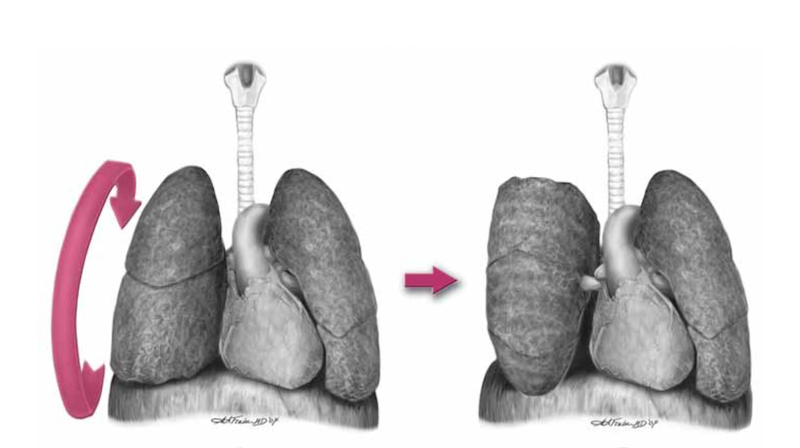
 

After cutting the ligament completely to the level of the inferior pulmonary vein, the lung is then twisted ‘lower lobe towards you’, ie. lower lobe is rotated anteriorly over the upper lobe until the lung is oriented ‘upside down’. The twisted vessels around the hilum become occluded and further haemorrhage from that side should be limited. Other priorities in the arrested patient will be aortic occlusion, internal cardiac massage, and blood products. Packs may be required to keep the lung from untwisting, and if return of spontaneous circulation is achieved, there is a risk of dysrhythmia, right heart failure, and refractory hypoxaemia.

I’ve only done this on pigs and human cadavers so am not speaking from any reassuring level of experience or competence. The literature is out there to read, and it’s up to you to decide how you want to expand or limit your options when you’ve cracked that chest in an arrested patient.

References

1. Flaris AN, Simms ER, Prat N, Reynard F, Caillot J-L, Voiglio EJ. Clamshell incision versus left anterolateral thoracotomy. Which one is faster when performing a resuscitative thoracotomy? The tortoise and the hare revisited. World J Surg. 2015 May;39(5):1306–11.

2. Simms ER, Flaris AN, Franchino X, Thomas MS, Caillot J-L, Voiglio EJ. Bilateral Anterior Thoracotomy (Clamshell Incision) Is the Ideal Emergency Thoracotomy Incision: An Anatomic Study. World J Surg. 2013 Feb 23;37(6):1277–85.

3. Wise D. Emergency thoracotomy: “how to do it.” Emerg Med J. 2005 Jan 1;22(1):22–4.

4. Davies GE, Lockey DJ. Thirteen Survivors of Prehospital Thoracotomy for Penetrating Trauma: A Prehospital Physician-Performed Resuscitation Procedure That Can Yield Good Results. The Journal of Trauma: Injury, Infection, and Critical Care. 2011 May;70(5):E75–8.

5. Wilson A, Wall MJ Jr., Maxson R, Mattox K. The pulmonary hilum twist as a thoracic damage control procedure. The American Journal of Surgery. 2003 Jul;186(1):49–52.

Needle decompression: it's still not going to work

A pet topic that keeps coming up here is management of tension pneumothorax. Plenty of studies demonstrate that traditionally taught needle thoracostomy may fail, and open, or ‘finger’ thoracostomy is recommended for the emergency management of tension pneumothorax in a patient who is being ventilated with positive pressure (including those patients in cardiac arrest).
A recent CT scan-based study of adult trauma patients makes the case that needle decompression with a standard iv cannula would be expected to fail in 42.5% of cases at the second intercostal space (ICS) compared with 16.7% at the fifth ICS at the anterior axillary line (AAL).
The authors add an important point: “As BMI increases, there is a stepwise increase in chest wall thickness, further compounding the difficulty of needle placement in all but the lowest BMI quartile for the second ICS.”
An accompanying editorial cautions that the proximity of the heart may confer a safety issue if a needle is inserted blindly into the left 5th ICS at the AAL.


Objective To compare the distance to be traversed during needle thoracostomy decompression performed at the second intercostal space (ICS) in the midclavicular line (MCL) with the fifth ICS in the anterior axillary line (AAL).

Design Patients were separated into body mass index (BMI) quartiles, with BMI calculated as weight in kilograms divided by height in meters squared. From each BMI quartile, 30 patients were randomly chosen for inclusion in the study on the basis of a priori power analysis (n = 120). Chest wall thickness on computed tomography at the second ICS in the MCL was compared with the fifth ICS in the AAL on both the right and left sides through all BMI quartiles.

Setting Level I trauma center.

Patients Injured patients aged 16 years or older evaluated from January 1, 2009, to January 1, 2010, undergoing computed tomography of the chest.

Results A total of 680 patients met the study inclusion criteria (81.5% were male and mean age was 41 years [range, 16-97 years]). Of the injuries sustained, 13.2% were penetrating, mean (SD) Injury Severity Score was 15.5 (10.3), and mean BMI was 27.9 (5.9) (range, 15.4-60.7). The mean difference in chest wall thickness between the second ICS at the MCL and the fifth ICS at the AAL was 12.9 mm (95% CI, 11.0-14.8; P < .001) on the right and 13.4 mm (95% CI, 11.4-15.3; P < .001) on the left. There was a stepwise increase in chest wall thickness across all BMI quartiles at each location of measurement. There was a significant difference in chest wall thickness between the second ICS at the MCL and the fifth ICS at the AAL in all quartiles on both the right and the left. The percentage of patients with chest wall thickness greater than the standard 5-cm decompression needle was 42.5% at the second ICS in the MCL and only 16.7% at the fifth ICS in the AAL.

Conclusions In this computed tomography–based analysis of chest wall thickness, needle thoracostomy decompression would be expected to fail in 42.5% of cases at the second ICS in the MCL compared with 16.7% at the fifth ICS in the AAL. The chest wall thickness at the fifth ICS AAL was 1.3 cm thinner on average and may be a preferred location for needle thoracostomy decompression.

Radiologic evaluation of alternative sites for needle decompression of tension pneumothorax
Arch Surg. 2012 Sep 1;147(9):813-8

Facilitated pericardial drainage

Drs Wyatt and Haugh describe a modified resuscitative thoracotomy technique which provided surgically facilitated pericardial drainage. A patient with a ruptured pseudoaneurysm of the right ventricular outflow tract presented in shock and arrested in the ED. She had had a prior history of idiopathic ventricular tachycardia and had undergone cardiac ablation of the posteroseptal wall of the right ventricular outflow tract. Sonographically identified tamponade was treated with pericardiocentesis which failed due to clotted blood, so a left lateral thoracotomy was performed by the emergency physician. Rather than fully expose the heart for repair in the ED, they elected to just make a 2cm incision in the pericardium which allowed drainage of blood and restoration of circulation. This was combined with blood product transfusion to buy time for the arrival of a cardiothoracic surgeon and transfer to the operating room.
Useful learning points from this paper are:

  • Ablation procedures are becoming more common
  • Serious complications such as atrioesophageal fistula, pseudoaneurysm, pericardial effusion, and cardiac tamponade occur approximately 3% of the time
  • When tamponade is suspected or confirmed ask patient about recent cardiac procedures such as catheterisations, surgery, and ablation procedures
  • Radiofrequency ablation procedures are often performed on the right side of the heart in areas that may be relatively inaccessible from a left-sided lateral thoracotomy approach.

Modified Emergency Department Thoracotomy for Postablation Cardiac Tamponade
Ann Emerg Med. 2012 Apr;59(4):265-7
[EXPAND Click for abstract]


Cardiac dysrhythmias are a common problem in the United States. Radiofrequency ablation is being used more frequently as a treatment for these diagnoses. Although rare, serious complications such as cardiac tamponade have been reported as a result of ablation procedures. Traditionally, emergency department (ED) thoracotomy has been reserved for cases of traumatic arrest only. We report a case of a successful modified ED thoracotomy in a patient with postablation cardiac tamponade and subsequent obstructive shock who failed intravenous fluid resuscitation, pressor administration, and multiple attempts at pericardiocentesis. In this case, a modified approach was used to incise the pericardium. Although this was associated with large blood loss, we believed that using the traditional method of completely removing the pericardium would have resulted in uncontrolled hemorrhage. Instead, our method led to successful resuscitation of the patient until definitive care was available. A smaller pericardial incision than is traditionally used during ED thoracotomy deserves further consideration and research to determine whether and when it may be most useful as a temporizing treatment of cardiac tamponade when other methods have failed.

[/EXPAND]

Is it time to abandon plain radiography in the trauma room?

For patients who will be having a chest CT, perhaps sonography could replace chest radiography in the resus room as the initial imaging step; this recent prospective study shows its superiority over the ‘traditional’ ATLS approach.

In haemodynamically stable patients with prophylactic pelvic splints in place, one could easily argue against plain pelvis films too (the caveat being rapid access to CT is necessary). The arguments against resus-room lateral cervical spine x-rays were made ages ago and these are now rarely done in the UK & Australia.

Is it time to abandon plain radiography altogether for stable major trauma patients?


Background: The accuracy of combined clinical examination (CE) and chest radiography (CXR) (CE + CXR) vs thoracic ultrasonography in the acute assessment of pneumothorax, hemothorax, and lung contusion in chest trauma patients is unknown.

Methods: We conducted a prospective, observational cohort study involving 119 adult patients admitted to the ED with thoracic trauma. Each patient, secured onto a vacuum mattress, underwent a subsequent thoracic CT scan after first receiving CE, CXR, and thoracic ultrasonography. The diagnostic performance of each method was also evaluated in a subgroup of 35 patients with hemodynamic and/or respiratory instability.

Results: Of the 237 lung fields included in the study, we observed 53 pneumothoraces, 35 hemothoraces, and 147 lung contusions, according to either thoracic CT scan or thoracic decompression if placed before the CT scan. The diagnostic performance of ultrasonography was higher than that of CE + CXR, as shown by their respective areas under the receiver operating characteristic curves (AUC-ROC): mean 0.75 (95% CI, 0.67-0.83) vs 0.62 (0.54-0.70) in pneumothorax cases and 0.73 (0.67-0.80) vs 0.66 (0.61-0.72) for lung contusions, respectively (all P < .05). In addition, the diagnostic performance of ultrasonography to detect pneumothorax was enhanced in the most severely injured patients: 0.86 (0.73-0.98) vs 0.70 (0.61-0.80) with CE + CXR. No difference between modalities was found for hemothorax.
Conclusions: Thoracic ultrasonography as a bedside diagnostic modality is a better diagnostic test than CE and CXR in comparison with CT scanning when evaluating supine chest trauma patients in the emergency setting, particularly for diagnosing pneumothoraces and lung contusions.

Diagnostic accuracy of ultrasonography in the acute assessment of common thoracic lesions after trauma
Chest. 2012 May;141(5):1177-83

Only when I laugh

A middle-aged martial arts enthusiast was training in Krav Maga, and participated in a high-contact punching and grappling sparring exercise in which his (younger, heavier) partner threw him to the ground and landed on him. During the throw the patient felt a ‘pop’ in his right side, and wondered whether he’d fractured a rib. During the subsequent five rounds against two additional sparring partners he noticed a clicking in the same area every time he was grappling, and pain in the right side when pushing up off the floor with his right arm. As a trained emergency physician, he assessed his own level of breathing comfort throughout the training to reassure himself he didn’t have a significant pneumothorax, and therefore elected to continue to fight in the interests of assessing his ability to defend himself while injured.
Pain on deep inspiration, coughing, and squeezing the chest suggested a fractured rib, so out of curiosity at work the next day he ultrasounded the area of maximum tenderness:

Discontinuity in cortex signifies rib fracture

Examination of the lung confirmed pleural sliding, B-lines, and ‘pearls on a string’, which excluded pneumothorax.
Sonography is more sensitive than radiography for the detection of rib fractures and may also detect costochondral junction injuries and disruption of costal cartilage1. This video from Hennepin County Medical Centre takes you through the simple procedure:

Although the management of rib fractures is no different from that of chest wall contusion, knowledge of the presence of fracture in this case is helpful to this patient in deciding when to return to the questionably sane ‘hobby’ of fighting bigger guys half his age.
The patient’s consent was obtained prior to the publication of the ultrasound image.

1. Sonography Compared with Radiography in Revealing Acute Rib Fracture
AJR Am J Roentgenol. 1999 Dec;173(6):1603-9.
Full text article
[EXPAND Click to read abstract]


OBJECTIVE: This study was undertaken to compare the sensitivities of sonography and radiography for revealing acute rib fracture.

SUBJECTS AND METHODS: Chest radiography and rib sonography were performed on 50 patients with suspected rib fractures. Sonography was performed with a 9- or 12-MHz linear transducer. Fractures were identified by a disruption of the anterior margin of the rib, costochondral junction, or costal cartilage. The incidence, location, and degree of displacement of fractures revealed by radiography and sonography were compared. Sonography was performed again after 3 weeks in 37 subjects.

RESULTS: At presentation, radiographs revealed eight rib fractures in six (12%) of 50 patients and sonography revealed 83 rib fractures in 39 (78%) of 50 patients. Seventy-four (89%) of the 83 sonographically detected fractures were located in the rib, four (5%) were located at the costochondral junction, and five (6%) in the costal cartilage. Repeated sonography after 3 weeks showed evidence of healing in all reexamined fractures. Combining sonography at presentation and after 3 weeks, 88% of subjects had sustained a fracture.

CONCLUSION: Sonography reveals more fractures than does radiography and will reveal fractures in most patients presenting with suspected rib fracture. Further scientific studies are needed to clarify the appropriate role for sonography in rib fracture detection.

[/EXPAND]

Posterior pericardiocentesis

Dr Emanuele Catena and colleagues report a case of an adult male who presented 7 days post cardiac surgery with simultaneous pleural and pericardial effucions causing dyspnoea, tachycardia and hypotension.

Old skool pericardiocentesis

His pericardial effusion was posterior which usually requires surgical drainage, but the adjacent left pleural effusion was associated with pulmonary atelectasis and displacement of the lung, allowing them to insert a needle using sonographic guidance first into the pleural space then the pericardial space.
They inserted through the fourth intercostal space 4 cm medially to the left posterior axillary line (with the patient positioned in the semireclining position). They used agitated saline bubbles to confirm first the pleural then the pericardial location of the needle tip. A 30-cm-long catheter was introduced into the posterior pericardium using the Seldinger technique, and serous-haemorrhagic fluid was drained. The catheter was then retracted allowing drainage of the pleural effusion.
The procedure resulted in haemodynamic and respiratory improvement.
The authors summarise:


This case reports the technique of a “back pericardiocentesis” performed under echographic guidance as a valid alternative to surgery in the peculiar situation characterized by the simultaneous presence of a large left pleural effusion. In the presence of a large left pleural effusion, pulmonary atelectasis and displacement of air-filled pulmonary tissue allows ultrasound transmission from a patient’s back to the heart through a liquid interface and needle insertion “from back” to reach the pericardial space.

 
Pericardiocentesis From Back Under Echographic Guidance An Approach for Posterior Pericardial Effusions
Circulation. 2011 Dec 13;124(24):e835-6

Complications after penetrating cardiac injury

Trauma specialists from Arizona and California describe patients with penetrating cardiac wounds, a quarter of whom survive to discharge. Survival post discharge is good, with a range of complications at follow up but no operative intervention was required for the complications.


HYPOTHESIS:
A significant rate of postdischarge complications is associated with penetrating cardiac injuries.

DESIGN: Retrospective trauma registry review.

SETTING: Level I trauma center.

PATIENTS: All patients sustaining penetrating cardiac injuries between January 2000 and June 2010. Patient demographics, clinical data, operative findings, outpatient follow-up, echocardiogram results, and outcomes were extracted.

MAIN OUTCOME MEASURES: Cardiac-related complications and mortality.

RESULTS: During the 10.5-year study period, 406 of 40,706 trauma admissions (1.0%) sustained penetrating cardiac injury. One hundred nine (26.9%) survived to hospital discharge. The survivors were predominantly male (94.4%), with a mean (SD) age of 30.8 (11.7) years, and 74.3% sustained a stab wound. Signs of life were present on admission in 92.6%. Cardiac chambers involved were the right ventricle (45.9%), left ventricle (40.3%), right atrium (10.1%), left atrium (0.9%), and combined (2.8%). In-hospital follow-up was available for a mean (SD) of 11.0 (9.8) days (median, 8 days; range, 3-65 days) and outpatient follow-up was available in 46 patients (42.2%) for a mean (SD) of 1.9 (4.1) months (median, 0.9 months; range, 0.2-12 months). Abnormal echocardiograms demonstrated pericardial effusions (9), abnormal wall motion (8), decreased ejection fraction (<45%) (8), intramural thrombus (4), valve injury (4), cardiac enlargement (2), conduction abnormality (2), pseudoaneurysm (1), aneurysm (1), and septal defect (1). No operative intervention was required for the complications. The 1-year and 9-year survival rates were 97% and 88%, respectively.
CONCLUSIONS: Penetrating cardiac injuries remain highly lethal. A significant rate of cardiac complications can be expected and follow-up echocardiographic evaluation is warranted prior to discharge. The majority of these, however, can be managed without the need for surgical intervention.

Postdischarge Complications After Penetrating Cardiac Injury: a survivable injury with a high postdischarge complication rate
Arch Surg. 2011 Sep;146(9):1061-6

Pre-hospital thoracotomy

The London Helicopter Emergency Medical Service provides a physician / paramedic team to victims of trauma. One of the interventions performed by their physicians is pre-hospital resuscitative thoracotomy to patients with cardiac arrest due to penetrating thoracic trauma. They have published the outcomes from this procedure over a 15 year period which show an 18% survival to discharge rate, with a high rate of neurologically intact survivors1.
The article was submitted for publication on February 1, 2010, and in the discussion mentions a further two survivors from the procedure performed after conducting the study. It is likely therefore in the year and a half since submission still more patients have been saved. It will be interesting to read future reports from this team as the numbers accumulate; penetrating trauma missions are sadly increasing in frequency.
Having worked for these guys and performed this procedure in the field a few times myself, I can attest to the training and governance surrounding this system. The technique of clamshell thoracotomy is well described 2 and one I would recommend for the non-surgeon.

BACKGROUND: Prehospital cardiac arrest associated with trauma almost always results in death. A case of survival after prehospital thoracotomy was published in 1994 and several others have followed. This article describes the result of prehospital thoracotomy in a physician-led system for patients with stab wounds to the chest who suffered cardiac arrest on scene.
METHODS: A 15-year retrospective prehospital trauma database review identified victims of stab wounds to the chest who suffered cardiac arrest on scene and had thoracotomy performed according to local standard operating procedures.
RESULTS: Overall, 71 patients met inclusion criteria. Thirteen patients (18%) survived to hospital discharge. Neurologic outcome was good in 11 patients and poor in 2. Presenting cardiac rhythm was asystole in four patients, pulseless electrical activity in five, and unrecorded in the remaining four. All survivors had cardiac tamponade. The medical team was present at the time of cardiac arrest for six survivors (good neurologic outcome): arrived in the first 5 minutes after arrest in three patients (all good neurologic outcome), arrived 5 minutes to 10 minutes after arrest in two patients (one poor neurologic outcome), and in one patient (poor neurologic outcome) the period was unknown. Of the survivors, seven thoracotomies were performed by emergency physicians and six by anesthesiologists.
CONCLUSIONS: Prehospital thoracotomy is a well-established procedure in this physician-led prehospital service. Results from this and other similar systems suggest that when performed for the subgroup of patients described, significant numbers of survivors with good neurologic outcome can be expected.

1. Thirteen Survivors of Prehospital Thoracotomy for Penetrating Trauma: A Prehospital Physician-Performed Resuscitation Procedure That Can Yield Good Results
J Trauma. 2011 May;70(5):E75-8
2. Emergency thoracotomy: “how to do it”
Emerg Med J. 2005 January; 22(1):22–24
Full text available here

E-FAST for pneumothorax

Some further evidence of the superiority of ultrasound over chest x-ray for the detection of pneumothorax (although it’s not perfect):

INTRODUCTION: Early identification of pneumothorax is crucial to reduce the mortality in critically injured patients. The objective of our study is to investigate the utility of surgeon performed extended focused assessment with sonography for trauma (EFAST) in the diagnosis of pneumothorax.
METHODS: We prospectively analysed 204 trauma patients in our level I trauma center over a period of 12 (06/2007-05/2008) months in whom EFAST was performed. The patients’ demographics, type of injury, clinical examination findings (decreased air entry), CXR, EFAST and CT scan findings were entered into the data base. Sensitivity, specificity, positive (PPV) and negative predictive values (NPV) were calculated.
RESULTS: Of 204 patients (mean age–43.01+/-19.5 years, sex–male 152, female 52) 21 (10.3%) patients had pneumothorax. Of 21 patients who had pneumothorax 12 were due to blunt trauma and 9 were due to penetrating trauma. The diagnosis of pneumothorax in 204 patients demonstrated the following: clinical examination was positive in 17 patients (true positive in 13/21, 62%; 4 were false positive and 8 were false negative), CXR was positive in 16 (true positive in 15/19, 79%; 1 false positive, 4 missed and 2 CXR not performed before chest tube) patients and EFAST was positive in 21 patients (20 were true positive [95.2%], 1 false positive and 1 false negative). In diagnosing pneumothorax EFAST has significantly higher sensitivity compared to the CXR (P=0.02).
CONCLUSIONS: Surgeon performed trauma room extended FAST is simple and has higher sensitivity compared to the chest X-ray and clinical examination in detecting pneumothorax.

Extended focused assessment with sonography for trauma (EFAST) in the diagnosis of pneumothorax: experience at a community based level I trauma center
Injury. 2011 May;42(5):511-4

Limits on resuscitative thoractomy in ED

Eighteen trauma centers contributed ED resuscitative thoracotomy data to a study that commenced enrollment in January 2003. During the ensuing 6 years, 56 patients survived to hospital discharge. Mean age was 31.3; the youngest was a 15-year-old female and the oldest was a 64-year-old male; 93% were male. Injury mechanism was stab wound (SW) in 30 patients, gunshot wound (GSW) in 21 patients, and blunt trauma in 5 patients.
The most common injury was a SW to a ventricle (n =17), accounting for 30% of survivors, followed by a GSW to the lung (n =9) in 16%. There were five survivors (9%) after blunt trauma. Two patients were revived with isolated head trauma who had deteriorated from extensive hemorrhage, one from an open blunt skull fracture (who had 5 minutes of prehospital CPR and left the hospital neurologically intact.) and the other from SWs to the scalp. Two patients also survived with isolated neck injuries: a SW to the vertebral artery and a GSW to the internal carotid artery.
34% of survivors underwent prehospital CPR. Corroborating the reported duration of CPR, the mean base deficit (BD) was 23.3 mequiv/L (range, 14–32 mequiv/L) in those undergoing CPR >5 minutes. In the SW group, the duration was 2 minutes to 10 minutes; the sole survivor after 10 minutes had ventricular wounds with pericardial tamponade. In the GSW group, prehospital CPR was from 1 minute to 15 minutes. The only patient surviving with 15 minutes of CPR also had a ventricular wound with pericardial tamponade but had a moderate neurologic deficit at discharge. In the blunt group, CPR ranged from 3 minutes to 9 minutes; the survivor with 9 minutes of CPR had an atrial rupture with pericardial tamponade.
Seven patients survived with asystole at ED arrival; of significance, all patients had pericardial tamponade. At the time of hospital discharge, three of these patients (43%) had functional neurologic recovery.
The authors state: ‘most recent edition of the ACSCOT advanced trauma life support manual continues to declare “patients sustaining blunt injuries who arrive pulseless but with myocardial electrical activity are not candidates for resuscitative thoracotomy”. But these statements are not congruent with most of the recent literature.

Recommended Limits of Resuscitative Thoracotomy in the ED


BACKGROUND: Since the promulgation of emergency department (ED) thoracotomy >40 years ago, there has been an ongoing search to define when this heroic resuscitative effort is futile. In this era of health care reform, generation of accurate data is imperative for developing patient care guidelines. The purpose of this prospective multicenter study was to identify injury patterns and physiologic profiles at ED arrival that are compatible with survival.
METHODS: Eighteen institutions representing the Western Trauma Association commenced enrollment in January 2003; data were collected prospectively.
RESULTS: During the ensuing 6 years, 56 patients survived to hospital discharge. Mean age was 31.3 years (15-64 years), and 93% were male. As expected, survival was predominant in those with thoracic injuries (77%), followed by abdomen (9%), extremity (7%), neck (4%), and head (4%). The most common injury was a ventricular stab wound (30%), followed by a gunshot wound to the lung (16%); 9% of survivors sustained blunt trauma, 34% underwent prehospital cardiopulmonary resuscitation (CPR), and the presenting base deficit was >25 mequiv/L in 18%. Relevant to futile care, there were survivors of blunt torso injuries with CPR up to 9 minutes and penetrating torso wounds up to 15 minutes. Asystole was documented at ED arrival in seven patients (12%); all these patients had pericardial tamponade and three (43%) had good functional neurologic recovery at hospital discharge.
CONCLUSION: Resuscitative thoracotomy in the ED can be considered futile care when (a) prehospital CPR exceeds 10 minutes after blunt trauma without a response, (b) prehospital CPR exceeds 15 minutes after penetrating trauma without a response, and (c) asystole is the presenting rhythm and there is no pericardial tamponade.
Defining the Limits of Resuscitative Emergency Department Thoracotomy: A Contemporary Western Trauma Association Perspective
J Trauma. 2011 Feb;70(2):334-339.