Tag Archives: thoracotomy

Hilar Twists & Human Error

An engaging scene from ‘Code Blue‘ demonstrated a Helicopter Emergency Medical Service team managing a patient with major thoracic haemorrhage. They did a right thoracotomy and wanted to clamp the hilum but there was some kit missing from the pack.

Unfortunately, the video is no longer available.

This scene had some great discussion points for prehospital professionals, even if the specific scenario is somewhat unlikely for most people’s practice:

  • Non-compressible haemorrhage is possibly the biggest single clinical challenge when you’re a long way from hospital
  • Agitated friends and family can be disruptive – allocate a rescuer to look after them
  • Having blood products to give is essential
  • Don’t rely on the memory of individuals, who are fallible, to pack your equipment. “I was sure I put them in” didn’t cut it when the team needed forceps to clamp the pulmonary hilum and stop the bleeding. Checklists are the in thing, for good reason.
  • Luckily, you don’t need to clamp the hilum (which is tricky) in massive unilateral thoracic haemorrhage. You can just twist the lung 180 degrees on the hilum so it’s upside down. This can prevent further haemorrhage and air embolism.

What’s a hilar twist then?

The hilar twist manoeuvre, as it’s called, is worth learning if you’re a clinician who is prepared to do resuscitative clamshell thoracotomy for penetrating traumatic cardiac arrest. The clamshell is quick and provides excellent exposure(1) and is preferred to lateral thoracotomy(2).

The primary purpose of clamshell thoracotomy in penetrating traumatic arrest is to relieve cardiac tamponade and control a cardiac wound(3). It is well described and continues to save lives in the prehospital setting(4).

However, sometimes you’ll open the chest and the pericardium will be empty (other than containing the heart of course), and there will be massive haemorrhage on one side of the chest. Although most of these patients will be unsalvageable outside a trauma centre’s operating room, it’s worth trying something once you’ve gone to all the trouble of opening the chest. The hilar twist(5) is probably the best option for the non-surgeon, especially when some muppet’s forgotten to pack a clamp.

In order to make the lung mobile enough to twist, it’s first necessary to cut through the inferior pulmonary ligament. This is also known as simply the pulmonary ligament (because there’s no superior equivalent) and sometimes the inferior hilar ligament. It’s not actually a ligament, but an extension of the parietal pleura extending downwards in a fold from the hilum. Some describe it as hanging down from the hilum like a ‘wizard’s sleeve’, which invariably gets a giggle from some of our trainees from the United Kingdom for some reason.
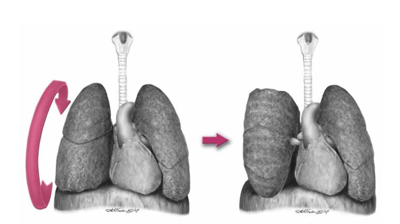
 

After cutting the ligament completely to the level of the inferior pulmonary vein, the lung is then twisted ‘lower lobe towards you’, ie. lower lobe is rotated anteriorly over the upper lobe until the lung is oriented ‘upside down’. The twisted vessels around the hilum become occluded and further haemorrhage from that side should be limited. Other priorities in the arrested patient will be aortic occlusion, internal cardiac massage, and blood products. Packs may be required to keep the lung from untwisting, and if return of spontaneous circulation is achieved, there is a risk of dysrhythmia, right heart failure, and refractory hypoxaemia.

I’ve only done this on pigs and human cadavers so am not speaking from any reassuring level of experience or competence. The literature is out there to read, and it’s up to you to decide how you want to expand or limit your options when you’ve cracked that chest in an arrested patient.

References

1. Flaris AN, Simms ER, Prat N, Reynard F, Caillot J-L, Voiglio EJ. Clamshell incision versus left anterolateral thoracotomy. Which one is faster when performing a resuscitative thoracotomy? The tortoise and the hare revisited. World J Surg. 2015 May;39(5):1306–11.

2. Simms ER, Flaris AN, Franchino X, Thomas MS, Caillot J-L, Voiglio EJ. Bilateral Anterior Thoracotomy (Clamshell Incision) Is the Ideal Emergency Thoracotomy Incision: An Anatomic Study. World J Surg. 2013 Feb 23;37(6):1277–85.

3. Wise D. Emergency thoracotomy: “how to do it.” Emerg Med J. 2005 Jan 1;22(1):22–4.

4. Davies GE, Lockey DJ. Thirteen Survivors of Prehospital Thoracotomy for Penetrating Trauma: A Prehospital Physician-Performed Resuscitation Procedure That Can Yield Good Results. The Journal of Trauma: Injury, Infection, and Critical Care. 2011 May;70(5):E75–8.

5. Wilson A, Wall MJ Jr., Maxson R, Mattox K. The pulmonary hilum twist as a thoracic damage control procedure. The American Journal of Surgery. 2003 Jul;186(1):49–52.

Open cardiac massage in asthmatic arrests?

This idea was provoked by a colleague some years ago who could not achieve a palpable pulse during CPR of an arrested asthmatic child. He wondered whether the severe hyperinflation was rendering external cardiac compressions ineffective and whether he should have done a (prehospital) thoracotomy.
The literature is not strong. The 2010 AHA Guidelines rightly focus on reducing hyperinflation by disconnecting the tracheal tube from the ventilator circuit, and they mention ECMO for refractory cases, but there is no mention of open chest CPR.
I can only find two papers discussing it, both pretty old. A case series in the British Medical Journal from 1968 describes three patients with asthma who had asystolic arrests but did not achieve femoral pulses with external compressions(1). In two, open cardiac massage was performed resulting in restoration of sinus rhythm and cardiac output, and one appeared to make a neurological recovery.
A case report in 1987 describes a 32 year old man in asystolic cardiac arrest due to asthma(2):

“Ventilation required very high inflation pressures and little air movement was heard within the chest despite the administration of Adrenaline 1 mg and Aminophylline 250mg intravenously, and Adrenaline 1mg via the endotracheal tube. This was followed by an intravenous infusion of 100 ml of 8.4% Sodium Bicarbonate solution. External cardiac massage failed to produce a palpable pulse in the carotid area. The chest was, therefore, opened through a left anterolateral thoracotomy. The lungs appeared hyperinflated, bulky and tense and did not collapse when the pleural cavity was opened. The pericardium was opened and asystole confirmed, following eight to ten compressions of the heart some intrinsic activity commenced, ventilation also became much easier.”

He achieved ROSC and became haemodynamically stable but failed to wake up and treatment was withdrawn some days later.
Neither reports include mention of disconnection strategies to reduce hyperinflation. The lack of neurological recovery is not surprising given the apparent prolonged state of arrest the patients were resuscitated from. However there does appear to be a survivor who may not have made it had standard resuscitation (at the time) been continued.
Does this mean I would open the chest in an arrested asthma patient?
Not straight away, no. I would treat dynamic hyperinflation with tube disconnection and external compressions. I would correct absolute and relative hypovolaemia with crystalloid. I would treat bronchospasm (and possible anaphylaxis) with intravenous adrenaline/epinephrine. And I would exclude pneumothorax, possibly with ultrasound or more likely with bilateral open thoracostomies. If however these measures resulted in no detectable carotid flow with external cardiac compressions, ECMO was not available, and the arrest was not prolonged, I would definitely consider doing internal cardiac massage via thoracotomy.
What about you?
1. Grant IW, Kennedy WP, Malone DN
Deaths from asthma
Br Med J. 1968 May 18;2(5602):429–30
2. Diament RH, Sloan JP
Failed resuscitation in acute severe asthma: a medical indication for emergency thoracotomy?
Arch Emerg Med. 1987 Dec;4(4):233–5

Another argument for ED thoracotomy

ICM-iconA team from Los Angeles (including the great Kenji Inaba) has published a study on penetrating cardiac wounds in the pediatric population[1]. This is one of the largest studies on this thankfully rare event.
The outcome was poor which may be due to the high proportion of patients arriving at hospital without signs of life (SOL).
What I like about the paper is the discussion of their liberal policy for the use of resuscitative ED thoracotomy:


…we do not rely heavily on prehospital data regarding the precise timing of loss of SOL. Thus, at the discretion of the attending trauma surgeon, every penetrating injury to the chest with SOL lost during patient transport will be considered for ED thoracotomy.

In cases when a perfusing cardiac rhythm is regained, the patient will receive all operative and critical care support as standard of care. If the patient progresses to brain death, aggressive donor management will be implemented in accordance with consent obtained by the organ procurement organization.

In a recent publication, we observed two pediatric patients who underwent ED thoracotomy that subsequently became organ donors after brain death was declared [2]. A total of nine organs were recovered for transplantation. This contemporary outcome measure is of paramount importance in the current era of significant organ shortage.

When such aggressive resuscitative procedures are attempted on arrested trauma patients, there is a temptation to justify inaction on the grounds of futility or the risk of ‘creating a vegetable’. This paper reminds us that other outcome benefits may arise from attempted resuscitation even if the patient does not survive.

These benefits include the saving of other lives through organ donation. In addition to this, there is the opportunity for family members to be with their loved one on the ICU, to hold their warm hand for the last time, to hear the news broken by a team they have gotten to know and trust, to enact any spiritual or religious rites that may provide a source of comfort and closure, and to be there during withdrawal of life sustaining therapies after diagnosis of brain stem death. That will never be pleasant, but on the bleak spectrum of parental torture it may be better than being told the devastating news in the ED relatives’ room by a stranger they’ve never met but will remember forever.

The ED thoracotomy may at the very least remove any doubt that everything that could have been done, was done.
1. Penetrating cardiac trauma in adolescents: A rare injury with excessive mortality
Journal of Pediatric Surgery (2013) 48, 745–749
[EXPAND Abstract]


Background Penetrating cardiac injuries in pediatric patients are rarely encountered. Likewise, the in-hospital outcome measures following these injuries are poorly described.

Methods All pediatric patients (<18years) sustaining penetrating cardiac injuries between 1/2000 and 12/2010 were retrospectively identified using the trauma registry of an urban level I trauma center. Demographic and admission variables, operative findings, and hospital course were extracted. Outpatient follow-up data were obtained through chart reviews and cardiac-specific imaging studies.

Results During the 11-year study period, 32 of the 4569 pediatric trauma admissions (0.7%) sustained penetrating cardiac injuries. All patients were male and the majority suffered stab wounds (81.2%). The mean systolic blood pressure on admission was 28.8±52.9mmHg and the mean ISS was 46.9±27.7. Cardiac chambers involved were the right ventricle (46.9%), the left ventricle (43.8%), and the right atrium (18.8%). Overall, 9 patients (28.1%) survived to hospital discharge. Outpatient follow-up echocardiography was available for 4 patients (44.4%). An abnormal echocardiography result was found in 1 patient, demonstrating hypokinesia and tricuspid regurgitation.

Conclusions Penetrating cardiac trauma is a rare injury in the pediatric population. Cardiac chambers predominantly involved are the right and left ventricles. This injury is associated with a low in-hospital survival (<30%).

[/EXPAND]
2. Organ donation: an important outcome after resuscitative thoracotomy
J Am Coll Surg. 2010 Oct;211(4):450-5
[EXPAND Abstract]


BACKGROUND: The persistent shortage of transplantable organs remains a critical issue around the world. The purpose of this study was to investigate outcomes, including organ procurement, in trauma patients undergoing resuscitative emergency department thoracotomy (EDT). Our hypothesis was that potential organ donor rescue is one of the important outcomes after traumatic arrest and EDT.

STUDY DESIGN: Retrospective study at Los Angeles County and University of Southern California Medical Center. Patients undergoing resuscitative EDT from January 1, 2006 through June 30, 2009 were analyzed. Primary outcomes measures included survival. Secondary outcomes included organ donation and the brain-dead potential organ donor.

RESULTS: During the 42-month study period, a total of 263 patients underwent EDT. Return of a pulse was achieved in 85 patients (32.3%). Of those patients, 37 (43.5%) subsequently died in the operating room and 48 (56.5%) survived to the surgical intensive care unit. Overall, 5 patients (1.9%) survived to discharge and 11 patients (4.2%) became potential organ donors. Five of the 11 potential organ donors had sustained a blunt mechanism injury. Of the 11 potential organ donors, 8 did not donate: 4 families declined consent, 3 because of poor organ function, and 1 expired due to cardiopulmonary collapse. Eventually 11 organs (6 kidneys, 2 livers, 2 pancreases, and 1 small bowel) were harvested from 3 donors. Two of the 3 donors had sustained blunt injury and 1 penetrating mechanism of injury.

CONCLUSIONS: Procurement of organs is one of the tangible outcomes after EDT. These organs have the potential to alter the survival and quality of life of more recipients than the number of survivors of the procedure itself.

[/EXPAND]

Thoracoabdominal trauma outcomes

In what the authors describe as ‘the largest and most rigorous description of blunt thoracoabdominal injury to date’, we learn some interesting things and are reminded of some others:

Most solid organ injuries in the abdomen are managed non-operatively

Thoracotomy – especially non-resuscitative thoracotomy – is rare

In patients with thoracoabdominal trauma, the overwhelming majority of injuries requiring operative intervention were found in the abdomen, therefore..

…excluding those patients in extremis requiring a resuscitative thoracotomy, the initial incision, without directive radiological information, belongs in the abdomen

Concomitant thoracic injury did not preclude nonoperative management of abdominal solid organ injury

This is a really interesting paper providing important data on the outcomes and management of a patient group that frequently produces management dilemmas in trauma centres.
The double jeopardy of blunt thoracoabdominal trauma
Arch Surg. 2012 Jun;147(6):498-504
[EXPAND Click for abstract]


OBJECTIVES: To examine the specific injuries, need for operative intervention, and clinical outcomes of patients with blunt thoracoabdominal trauma.

DESIGN: Trauma registry and medical record review.

SETTING: Level I trauma center in Los Angeles, California.

PATIENTS: All patients with thoracoabdominal injuries from January 1996 to December 2010.

MAIN OUTCOME MEASURES: Injuries, incidence and type of operative intervention, clinical outcomes, and risk factors for mortality.

RESULTS: Blunt thoracoabdominal injury occurred in 1661 patients. Overall, 474 (28.5%) required laparotomy, 31 (1.9%) required thoracotomy (excluding resuscitative thoracotomy), and 1146 (69.0%) required no thoracic or abdominal operation. Overall incidence of intraabdominal solid organ injury was 59.7% and hollow viscus injury, 6.0%. Blunt cardiac trauma occurred in 6.3%; major thoracic vessel injury, in 4.6%; and diaphragmatic trauma, in 6.0%. The majority of solid organ injuries were managed nonoperatively (liver, 83.9%; spleen, 68.3%; and kidney, 91.2%). Excluding patients with severe head trauma, mortality ranged from 4.5% with nonoperative management to 18.1% and 66.7% in those requiring laparotomy and dual cavitary exploration, respectively. Age 55 years or older, Injury Severity Score of 25 or more, Glasgow Coma Scale score of 8 or less, initial hypotension, massive transfusion, and liver, cardiac, or abdominal vascular trauma were all independent risk factors for mortality.

CONCLUSIONS: Most patients with blunt thoracoabdominal trauma are managed nonoperatively. The need for non-resuscitative thoracotomy or combined thoracoabdominal operation is rare. The abdomen contains the overwhelming majority of injuries requiring operative intervention and should be the initial cavity of exploration in the patient requiring emergent surgery without directive radiologic data.

[/EXPAND]

Facilitated pericardial drainage

Drs Wyatt and Haugh describe a modified resuscitative thoracotomy technique which provided surgically facilitated pericardial drainage. A patient with a ruptured pseudoaneurysm of the right ventricular outflow tract presented in shock and arrested in the ED. She had had a prior history of idiopathic ventricular tachycardia and had undergone cardiac ablation of the posteroseptal wall of the right ventricular outflow tract. Sonographically identified tamponade was treated with pericardiocentesis which failed due to clotted blood, so a left lateral thoracotomy was performed by the emergency physician. Rather than fully expose the heart for repair in the ED, they elected to just make a 2cm incision in the pericardium which allowed drainage of blood and restoration of circulation. This was combined with blood product transfusion to buy time for the arrival of a cardiothoracic surgeon and transfer to the operating room.
Useful learning points from this paper are:

  • Ablation procedures are becoming more common
  • Serious complications such as atrioesophageal fistula, pseudoaneurysm, pericardial effusion, and cardiac tamponade occur approximately 3% of the time
  • When tamponade is suspected or confirmed ask patient about recent cardiac procedures such as catheterisations, surgery, and ablation procedures
  • Radiofrequency ablation procedures are often performed on the right side of the heart in areas that may be relatively inaccessible from a left-sided lateral thoracotomy approach.

Modified Emergency Department Thoracotomy for Postablation Cardiac Tamponade
Ann Emerg Med. 2012 Apr;59(4):265-7
[EXPAND Click for abstract]


Cardiac dysrhythmias are a common problem in the United States. Radiofrequency ablation is being used more frequently as a treatment for these diagnoses. Although rare, serious complications such as cardiac tamponade have been reported as a result of ablation procedures. Traditionally, emergency department (ED) thoracotomy has been reserved for cases of traumatic arrest only. We report a case of a successful modified ED thoracotomy in a patient with postablation cardiac tamponade and subsequent obstructive shock who failed intravenous fluid resuscitation, pressor administration, and multiple attempts at pericardiocentesis. In this case, a modified approach was used to incise the pericardium. Although this was associated with large blood loss, we believed that using the traditional method of completely removing the pericardium would have resulted in uncontrolled hemorrhage. Instead, our method led to successful resuscitation of the patient until definitive care was available. A smaller pericardial incision than is traditionally used during ED thoracotomy deserves further consideration and research to determine whether and when it may be most useful as a temporizing treatment of cardiac tamponade when other methods have failed.

[/EXPAND]

Modified ED thoracotomy to avoid exsanguination

Emergency physicians from Minnesota (and graduates of the amazing Hennepin Emergency Medicine Residency) describe a patient who developed cardiac tamponade after an ablation procedure for dysrhythmia. Attempts at pericardiocentesis by both emergency and cardiology staff were hindered by clotted blood, and so a left lateral thoracotomy was performed in the ED due to loss of pulse and lack of immediate availability of an operating room. A limited pericardial incision was made to allow drainage of sufficient blood to relieve tamponade while avoiding catastrophic blood loss from the underlying lesion, which turned out to be a 1.5-cm hole in the right ventricular outflow tract. The patient made a full recovery.

Cardiac dysrhythmias are a common problem in the United States. Radiofrequency ablation is being used more frequently as a treatment for these diagnoses. Although rare, serious complications such as cardiac tamponade have been reported as a result of ablation procedures. Traditionally, emergency department (ED) thoracotomy has been reserved for cases of traumatic arrest only. We report a case of a successful modified ED thoracotomy in a patient with postablation cardiac tamponade and subsequent obstructive shock who failed intravenous fluid resuscitation, pressor administration, and multiple attempts at pericardiocentesis. In this case, a modified approach was used to incise the pericardium. Although this was associated with large blood loss, we believed that using the traditional method of completely removing the pericardium would have resulted in uncontrolled hemorrhage. Instead, our method led to successful resuscitation of the patient until definitive care was available. A smaller pericardial incision than is traditionally used during ED thoracotomy deserves further consideration and research to determine whether and when it may be most useful as a temporizing treatment of cardiac tamponade when other methods have failed.

Modified Emergency Department Thoracotomy for Postablation Cardiac Tamponade
Annals of Emergency Medicine In Press – Full Text Available here from Annals site at time of blogging

Pre-hospital thoracotomy

The London Helicopter Emergency Medical Service provides a physician / paramedic team to victims of trauma. One of the interventions performed by their physicians is pre-hospital resuscitative thoracotomy to patients with cardiac arrest due to penetrating thoracic trauma. They have published the outcomes from this procedure over a 15 year period which show an 18% survival to discharge rate, with a high rate of neurologically intact survivors1.
The article was submitted for publication on February 1, 2010, and in the discussion mentions a further two survivors from the procedure performed after conducting the study. It is likely therefore in the year and a half since submission still more patients have been saved. It will be interesting to read future reports from this team as the numbers accumulate; penetrating trauma missions are sadly increasing in frequency.
Having worked for these guys and performed this procedure in the field a few times myself, I can attest to the training and governance surrounding this system. The technique of clamshell thoracotomy is well described 2 and one I would recommend for the non-surgeon.

BACKGROUND: Prehospital cardiac arrest associated with trauma almost always results in death. A case of survival after prehospital thoracotomy was published in 1994 and several others have followed. This article describes the result of prehospital thoracotomy in a physician-led system for patients with stab wounds to the chest who suffered cardiac arrest on scene.
METHODS: A 15-year retrospective prehospital trauma database review identified victims of stab wounds to the chest who suffered cardiac arrest on scene and had thoracotomy performed according to local standard operating procedures.
RESULTS: Overall, 71 patients met inclusion criteria. Thirteen patients (18%) survived to hospital discharge. Neurologic outcome was good in 11 patients and poor in 2. Presenting cardiac rhythm was asystole in four patients, pulseless electrical activity in five, and unrecorded in the remaining four. All survivors had cardiac tamponade. The medical team was present at the time of cardiac arrest for six survivors (good neurologic outcome): arrived in the first 5 minutes after arrest in three patients (all good neurologic outcome), arrived 5 minutes to 10 minutes after arrest in two patients (one poor neurologic outcome), and in one patient (poor neurologic outcome) the period was unknown. Of the survivors, seven thoracotomies were performed by emergency physicians and six by anesthesiologists.
CONCLUSIONS: Prehospital thoracotomy is a well-established procedure in this physician-led prehospital service. Results from this and other similar systems suggest that when performed for the subgroup of patients described, significant numbers of survivors with good neurologic outcome can be expected.

1. Thirteen Survivors of Prehospital Thoracotomy for Penetrating Trauma: A Prehospital Physician-Performed Resuscitation Procedure That Can Yield Good Results
J Trauma. 2011 May;70(5):E75-8
2. Emergency thoracotomy: “how to do it”
Emerg Med J. 2005 January; 22(1):22–24
Full text available here

Limits on resuscitative thoractomy in ED

Eighteen trauma centers contributed ED resuscitative thoracotomy data to a study that commenced enrollment in January 2003. During the ensuing 6 years, 56 patients survived to hospital discharge. Mean age was 31.3; the youngest was a 15-year-old female and the oldest was a 64-year-old male; 93% were male. Injury mechanism was stab wound (SW) in 30 patients, gunshot wound (GSW) in 21 patients, and blunt trauma in 5 patients.
The most common injury was a SW to a ventricle (n =17), accounting for 30% of survivors, followed by a GSW to the lung (n =9) in 16%. There were five survivors (9%) after blunt trauma. Two patients were revived with isolated head trauma who had deteriorated from extensive hemorrhage, one from an open blunt skull fracture (who had 5 minutes of prehospital CPR and left the hospital neurologically intact.) and the other from SWs to the scalp. Two patients also survived with isolated neck injuries: a SW to the vertebral artery and a GSW to the internal carotid artery.
34% of survivors underwent prehospital CPR. Corroborating the reported duration of CPR, the mean base deficit (BD) was 23.3 mequiv/L (range, 14–32 mequiv/L) in those undergoing CPR >5 minutes. In the SW group, the duration was 2 minutes to 10 minutes; the sole survivor after 10 minutes had ventricular wounds with pericardial tamponade. In the GSW group, prehospital CPR was from 1 minute to 15 minutes. The only patient surviving with 15 minutes of CPR also had a ventricular wound with pericardial tamponade but had a moderate neurologic deficit at discharge. In the blunt group, CPR ranged from 3 minutes to 9 minutes; the survivor with 9 minutes of CPR had an atrial rupture with pericardial tamponade.
Seven patients survived with asystole at ED arrival; of significance, all patients had pericardial tamponade. At the time of hospital discharge, three of these patients (43%) had functional neurologic recovery.
The authors state: ‘most recent edition of the ACSCOT advanced trauma life support manual continues to declare “patients sustaining blunt injuries who arrive pulseless but with myocardial electrical activity are not candidates for resuscitative thoracotomy”. But these statements are not congruent with most of the recent literature.

Recommended Limits of Resuscitative Thoracotomy in the ED


BACKGROUND: Since the promulgation of emergency department (ED) thoracotomy >40 years ago, there has been an ongoing search to define when this heroic resuscitative effort is futile. In this era of health care reform, generation of accurate data is imperative for developing patient care guidelines. The purpose of this prospective multicenter study was to identify injury patterns and physiologic profiles at ED arrival that are compatible with survival.
METHODS: Eighteen institutions representing the Western Trauma Association commenced enrollment in January 2003; data were collected prospectively.
RESULTS: During the ensuing 6 years, 56 patients survived to hospital discharge. Mean age was 31.3 years (15-64 years), and 93% were male. As expected, survival was predominant in those with thoracic injuries (77%), followed by abdomen (9%), extremity (7%), neck (4%), and head (4%). The most common injury was a ventricular stab wound (30%), followed by a gunshot wound to the lung (16%); 9% of survivors sustained blunt trauma, 34% underwent prehospital cardiopulmonary resuscitation (CPR), and the presenting base deficit was >25 mequiv/L in 18%. Relevant to futile care, there were survivors of blunt torso injuries with CPR up to 9 minutes and penetrating torso wounds up to 15 minutes. Asystole was documented at ED arrival in seven patients (12%); all these patients had pericardial tamponade and three (43%) had good functional neurologic recovery at hospital discharge.
CONCLUSION: Resuscitative thoracotomy in the ED can be considered futile care when (a) prehospital CPR exceeds 10 minutes after blunt trauma without a response, (b) prehospital CPR exceeds 15 minutes after penetrating trauma without a response, and (c) asystole is the presenting rhythm and there is no pericardial tamponade.
Defining the Limits of Resuscitative Emergency Department Thoracotomy: A Contemporary Western Trauma Association Perspective
J Trauma. 2011 Feb;70(2):334-339.

Military pre-hospital thoracotomy

Military doctors in Afghanistan reviewed their experience of thoracotomy done within 24 hours of admission to their hospital. The ballistic nature of thoracic penetrating trauma (mainly Afghan civilians without body armour) differs from the typical knife-wound related injury seen in survivors of thoracotomy reported in the pre-hospital literature.
Six of the patients presented in cardiac arrest – four PEA and two asystole. One of the PEA patients survived; this patient had sustained a thoracoabdominal GSW and had arrested 8 minutes from hospital. Following emergency thoracotomy, aortic control, and concomitant massive transfusion, return of spontaneous circulation (ROSC) was achieved and damage control surgery undertaken in both chest and abdomen.
The two patients in asystole had sustained substantial pulmonary and hilar injuries, and ROSC was never achieved. The patients in PEA all had arrested as a consequence of hypovolaemia from solid intra-abdominal visceral haemorrhage. All patients in PEA had ROSC achieved, albeit temporarily.
Following thoracotomy, patients required surgical manoeuvres such as pulmonary hilar clamping, packing and temporary aortic occlusion; hypovolaemia was the leading underlying cause of the cardiac arrest. These factors lead the authors to conclude that although isolated cardiac wounds do feature in war, they are unusual and the injury pattern of casualties in conflict zones are often complex and multifactorial.
Is pre-hospital thoracotomy necessary in the military environment?
Injury. 2010 Jul;41(7):1008-12

Pre-hospital thoracotomy and aortic clamping in blunt trauma

This is one of those ‘wow they really do that!?‘ papers…Patients undergoing thoracotomy and aortic clamping for pre-hospital blunt traumatic arrest either in the field or in the ED were evaluated for the outcome of survival to ICU admission. None of the 81 patients who underwent this intervention survived to discharge.
Field thoracotomy resulted in shorter times from arrival of the emergency medical team to performance of the thoracotomy (19.2 vs 30.7 mins). Patients who arrested in front of the team had a greater ICU admission rate than those who were already in cardiac arrest when the team arrived (70% vs 8%).
One may argue against an intervention that seems to have resulted in no benefit to the patient. However a counterargument might be that an ICU admission allows for better end-of-life management for grieving families, and for the possibility of organ donation.
Interestingly, there were some neurologically intact survivors of emergency thoracotomy for blunt trauma by this service, although they were excluded from the study for either (i) receiving the field thoracotomy before full arrest or (ii) arresting after arrival in the ED.
Role of resuscitative emergency field thoracotomy in the Japanese helicopter emergency medical service system
Resuscitation. 2009 Nov;80(11):1270-4