Tag Archives: ACLS

Inhaled nitric oxide: a tool for all resuscitationists?

The use of inhaled nitric oxide is established in certain groups of patients: it improves oxygenation (but not survival) in patients with acute respiratory distress syndrome(1), and it is used in neonatology for management of persistent pulmonary hypertension of the newborn(2). But it can be applied in other resuscitation settings: in arrested or peri-arrest patients with pulmonary hypertension.

Read this (modified) description of a case managed by one of my resuscitationist friends from an overseas location:

A young lady suffered a placental abruption requiring emergency hysterectomy. She arrested twice in the operating room after suspected amniotic fluid embolism. She had fixed dilated pupils.
She developed extreme pulmonary hypertension with suprasystemic pulmonary artery pressures, and she went down the pulmonary HT spiral as I stood there. On ultrasound her distended RV was making her LV totally collapse. She arrested. Futile CPR was started.
I have never had an extreme pulmonary HT survive an arrest. I grabbed a bag and rapidly set up a manual inhaled Nitric Oxide system and bagged and begged…
She achieved ROSC after some minutes. A repeat ultrasound showed a well functioning LV and less dilated RV.
Today, after 12 hours she is opening her eyes and obeying commands. Still a long way to go, but alive.

It sounds impressive. I don’t have more case details, and don’t know how confident they could be about the diagnosis of amniotic fluid embolism but the presentation certainly fits with acute pulmonary hypertension with RV failure. The use of inhaled nitric oxide has certainly been described for similar scenarios before(3). But it raises bigger questions: is this something we should all be capable of? Are there cardiac arrests involving or caused by pulmonary hypertension that will not respond to resuscitation without nitric oxide?

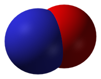
Nitric oxide
Inhaled nitric oxide is a pulmonary vasodilator. It decreases right-ventricular afterload and improves cardiac index by selectively decreasing pulmonary vascular resistance without causing systemic hypotension(4).

RV failure and pulmonary hypertension
Patients may become shocked or suffer cardiac arrest due to acute right ventricular dysfunction. This may be due to a primary cardiac cause such as right ventricular infarction (always consider this in a hypotensive patient with inferior STEMI, and confirm with a right ventricular ECG and/or echo). Alternatively it could be due to a pulmonary or systemic cause resulting in severe pulmonary hypertension, causing secondary right ventricular dysfunction. The commonest causes of acute pulmonary hypertension are massive PE, sepsis, and ARDS(5).

The haemodynamic consequences of RV failure are reduced pulmonary blood flow and inadequate left ventricular filling, leading to decreased cardiac output, shock, and arrest. In severe acute pulmonary hypertension the RV distends, resulting in a shift of the interventricular septum which compresses the LV and further inhibits LV filling (the concept of ventricular interdependence).

What’s wrong with standard ACLS?
In some patients with PHT who arrest, CPR may be ineffective due to a failure to achieve adequate pulmonary blood flow and ventricular filling. In one study of patients with known chronic PHT who arrested in the ICU, survival rates even for ventricular fibrillation were extremely poor and when measured end tidal carbon dioxide levels were very low. In the same study it was noted that some of the survivors had received an intravenous bolus administration of iloprost, a prostacyclin analogue (and pulmonary vasodilator) during CPR(6).

CPR may therefore be ineffective. Intubation and positive pressure ventilation may also be associated with haemodynamic deterioration in PHT patients(7), and intravenous epinephrine (adrenaline) has variable effects on the pulmonary circulation which could be deleterious(8).

If inhaled nitric oxide (iNO) can improve pulmonary blood flow and reduce right ventricular afterload, it could theoretically be of value in cases of shock or arrest with RV failure, especially in cases of pulmonary hypertension; these are patients who otherwise have poor outcomes and may not benefit from CPR.

Is the use of iNO described in shock or arrest?
Numerous case reports and series demonstrate recovery from shock or arrest following nitric oxide use in various situations of decompensated right ventricular failure from pulmonary hypertension secondary to pulmonary fibrotic disease(9), pneumonectomy surgery(10), and pulmonary embolism(11) including post-embolectomy(12).

Acute hemodynamic improvement was demonstrated following iNO therapy in a series of right ventricular myocardial infarction patients with cardiogenic shock(13).

A recent systematic review of inhaled nitric oxide in acute pulmonary embolism documented improvements in oxygenation and hemodynamic variables, “often within minutes of administration of iNO”. The authors state that these case reports underscore the need for randomised controlled trials to establish the safety and efficacy of iNO in the treatment of massive acute PE(14).

Why aren’t they telling us to use it?
If iNO may be helpful in certain cardiac arrest patients, why isn’t ILCOR recommending it? Actually it is mentioned – in the context of paediatric life support. The European Resuscitation Council states:

ERC Guideline: (Paediatric) Pulmonary hypertension
There is an increased risk of cardiac arrest in children with pulmonary hypertension.
Follow routine resuscitation protocols in these patients with emphasis on high FiO2 and alkalosis/hyperventilation because this may be as effective as inhaled nitric oxide in reducing pulmonary vascular resistance.
Resuscitation is most likely to be successful in patients with a reversible cause who are treated with intravenous epoprostenol or inhaled nitric oxide.
If routine medications that reduce pulmonary artery pressure have been stopped, they should be restarted and the use of aerosolised epoprostenol or inhaled nitric oxide considered.
Right ventricular support devices may improve survival

Should we use it?
So if acute (or acute on chronic) pulmonary hypertension can be suspected or demonstrated based on history, examination, and echo findings, and the patient is in extremis, it might be anticipated that standard ACLS approaches are likely to be futile (as they often are if the underlying cause is not addressed). One might consider attempts to induce pulmonary vasodilation to improve pulmonary blood flow and LV filling, improving oxygenation, and reducing RV afterload as means of reversing acute cor pulmonale.

Are there other pulmonary vasodilators we can use?
iNO is not the only means of inducing pulmonary vasodilation. Oxygen, hypocarbia (through hyperventilation)(15), and alkalosis are all known pulmonary vasodilators, the latter providing an argument for intravenous bicarbonate therapy from some quarters(16). Prostacyclin is a cheaper alternative to iNO(17) and can be given by inhalation or intravenously, although is more likely to cause systemic hypotension than iNO. Some inotropic agents such as milrinone and levosimendan can lower pulmonary vascular resistance(18).

What’s the take home message?
The take home message for me is that acute pulmonary hypertension provides yet another example of a condition that requires the resuscitationist to think beyond basic ACLS algorithms and aggressively pursue and manage the underlying cause(s) of shock or arrest. Inhaled pulmonary vasodilators may or may not be available but, as always, whatever resources and drugs are used, they need to be planned for well in advance. What’s your plan?

References
1. Adhikari NKJ, Dellinger RP, Lundin S, Payen D, Vallet B, Gerlach H, et al.
Inhaled Nitric Oxide Does Not Reduce Mortality in Patients With Acute Respiratory Distress Syndrome Regardless of Severity.
Critical Care Medicine. 2014 Feb;42(2):404–12
2. Steinhorn RH.
Neonatal pulmonary hypertension.
Pediatric Critical Care Medicine. 2010 Mar;11:S79–S84 Full text
3. McDonnell NJ, Chan BO, Frengley RW.
Rapid reversal of critical haemodynamic compromise with nitric oxide in a parturient with amniotic fluid embolism.
International Journal of Obstetric Anesthesia. 2007 Jul;16(3):269–73
4. Creagh-Brown BC, Griffiths MJ, Evans TW.
Bench-to-bedside review: Inhaled nitric oxide therapy in adults.
Critical Care. 2009;13(3):221 Full text
5. Tsapenko MV, Tsapenko AV, Comfere TB, Mour GK, Mankad SV, Gajic O.
Arterial pulmonary hypertension in noncardiac intensive care unit.
Vasc Health Risk Manag. 2008;4(5):1043–60 Full text
6. Hoeper MM, Galié N, Murali S, Olschewski H, Rubenfire M, Robbins IM, et al.
Outcome after cardiopulmonary resuscitation in patients with pulmonary arterial hypertension.
American Journal of Respiratory and Critical Care Medicine. 2002 Feb 1;165(3):341–4.
Full text
7. Höhn L, Schweizer A, Morel DR, Spiliopoulos A, Licker M.
Circulatory failure after anesthesia induction in a patient with severe primary pulmonary hypertension.
Anesthesiology. 1999 Dec;91(6):1943–5 Full text
8. Witham AC, Fleming JW.
The effect of epinephrine on the pulmonary circulation in man.
J Clin Invest. 1951 Jul;30(7):707–17 Full text
9. King R, Esmail M, Mahon S, Dingley J, Dwyer S.
Use of nitric oxide for decompensated right ventricular failure and circulatory shock after cardiac arrest.
Br J Anaesth. 2000 Oct;85(4):628–31. Full text
10. Fernández-Pérez ER, Keegan MT, Harrison BA.
Inhaled nitric oxide for acute right-ventricular dysfunction after extrapleural pneumonectomy.
Respir Care. 2006 Oct;51(10):1172–6 Full text
11. Summerfield DT, Desai H, Levitov A, Grooms DA, Marik PE.
Inhaled Nitric Oxide as Salvage Therapy in Massive Pulmonary Embolism: A Case Series.
Respir Care. 2012 Mar 1;57(3):444–8 Full text
12. Schenk P, Pernerstorfer T, Mittermayer C, Kranz A, Frömmel M, Birsan T, et al.
Inhalation of nitric oxide as a life-saving therapy in a patient after pulmonary embolectomy.
Br J Anaesth. 1999 Mar;82(3):444–7 Full text
13. Inglessis I, Shin JT, Lepore JJ, Palacios IF, Zapol WM, Bloch KD, et al.
Hemodynamic effects of inhaled nitric oxide in right ventricular myocardial infarction and cardiogenic shock.
Journal of the American College of Cardiology. 2004 Aug;44(4):793–8 Full text
14. Bhat T, Neuman A, Tantary M, Bhat H, Glass D, Mannino W, Akhtar M, Bhat A, Teli S, Lafferty J.
Inhaled nitric oxide in acute pulmonary embolism: a systematic review.
Rev Cardiovasc Med 2015;16(1):1–8.
15. Mahdi M, Joseph NJ, Hernandez DP, Crystal GJ, Baraka A, Salem MR.
Induced hypocapnia is effective in treating pulmonary hypertension following mitral valve replacement.
Middle East J Anaesthesiol. 2011 Jun;21(2):259-67
16. Evans S, Brown B, Mathieson M, Tay S.
Survival after an amniotic fluid embolism following the use of sodium bicarbonate.
BMJ Case Rep. 2014;2014
17. Fuller BM, Mohr NM, Skrupky L, Fowler S, Kollef MH, Carpenter CR.
The Use of Inhaled Prostaglandins in Patients With ARDS: A Systematic Review and Meta-analysis.
Chest. 2015 Jun;147(6):1510–22 Full text
18. LITFL: Right Ventricular Failure
Further reading
Life In The Fast Lane iNO info
LITFL on Pulmonary Hypertension

Esmolol for refractory VF

Already well publicised on social media, the team at Hennepin County published a retrospective comparison between patients with refractory VF who received esmolol with those who did not(1). The results are impressive and I look forward to further studies on this.
I work in an ED in a hospital with no cath lab and no access to extracorporeal life support, limiting our options for patients who remain in shockable rhythms despite ACLS interventions. We now have esmolol available in our resus room. You might want to keep it in your list of options for ACLS-refractory VF, which might also include double sequential external defibrillation(2) and even stellate ganglion block.
The dose of esmolol used was: loading dose 500 mcg/kg, followed by infusions of 0, 50, or 100 mcg/kg/min
An important point to note in the esmolol study is that almost all patients received high-quality mechanical CPR with the combined use of an impedence threshold device to augment venous return and cardiac output. The authors “speculate that this additional hemodynamic support may be essential given the hypotensive effects of esmolol.”
1. Use of esmolol after failure of standard cardiopulmonary resuscitation to treat patients with refractory ventricular fibrillation
Resuscitation. 2014 Oct;85(10):1337-41
[EXPAND Abstract]


INTRODUCTION: We compare the outcomes for patients who received esmolol to those who did not receive esmolol during refractory ventricular fibrillation (RVF) in the emergency department (ED).

METHODS: A retrospective investigation in an urban academic ED of patients between January 2011 and January 2014 of patients with out-of-hospital or ED cardiac arrest (CA) with an initial rhythm of ventricular fibrillation (VF) or ventricular tachycardia (VT) who received at least three defibrillation attempts, 300mg of amiodarone, and 3mg of adrenaline, and who remained in CA upon ED arrival. Patients who received esmolol during CA were compared to those who did not.

RESULTS: 90 patients had CA with an initial rhythm of VF or VT; 65 patients were excluded, leaving 25 for analysis. Six patients received esmolol during cardiac arrest, and nineteen did not. All patients had ventricular dysrhythmias refractory to many defibrillation attempts, including defibrillation after administration of standard ACLS medications. Most received high doses of adrenaline, amiodarone, and sodium bicarbonate. Comparing the patients that received esmolol to those that did not: 67% and 42% had temporary return of spontaneous circulation (ROSC); 67% and 32% had sustained ROSC; 66% and 32% survived to intensive care unit admission; 50% and 16% survived to hospital discharge; and 50% and 11% survived to discharge with a favorable neurologic outcome, respectively.

CONCLUSION: Beta-blockade should be considered in patients with RVF in the ED prior to cessation of resuscitative efforts.

[/EXPAND]
2. Double Sequential External Defibrillation in Out-of-Hospital Refractory Ventricular Fibrillation: A Report of Ten Cases.
Prehosp Emerg Care. 2015 January-March;19(1):126-130
[EXPAND Abstract]


Background. Ventricular fibrillation (VF) is considered the out-of-hospital cardiac arrest (OOHCA) rhythm with the highest likelihood of neurologically intact survival. Unfortunately, there are occasions when VF does not respond to standard defibrillatory shocks. Current American Heart Association (AHA) guidelines acknowledge that the data are insufficient in determining the optimal pad placement, waveform, or energy level that produce the best conversion rates from OOHCA with VF.

Objective. To describe a technique of double sequential external defibrillation (DSED) for cases of refractory VF (RVF) during OOHCA resuscitation.

Methods. A retrospective case series was performed in an urban/suburban emergency medical services (EMS) system with advanced life support care and a population of 900,000. Included were all adult OOHCAs having RVF during resuscitation efforts by EMS providers. RVF was defined as persistent VF following at least 5 unsuccessful single shocks, epinephrine administration, and a dose of antiarrhythmic medication. Once the patient was in RVF, EMS personnel applied a second set of pads and utilized a second defibrillator for single defibrillation with the new monitor/pad placement. If VF continued, EMS personnel then utilized the original and second monitor/defibrillator charged to maximum energy, and shocks were delivered from both machines simultaneously. Data were collected from electronic dispatch and patient care reports for descriptive analysis.

Results. From 01/07/2008 to 12/31/2010, a total of 10 patients were treated with DSED. The median age was 76.5 (IQR: 65-82), with median resuscitation time of 51minutes (IQR: 45-62). The median number of single shocks was 6.5 (IQR: 6-11), with a median of 2 (IQR: 1-3) DSED shocks delivered. VF broke after DSED in 7 cases (70%). Only 3 patients (30%) had ROSC in the field, and none survived to discharge.

Conclusion. This case series demonstrates that DSED may be a feasible technique as part of an aggressive treatment plan for RVF in the out-of-hospital setting. In this series, RVF was terminated 70% of the time, but no patient survived to discharge. Further research is needed to better understand the characteristics of and treatment strategies for RVF.

[/EXPAND]

Open cardiac massage in asthmatic arrests?

This idea was provoked by a colleague some years ago who could not achieve a palpable pulse during CPR of an arrested asthmatic child. He wondered whether the severe hyperinflation was rendering external cardiac compressions ineffective and whether he should have done a (prehospital) thoracotomy.
The literature is not strong. The 2010 AHA Guidelines rightly focus on reducing hyperinflation by disconnecting the tracheal tube from the ventilator circuit, and they mention ECMO for refractory cases, but there is no mention of open chest CPR.
I can only find two papers discussing it, both pretty old. A case series in the British Medical Journal from 1968 describes three patients with asthma who had asystolic arrests but did not achieve femoral pulses with external compressions(1). In two, open cardiac massage was performed resulting in restoration of sinus rhythm and cardiac output, and one appeared to make a neurological recovery.
A case report in 1987 describes a 32 year old man in asystolic cardiac arrest due to asthma(2):

“Ventilation required very high inflation pressures and little air movement was heard within the chest despite the administration of Adrenaline 1 mg and Aminophylline 250mg intravenously, and Adrenaline 1mg via the endotracheal tube. This was followed by an intravenous infusion of 100 ml of 8.4% Sodium Bicarbonate solution. External cardiac massage failed to produce a palpable pulse in the carotid area. The chest was, therefore, opened through a left anterolateral thoracotomy. The lungs appeared hyperinflated, bulky and tense and did not collapse when the pleural cavity was opened. The pericardium was opened and asystole confirmed, following eight to ten compressions of the heart some intrinsic activity commenced, ventilation also became much easier.”

He achieved ROSC and became haemodynamically stable but failed to wake up and treatment was withdrawn some days later.
Neither reports include mention of disconnection strategies to reduce hyperinflation. The lack of neurological recovery is not surprising given the apparent prolonged state of arrest the patients were resuscitated from. However there does appear to be a survivor who may not have made it had standard resuscitation (at the time) been continued.
Does this mean I would open the chest in an arrested asthma patient?
Not straight away, no. I would treat dynamic hyperinflation with tube disconnection and external compressions. I would correct absolute and relative hypovolaemia with crystalloid. I would treat bronchospasm (and possible anaphylaxis) with intravenous adrenaline/epinephrine. And I would exclude pneumothorax, possibly with ultrasound or more likely with bilateral open thoracostomies. If however these measures resulted in no detectable carotid flow with external cardiac compressions, ECMO was not available, and the arrest was not prolonged, I would definitely consider doing internal cardiac massage via thoracotomy.
What about you?
1. Grant IW, Kennedy WP, Malone DN
Deaths from asthma
Br Med J. 1968 May 18;2(5602):429–30
2. Diament RH, Sloan JP
Failed resuscitation in acute severe asthma: a medical indication for emergency thoracotomy?
Arch Emerg Med. 1987 Dec;4(4):233–5

London Cardiac Arrest Symposium 2014

The focus of the entire day is cardiac arrest and this is the second day of the London Cardiac Arrest Symposium.

Professor Niklas Nielsen kicked off with a presentation of his Targeted Temperature Management trial.  It seems that even now there is uncertainty in the interpretation of this latest study. I take heart from the knowledge that Prof Nielsen has changed the practice of his institution to reflect the findings of his study – I have certainly changed my practice. But we need to remain aware that there is more work to be done to answer the multiple questions that remain and the need for further RCTs is recognised.

The management of Cardiac arrest after avalanche is not a clinical scenario that I imagine I’ll ever find myself in. The management is well documented in the ICAR MEDCOM guidelines 2012. Dr Peter Paal reminded us that you’re not dead until you’re rewarmed and dead unless: with asystole, CPR may be terminated (or withheld) if a patient is lethally injured or completely frozen, the airway is blocked and duration of burial >35 min, serum potassium >12 mmol L(-1), risk to the rescuers is unacceptably high or a valid do-not-resuscitate order exists.

The age old question about prognostication after cardiac arrest was tackled by Prof Mauro Oddo. He covered the evidence for clinical examination, SSPE, EEG, and neurone specific enolase. Bottom line, all of these modalities are useful but none are specific enough to be used as a stand alone test so multiple modalities are required.

SAMU is leading the way with prehospital ECMO. They have mastered the art of cannulation (in the Louvre no less!) but there haven’t enough cases to demonstrate a mortality benefit. The commencement of ECMO prehospital reduces low flow time and theoretically should improve outcomes. This is begging for a RCT.

The experience of the Italians with in hospital ECMO shoes a better survival rate for in-hospital rather than out of hospital cardiac arrests, explained Dr Tomasso Mauri. They treat patients with a no flow time of <6min and low flow rate of <45min and had a 31% ICU survival rate. If you want to learn more about ED ECMO go to http://edecmo.org.

VA-ECMO

The Douglas Chamberlain lecture this year was Selective aortic arch perfusion presented by Prof James Manning. He spoke about the use of this technique in cardiac arrest and also in trauma (where it is known to you as Zone 1 REBOA).

image-1

In cardiac arrest the aim is to improve coronary perfusion, to preserve perfusion to the heart and the brain, offer a route of rapid temperature control and offer a direct route of administration of adrenaline. Coronary perfusion is seen to be supra normal after SAAP. And the suggested place for SAAP is prior to ECMO.

image-5

It’s more familiar ground talking about SAAP in trauma. This Zone 1 occlusion preserves cerebral and cardiac perfusion while blood loss is limited and rapid fluid resuscitation can occur.

image-3

You can hear Prof Manning on SAAP over at EMCrit (of course!). 

It’s been another great conference. Put the dates for next year’s London Trauma & Cardiac Arrest Conferences in your diary: 8th-10th December 2015!

Happy Holidays & Keep Well

Louisa Chan


Left Ventricular Assist Device for Cardiac Arrest?

LVADguyiconAn interesting case report by Dr Heidlebaugh and colleagues from the Department of Emergency Medicine at the William Beaumont Hospital describes a 72 year old marathon runner who arrested during cardiac catheterisation. It suggests a possible novel alternative to ECMO for cardiac arrest.
The patient became bradycardic then asystolic during catheterisation of his right coronary artery. High quality CPR was initiated and an Impella LV assist device was placed. This restored cardiac output which was followed by episodes of venticular fibrillation and then ROSC. His initial low ejection fraction of 15% recovered after targeted temperature management on ICU to 50% and he fully recovered neurologically.
This patient already had femoral arterial access for introduction of the Impella, since he was in a cath lab. He also had immediate CPR on arresting, and was an abnormally fit 72 year old. It remains to be seen whether this procedure can be applied to other patients in cardiac arrest. The authors state:

..until ECLS is readily available, poor survival and neurological outcome after cardiac arrest might be avoided in many patients by the use of pLVAD to offload the LV and enhance perfusion. Furthermore, there may be a subset of patients, in whom the support that pLVAD offers is sufficient to optimize hemodynamic parameters and bridge to ROSC, thus reducing the need for ECLS.

This video by Dr. I-Wen Wang from the Barnes-Jewish Hospital explains how the Impella is inserted and how it works.


Full Neurologic Recovery and Return of Spontaneous Circulation Following Prolonged Cardiac Arrest Facilitated by Percutaneous Left Ventricular Assist Device
Ther Hypothermia Temp Manag. 2014 Sep 3. [Epub ahead of print]
[EXPAND Abstract]

Sudden cardiac arrest is associated with high early mortality, which is largely related to postcardiac arrest syndrome characterized by an acute but often transient decrease in left ventricular (LV) function. The stunned LV provides poor cardiac output, which compounds the initial global insult from hypoperfusion. If employed early, an LV assist device (LVAD) may improve survival and neurologic outcome; however, traditional methods of augmenting LV function have significant drawbacks, limiting their usefulness in the periarrest period. Full cardiac support with cardiopulmonary bypass is not always readily available but is increasingly being studied as a tool to intensify resuscitation. There have been no controlled trials studying the early use of percutaneous LVADs (pLVADs) in pericardiac arrest patients or intra-arrest as a bridge to return of spontaneous circulation. This article presents a case study and discussion of a patient who arrested while undergoing an elective coronary angioplasty and suffered prolonged cardiopulmonary resuscitation. During resuscitation, treatment included placement of a pLVAD and initiation of therapeutic hypothermia. The patient made a rapid and full recovery.

[/EXPAND]

Image is of M. Joshua Morris, a happy LVAD recipient (not the patient in the described study) who kindly alerted me to this article. Used with permission.

Down with "down" time!

A man in his 40s has a witnessed collapse and CPR is immediately started. Paramedics are on scene within 5 minutes and initiate advanced cardiac life support. He has refractory ventricular fibrillation which degenerates to asystole. He arrives in an emergency department where, with good ongoing CPR, he appears reasonably well perfused and even demonstrates some spontaneous movements and reactive pupils. He is placed on a mechanical CPR device and activation of the cardiac cath lab is requested. The patient has been in cardiac arrest now for 32 minutes. The cardiology fellow appears and asks: ‘what’s the down time?’

What’s the right answer? Would you say ‘half an hour’? ’32 minutes’?
And does it matter? Why is the cardiology fellow asking? Does she have an arbitrary cut off in mind, over which emergency coronary reperfusion will be denied?

I think there are several problems with conversations like these.
The first, is what does ‘down time’ even mean?
The second, is how relevant is a cardiac arrest time interval to prognosis in an individual patient?
The third, is what is the significance of any time interval in a patient who at the time of assessment has some signs that CPR is providing some perfusion and there is some evidence of brain function?

Let’s take the first. The definition of ‘down time’ does not appear to be standardised:
In this publication it appears to refer to the time before resuscitation is commenced, where it is demonstrated to be prognostically important.
Similarly, in this medical dictionary, it is defined as the ‘temporal duration from cardiac arrest until beginning cardiopulmonary resuscitation or advanced cardiac life support.
However, a post in Life in the Fast Lane defines it as ‘time to return of spontaneous circulation
This appears to agree with The New South Wales Government’s Intensive Care Monitoring and Coordination Unit who define it as ‘the time from when a person’s heart stops beating to the time it starts beating again
Yet another definition is used in King County, Washington, where it is defined as ‘the time interval from collapse to call 911‘.

So the first thing is to clarify what we’re talking about: “This patient received immediate bystander CPR. He has had resuscitation for 32 minutes”.

My friend in the UK, nurse resuscitationist Fernando Candal Carballido, coined the term ‘Time of Supported Circulation‘, or TOSC. I quite like this and think it could catch on.

The next question is so what? What if it was 90 minutes? At what point do we declare futility? This is where I believe the game has changed. Multiple survivors of prolonged resuscitation are springing up in the news and in the literature. Particularly in the subgroup of patients with minimal comorbidity, early CPR, and who receive circulatory support via ECMO or mechanical CPR while they undergo coronary reperfusion.

For a great example of a prolonged CPR survivor, check out paramedic Wayne Schneider’s story,
…or listen to Steven Bernard describe amazing results from ECMO used in Melbourne in the CHEER study, which includes survivors of over two hours of CPR.

So, in summary:

  • Be clear on your definitions when communicating with colleagues. ‘Down time’ does not appear to have a standard definition, so I would avoid its use.
  • Some patients without comorbidities who have had early bystander CPR may survive despite long periods of CPR (or ‘TOSC’), provided the underlying cause can be treated or is reversible.
  • ECMO and even more widely available mechanical CPR devices are extending the period in which these causes can be addressed.

Update 2016: I now use the terms ‘No Flow Time’ (time from arrest to first basic life support) and ‘Low Flow Time’ (time receiving CPR, which stops with ROSC). This is prognostically very important, with increasing numbers of reports of survivors who have had very long periods of low flow time.

Is 4 Joules per kg enough in kids?

glash-sim-paed-face-smResearchers from the Iberian-American Paediatric Cardiac Arrest Study Network challenge the evidence base behind defibrillation shock dose recommendations in children.
In a study of in-hospital pediatric cardiac arrest due to VT or VF, clinical outcome was not related to the cause or location of arrest, type of defibrillator and waveform, energy dose per shock, number of shocks, or cumulative energy dose, although there was a trend to better survival with higher doses per shock. 50% of children required more than the recommended 4J per kg and in over a quarter three or more shocks were needed to achieve defibrillation.
 
Shockable rhythms and defibrillation during in-hospital pediatric cardiac arrest
Resuscitation. 2014 Mar;85(3):387-91
[EXPAND Abstract]


OBJECTIVE: To analyze the results of cardiopulmonary resuscitation (CPR) that included defibrillation during in-hospital cardiac arrest (IH-CA) in children.

METHODS: A prospective multicenter, international, observational study on pediatric IH-CA in 12 European and Latin American countries, during 24 months. Data from 502 children between 1 month and 18 years were collected using the Utstein template. Patients with a shockable rhythm that was treated by electric shock(s) were included. The primary endpoint was survival at hospital discharge. Univariate logistic regression analysis was performed to find outcome factors.

RESULTS: Forty events in 37 children (mean age 48 months, IQR: 7-15 months) were analyzed. An underlying disease was present in 81.1% of cases and 24.3% had a previous CA. The main cause of arrest was a cardiac disease (56.8%). In 17 episodes (42.5%) ventricular fibrillation (VF) or pulseless ventricular tachycardia (pVT) was the first documented rhythm, and in 23 (57.5%) it developed during CPR efforts. In 11 patients (27.5%) three or more shocks were needed to achieve defibrillation. Return of spontaneous circulation (ROSC) was obtained in 25 cases (62.5%), that was sustained in 20 (50.0%); however only 12 children (32.4%) survived to hospital discharge. Children with VF/pVT as first documented rhythm had better sustained ROSC (64.7% vs. 39.1%, p=0.046) and survival to hospital discharge rates (58.8% vs. 21.7%, p=0.02) than those with subsequent VF/pVT. Survival rate was inversely related to duration of CPR. Clinical outcome was not related to the cause or location of arrest, type of defibrillator and waveform, energy dose per shock, number of shocks, or cumulative energy dose, although there was a trend to better survival with higher doses per shock (25.0% with <2Jkg(-1), 43.4% with 2-4Jkg(-1) and 50.0% with >4Jkg(-1)) and worse with higher number of shocks and cumulative energy dose.

CONCLUSION: The termination of pediatric VF/pVT in the IH-CA setting is achieved in a low percentage of instances with one electrical shock at 4Jkg(-1). When VF/pVT is the first documented rhythm, the results of defibrillation are better than in the case of subsequent VF/pVT. No clear relationship between defibrillation protocol and ROSC or survival has been observed. The optimal pediatric defibrillation dose remains to be determined; therefore current resuscitation guidelines cannot be considered evidence-based, and additional research is needed.

[/EXPAND]

Not finding a difference doesn't prove equivalence

Image from http://www.physio-control.com/
The recent LINC trial was a randomised controlled trial comparing a mechanical chest compression device (LUCAS) with manual CPR(1). “No significant difference” was found for any of the main outcome measures considered.
So do you think the LINC trial demonstrated that mechanical CPR using the LUCAS device is equivalent, or at least not inferior, to manual CPR?
This was an interesting and important trial for those of us who manage prehospital cardiac arrest patients. In some social media discussions, it appears to have been interpreted by some as evidence that they are equivalent resuscitative techniques or that LUCAS is not inferior to manual CPR.

LINCdata

However, unless you see a p-value less than 0.05 in the table above, (issues of multiple hypotheses testing aside) no evidence of anything was demonstrated; not of difference and certainly not of equivalence. When faced with 2-sided p values >5%, investigators often conclude that there is “no difference” between the treatments, leading to an assumption among readers that the treatments are equivalent. A better conclusion is that there is “no evidence” of a difference between treatments (see opinion piece by Sackett, 2004(2)). In order to determine if treatments are equivalent, equivalence must be tested directly.
How can we test for equivalence?
First, we must define equivalence. It is crucial that this definition is provided a priori i.e. defined before the data are examined. As the focus of the LINC study was on superiority the investigators did not offer an a priori definition of equivalence. However, the CIRC study(3), conducted some time earlier and similar in design, did. (This study examined an alternative mechanical CPR device, the Zoll AutoPulse).
When establishing equivalence between treatments, instead of the more customary null hypothesis of no difference between treatments, the hypothesis that the true difference is equal to a specified ‘delta’ (δ) is tested (4).
To analyse the LINC results to look for equivalence, we can derive our delta values from the CIRC study, which as we’ve said did offer an a priori definition of equivalence. For the purpose of illustration, we will use the risk-difference stopping boundaries calculated for the CIRC study, rather than the odds ratio based equivalence margins, on the grounds of greater simplicity and clinical appropriateness. Therefore, we set our equivalence margins at -δ=-1.4% and δ=1.6%, meaning, where LUCAS fared no worse than manual CPR by 1.4% and no better by 1.6%, we will consider the two techniques equally efficacious. Thus, we will declare equivalence between LUCAS and manual CPR if the 2-sided 95% CI for the treatment difference lies entirely within -1.4% and 1.6%, and noninferiority if the one-sided 97.5% CI for the treatment difference (equivalent to the lower limit of the two-sided 95% CI) lies above -1.4%. (5).
These concepts and how they differ from a traditional comparison are more readily appreciated graphically (Fig. 1).
Figure 1. Two one-sided test procedure and the equivalence margin in equivalence/noninferiority testing between LUCAS and manual CPR
1a Traditional comparative study, such as the LINC trial, shows results with confidence intervals that show no evidence of a difference as they encompass 0.

LINCtradcomp

1b. Using equivalence margins (-δ and δ) derived from a similar study (CIRC), we show that the LINC trial does not demonstrate that LUCAS and manual CPR are equally efficacious, since the 95% CI do not lie completely within the equivalence margins.

LINCequiv
1c. The one sided CI lies above -δ for some outcomes, allowing us to declare non-inferiority on those measures.
LINCnoninf

Conclusion
The presentation of the LINC trial’s results shows no evidence of a difference in outcomes between mechanical and manual CPR, which is not the same as showing they are equivalent or that mechanical CPR is non-inferior. However if we re-examine their data using equivalence margins (-δ, δ) derived from a similar study (CIRC), there is some evidence that the LUCAS device is not inferior to manual CPR (but not necessarily equivalent) with respect to longer term good neurological outcome.

References
1. Rubertsson S, Lindgren E, Smekal D, er al. Mechanical Chest Compressions and Simultaneous Defibrillation vs Conventional Cardiopulmonary Resuscitation in Out-of-Hospital Cardiac Arrest
JAMA. 2014 Jan 1;311(1):53-61
2. Sackett D. Superiority trials, non-inferiority trials, and prisoners of the 2-sided null hypothesis
Evid Based Med 2004;9:38-39 [Open Access]
3. Lerner EB, Persse D, Souders CM, et al. Design of the Circulation Improving Resuscitation Care (CIRC) Trial: a new state of the art design for out-of-hospital cardiac arrest research
Resuscitation. 2011 Mar;82(3):294-9
4. Dunnett CW, Gent M. Significance testing to establish equivalence between treatments, with special reference to data in the form of 2X2 tables. Biometrics. 1977 Dec;33(4):593-602
5. Piaggio G, Elbourne DR, Pocock SJ, et al. Reporting of noninferiority and equivalence randomized trials: extension of the CONSORT 2010 statement. JAMA. 2012;308(24):2594-604. [Open Access]

London Trauma Conference Day 2

London Trauma Conference 2013 – Day 2  by Dr Louisa Chan
So I find myself torn today: do I join the the main track with a Major incident theme or the Cardiac Masterclass? I never liked the thought of missing out on anything so I went to a bit of both.
 
Cardiac Masterclass
A lot of people probably think that managing cardiac arrest isn’t challenging and a bit dull because the patient is dead. But the Cardiac Masterclass would inspire you to think of a bright future for cardiac arrest management.
Mark Whitbread reminded us of how important dispatch is in the chain of survival. How much focus do we put on improving bystander CPR rates? Dispatcher assisted CPR has been shown to improve outcomes and needs to be skilfully done.
Ajay Jain pushes for all OHCA patients to be taken to a Cardiac Arrest centre for PCI. Why? Because the results he has from his centre for PCI in OHCA patients results in 77% (101/132) patients surviving to hosp discharge, 65% neurologically intact.
He also tells us that the ECG post arrest is a very poor predictor of PCI findings (although STEMI predicts a positive result) so they all should have PCI.
Lyon-survivors
 
More data from TOPCAT shows us that non survivors of OHCA are easy to cool.
 
LTC-mice
 
 
And maybe we should be cooling DURING cardiac arrest to minimise the reperfusion injury.
 
 
For persistent VF Prof Redwood says revascularisation is the key; when that doesn’t work then reducing LV volume may help so aspiration or an Impella may work. Failing that – ECMO.
 
Major Incidents
Major Incidents by their nature do not happen every day, so experience in these incidents is limited. The challenge then is how can we learn from incidents?
A standardised reporting system for a major incident database would be a good idea – www.majorincidentreporting.org – is where you will find the standard report form and open access database.
And then all I can suggest is that you need to come to the LTC and listen to the accounts of those who have been there. We heard about the Tokyo Sarin attack, Mumbai, and a very compelling story of multiple drownings from Steen Barnung.
Lessons from Tokyo – Sarin attack:

It will happen again
It will be chaos
Crowds cannot be controlled
Comms will fail
Clinical diagnosis – need a senior clinician
Treatment must be immediately available – 3min to absorb sarin
Decontamination – get naked, 90% decon with clothes removal.
Stream casualties
Empower the man on the ground.

 
Gadgets
LTC-MSUThe great thing about the London Trauma Conference is that it’s not just about the content of the tracks, there’s the networking and the opportunity to see new pieces of equipment.
The Norwegians won on the equipment front with their Mobile Stroke Unit. It’s due to go on line in 2014.
So TTFN and more from me on Day 3 of #LTC2013

London Trauma Conference 2013

FDIA_ImageOur inside reporter Dr Louisa Chan provides an update from Day One of the London Trauma Conference:
At risk of sounding like a resuscisaurus, last year was my first foray into the world of blogging. I’m proud to say that the genetic make up of most emergency physicians allows us to adapt so that others do not die! And so here I am again, making my way into the big smoke to report on the great developments of 2013.
I’ve struggled in the past to prise myself away from the main trauma track, it is after all the London Trauma Conference, which has left me curious as to the content of the Cardiac arrest symposium, this year it has been integrated, so I finally get to scratch that itch.
 
Prehospital Cardiac Arrest Management in Scotland
The conference was kicked off by Richard Lyon‘s inspirational description of his TOPCAT study.
In Scotland, of 50 cardiac arrests, 6 will survive to hospital and only 1 will survive to hospital discharge. The survival to hospital discharge in the UK is getting worse (4.8% 1995- 0.7% 2007)
Spurred on by these dreadful figures and a personal quest to improve cardiac arrest care (his father succumbed to a cardiac arrest in his forties)
All in all he has studied 400 cardiac arrest patients pre hospital. So what has he learnt?

  • Precise application of the chain of survival to your own system is vital in the delivery of Quality CPR.
  • He started in the ambulance control room analysing calls (CPR starts at step 11 so more experienced dispatchers skip thee quicker) and worked his way through the chain of survival.
  • The TOPCAT study revealed a 3 min delay to compressions where early intubation and cannulation were performed. Through an education program delivering knowledge and skills with individualised feedback they were able to increase on-chest time.
  • LEADERSHIP was a big factor. Having a clinician dedicated to managing the team improved on chest time and is now delivered by paramedics manning a car response in Edinburgh.
  • Breaks in CPR during movement are overcome by a mechanical chest compression device on carry sheet.
  • Non technical skills are monitored by camera feed
  • These changes have led to a survival to hospital discharge rate of 38% for patients in VF
  • This could translate into an extra 300 lives saved in Scotland when these changes are rolled out nationally.
  • And now there is a move to transport patients who are in VF after the third shock then straight to cath lab.

 
Echocardiography in cardiac arrest
Prof Tim Harris spoke about his passion – echocardiography in resuscitation. If you were in any doubt before then you would leave convinced.
Of course echo should not interfere with CPR so it should be done during the rhythm check with a 10 sec count down.
He covered the usual uses; PEA vs EMD in prognostication (92% sensitivity and 82% specificity to ROSC), Circulation assessment and an estimation of EF (Normal function – anterior mitral valve leaflet hits the septum or is within 5mm , EF 30-45% between 5mm- 18mm and >18mm ant mitral valve leaflets – 30% EF)
 
Cardiogenic shock after cardiac arrest
Professor Deakin: optimising cardiac function after ROSC revolves around the three elements of preload, SVR and myocardial contractility. For those who can still remember how, he recommends preload should be optimised to a LA pressure 15-20mmHg (2-12 normal) with a Swan Ganz catheter.
SVR and contractility can be manipulated thereafter using traditional vasopressors and inotropes or more novel agents like Levosimendan.
Mechanical devices such as IABP, Impella, TandemSupport are useful if available.
Where does the future lie? Perhaps synchronised pacing, hypothermia, extrathoracic ventilation and gene therapy.
LTC-BrohiOpen chest cardiac massage
Prof Karim Brohi: external chest compressions have been around since the 1960′s. Without a doubt external compressions generate a cardiac output, but is this the best way?
Over the last 10 years the priorities in traumatic cardiac arrest have changed – chest compressions are not instituted until after reversible causes have been addressed.
In non traumatic arrest how could we improve?
In canine models coronary perfusion pressure is five times better with internal cardiac massage, providing better survival rates with intact neurology.
There are a few human studies showing marked differences in cardiac index: 1.31 in the open group vs 0.61 in the closed group. In a Japanese study (1993), ROSC was achieved in 58% in open vs 1% closed.
The technique is two handed and the same as that taught in thoracotomy training. The difference is that in medical cardiac arrest you can use a smaller incision ( left lateral).
Who should we use open cardiac massage on? Perhaps in tamponade and pulmonary embolism?
How about when? When 10-15min with “standard care” has failed?
Perhaps it is time for a trial?
Post cardiac arrest syndrome and neuro protective measures
Prof Simon Redwood and Matt Thomas had overlapping talks on this . The bottom line is don’t have too much or too little CO2 or O2. The therapeutic hypothermia debate continues, what is evident is that there should be temperature control to avoid hyperthermia but what temperature? And there may be other benefits to hypothermia eg. limitation of infarct size.
What has been evident from all the speakers today is that it is an integrated system that saves lives and in order to guide the development of your system you need data and the belief that you can improve cardiac arrest outcomes.
More from me tomorrow!
Louisa Chan