Tag Archives: shock

It’s Tamponade – Now What?

You ultrasound the chest of your shocked patient in resus with fluid refractory hypotension. You see fluid around the heart. The right ventricle keeps bowing inwards, which you recall being described as ‘a little invisible man jumping up and down using the RV as a trampoline’, and you know this is in fact a sign of right ventricular diastolic collapse.


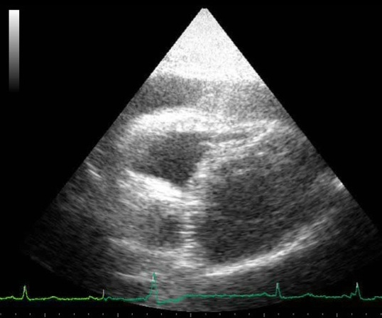
The collapse of the right side of the heart during diastole is the mechanism for shock and cardiac arrest due to tamponade, because the high pericardial pressures prevent the right heart from filling in diastole. This patient therefore has ‘tamponade physiology’ on ultrasound. A quick scan of the IVC shows it is dilated and does not collapse with respiration. This confirms a high central venous pressure (as do the patient’s distended neck veins), also consistent with tamponade physiology.


A formal echo done in resus confirms your suspicion of a dliated aortic root and visible dissection flap, so the diagnosis is now clear. This is type A aortic dissection with tamponade. The patient remains hypotensive and mottled with increasing drowsiness. Cardiothoracic surgery is based at another hospital site 30 minutes away by ambulance.


As the critical care clinician responsible for, or assisting with this patient’s care (emergency physician, intensivist, anaesthetist, rural GP, physician’s assistant, etc.), how do we get this patient to definitive care and mitigate the risk of deterioration en route? Let’s discuss the options using real life case examples, and consider the physiology, the evidence, and the dogma.


Here are four key questions to consider:
1. To drain or not to drain the pericardium?
2. To intubate or not to intubate?
3. If they arrest – CPR or no CPR?
4. How to transfer – physician escort or just send in an ambulance on lights and sirens?


Here are three scenarios that follow the intial assessment of the above patient. They are based on similar cases shared with me by participants on the Critical Care in the Emergency Department course.


Case 1

The patient is obtunded with profound shock and too unstable for transfer. The resus team undertakes pericardiocentesis and aspirates 30 ml of blood. The patient becomes conscious and cooperative and the systolic blood pressure (SBP) is 95 mmHg. The patient is transferred by paramedic ambulance to the cardothoracic centre where he is successfully operated on, resulting in a full recovery.


Case 2

As the patient is unconscious and requires interhospital transfer, the decision is made to intubate him for airway protection. He undergoes rapid sequence induction with ketamine, fentanyl, and rocuronium in the resus room. After capnographic confirmation of tracheal intubation he is manually ventilated via a self-inflating bag. The ED nurse reports a loss of palpable pulse and CPR is started. A team member suggests pericardiocentesis but a senior critical care physician says there is no point because ‘it won’t fix the underlying problem of aortic dissection’ and ’the blood will be clotted anyway’. After a brief attempt at standard ACLS, resuscitation efforts are discontinued and the patient is declared dead.


Case 3

The patient is hypotensive with a SBP of 90mmHg and drowsy but cooperative. The receiving centre has accepted the referral and an ambulance has been requested. The critical care physician responsible for patient transfers is requested to accompany the patient but declines, on the basis that ‘these cases are just like abdominal aortic aneurysms – they just need to get there asap. If they deteriorate en route we’re not going to do anything.’


The patient is transferred but 15 minutes into the journey he becomes unresponsive and loses his cardiac output. The transporting paramedics provide chest compressions and adrenaline/epinephrine but are unable to resuscitate him.
These cases illustrate some of the pitfalls and fallacies associated with tamponade due to type A dissection.


Pericardiocentesis

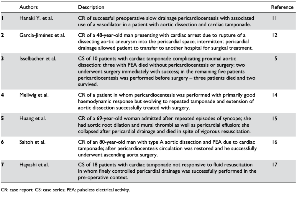
Pericardiocentesis can definitely be life-saving, restoring vital organ perfusion and buying time to get the patient to definitive surgery. Numerous case reports and case series provide evidence of its utility, even in patients in PEA cardiac arrest(1). The authors of the two largest cases series both used 8F pigtail drainage catheters(1,2).


Reports in the literature regarding the pericardiocentesis of critical cardiac tamponade complicating aortic dissection from Cruz et al


One key component of procedural success was controlled pericardial drainage, removing small volumes and reassessing the blood pressure, aiming for a SBP of 90 mmHg. The danger is overshooting, resulting in hypertension and extending the underlying aortic dissection which can be fatal (3).


Those still unconvinced by the evidence may be swayed by guidelines. The 2015 European Society of Cardiology Guidelines for the diagnosis and management of pericardial diseases (4) state:


“In the setting of aortic dissection with haemopericardium and suspicion of cardiac tamponade, emergency transthoracic echocardiography or a CT scan should be performed to confirm the diagnosis. In such a scenario, controlled pericardial drainage of very small amounts of the haemopericardium can be attempted to temporarily stabilize the patient in order to maintain blood pressure at 90 mmHg. (Class IIa, Level C)”


Intubation

Deterioration of tamponade patients following intubation is well described in the literature and the risk is well appreciated by cardiothoracic anaesthetists(5).


Once positive pressure ventilation is started, positive pleural pressure is transmitted to the pericardium, where pressures can exceed right ventricular diastolic pressure and prevent cardiac filling. The result is a fall in and possible loss of cardiac output. This is further exacerbated by the addition of PEEP(6).


One suggested approach if the patient must be intubated for airway protection but is not yet in the operating room with a surgeon ready to cut, is to consider intubation under local anaesthesia and allow the patient to breathe spontaneously (maintaining negative pleural pressure) through the tube until the surgeon is ready to open the chest(5).


Alternatively preload with fluid, use cautious doses of induction agent, and ventilate with low tidal volumes and zero PEEP. However the patient can still crash, so remember that these effects of ventilation on cardiac output in tamponade can be mitigated by the removal of a relatively small volume of pericardial fluid(6).


Cardiac Arrest

In cardiac arrest, external chest compressions are unlikely to be of benefit. In a study on baboons subjected to cardiac tamponade, closed chest massage resulted in an increase in intrapericardial pressure. There was an increase in systolic pressure, but a marked decrease in diastolic pressure, with an overall decrease in mean arterial pressure(7).

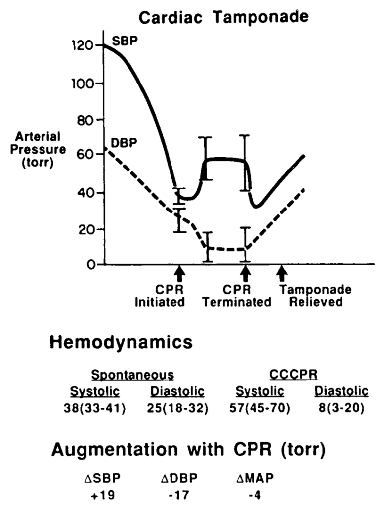
Pressure changes from CPR during tamponade in baboons. From Luna et al (7)
This would lead to impaired coronary perfusion and would be very unlikely to result in return of spontaneous circulation (ROSC). In the clinical situation described above, it is only relief of tamponade that is going to provide an arrested patient with a chance of recovery.


Transport

For patients with cardiac tamponade requiring interhospital (or intrahospital) transfer, it would seem vital therefore that the patient is accompanied by a clinician willing and capable to perform pericardiocentesis in the event of severe deterioriation or arrest en route. This simple life-saving intervention to deliver the patient alive to the operating room should be made available should the need arise.


Summary

  • Patients presenting in shock from cardiac tamponade often have treatable underlying causes and represent a situation where the planning and actions of the resuscitationist can be truly life-saving.
  • Pericardiocentesis is recommended in profound shock to buy time for definitive intervention. Controlled pericardiocentesis should be performed paying strict attention to SBP to avoid ‘overshooting’ to a hypertensive state in type A aortic dissection. In cardiac arrest, chest compressions are likely to be ineffective and pericardiocentesis is mandatory for ROSC.
  • The institution of positive pressure ventilation often results in worsened shock or cardiac arrest, and this is exacerbated by PEEP. Where possible, avoid intubation until the patient is in the operating room, or use low tidal volumes and no PEEP. Even then pericardiocentesis may be necessary to maintain or restore cardiac output.
  • Patients requiring transport who have tamponade should be accompanied by a clinician able to perform pericardiocentesis in the event of en route deterioration.


References

  1. Cruz I, Stuart B, Caldeira D, Morgado G, Gomes AC, Almeida AR, et al. Controlled pericardiocentesis in patients with cardiac tamponade complicating aortic dissection: Experience of a centre without cardiothoracic surgery. European Heart Journal: Acute Cardiovascular Care. 2015 Mar 19;4(2):124–8.
  2. Hayashi T, Tsukube T, Yamashita T, Haraguchi T, Matsukawa R, Kozawa S, et al. Impact of Controlled Pericardial Drainage on Critical Cardiac Tamponade With Acute Type A Aortic Dissection. Circulation. 2012 Sep 10;126(11_suppl_1):S97–S101.
  3. Isselbacher EM, Cigarroa JE, Eagle KA. Cardiac tamponade complicating proximal aortic dissection. Is pericardiocentesis harmful? Circulation. 1994 Nov 1;90(5):2375–8.
  4. Adler Y, Charron P, Imazio M, Badano L, Barón-Esquivias G, Bogaert J, et al. 2015 ESC Guidelines for the diagnosis and management of pericardial diseases. European Heart Journal. 2015 Nov 7;36(42):2921–64.
  5. Ho AMH, Graham CA, Ng CSH, Yeung JHH, Dion PW, Critchley LAH, et al. Timing of tracheal intubation in traumatic cardiac tamponade: A word of caution. Resuscitation. 2009 Feb;80(2):272–4.
  6. Möller CT, Schoonbee CG, Rosendorff C. Haemodynamics of cardiac tamponade during various modes of ventilation. Br J Anaesth. 1979 May;51(5):409–15.
  7. Luna GK, Pavlin EG, Kirkman T, Copass MK, Rice CL. Hemodynamic effects of external cardiac massage in trauma shock. The Journal of Trauma: Injury, Infection, and Critical Care. 1989 Oct;29(10):1430–3.

Inhaled nitric oxide: a tool for all resuscitationists?

The use of inhaled nitric oxide is established in certain groups of patients: it improves oxygenation (but not survival) in patients with acute respiratory distress syndrome(1), and it is used in neonatology for management of persistent pulmonary hypertension of the newborn(2). But it can be applied in other resuscitation settings: in arrested or peri-arrest patients with pulmonary hypertension.

Read this (modified) description of a case managed by one of my resuscitationist friends from an overseas location:

A young lady suffered a placental abruption requiring emergency hysterectomy. She arrested twice in the operating room after suspected amniotic fluid embolism. She had fixed dilated pupils.
She developed extreme pulmonary hypertension with suprasystemic pulmonary artery pressures, and she went down the pulmonary HT spiral as I stood there. On ultrasound her distended RV was making her LV totally collapse. She arrested. Futile CPR was started.
I have never had an extreme pulmonary HT survive an arrest. I grabbed a bag and rapidly set up a manual inhaled Nitric Oxide system and bagged and begged…
She achieved ROSC after some minutes. A repeat ultrasound showed a well functioning LV and less dilated RV.
Today, after 12 hours she is opening her eyes and obeying commands. Still a long way to go, but alive.

It sounds impressive. I don’t have more case details, and don’t know how confident they could be about the diagnosis of amniotic fluid embolism but the presentation certainly fits with acute pulmonary hypertension with RV failure. The use of inhaled nitric oxide has certainly been described for similar scenarios before(3). But it raises bigger questions: is this something we should all be capable of? Are there cardiac arrests involving or caused by pulmonary hypertension that will not respond to resuscitation without nitric oxide?

Nitric oxide
Inhaled nitric oxide is a pulmonary vasodilator. It decreases right-ventricular afterload and improves cardiac index by selectively decreasing pulmonary vascular resistance without causing systemic hypotension(4).

RV failure and pulmonary hypertension
Patients may become shocked or suffer cardiac arrest due to acute right ventricular dysfunction. This may be due to a primary cardiac cause such as right ventricular infarction (always consider this in a hypotensive patient with inferior STEMI, and confirm with a right ventricular ECG and/or echo). Alternatively it could be due to a pulmonary or systemic cause resulting in severe pulmonary hypertension, causing secondary right ventricular dysfunction. The commonest causes of acute pulmonary hypertension are massive PE, sepsis, and ARDS(5).

The haemodynamic consequences of RV failure are reduced pulmonary blood flow and inadequate left ventricular filling, leading to decreased cardiac output, shock, and arrest. In severe acute pulmonary hypertension the RV distends, resulting in a shift of the interventricular septum which compresses the LV and further inhibits LV filling (the concept of ventricular interdependence).

What’s wrong with standard ACLS?
In some patients with PHT who arrest, CPR may be ineffective due to a failure to achieve adequate pulmonary blood flow and ventricular filling. In one study of patients with known chronic PHT who arrested in the ICU, survival rates even for ventricular fibrillation were extremely poor and when measured end tidal carbon dioxide levels were very low. In the same study it was noted that some of the survivors had received an intravenous bolus administration of iloprost, a prostacyclin analogue (and pulmonary vasodilator) during CPR(6).

CPR may therefore be ineffective. Intubation and positive pressure ventilation may also be associated with haemodynamic deterioration in PHT patients(7), and intravenous epinephrine (adrenaline) has variable effects on the pulmonary circulation which could be deleterious(8).

If inhaled nitric oxide (iNO) can improve pulmonary blood flow and reduce right ventricular afterload, it could theoretically be of value in cases of shock or arrest with RV failure, especially in cases of pulmonary hypertension; these are patients who otherwise have poor outcomes and may not benefit from CPR.

Is the use of iNO described in shock or arrest?
Numerous case reports and series demonstrate recovery from shock or arrest following nitric oxide use in various situations of decompensated right ventricular failure from pulmonary hypertension secondary to pulmonary fibrotic disease(9), pneumonectomy surgery(10), and pulmonary embolism(11) including post-embolectomy(12).

Acute hemodynamic improvement was demonstrated following iNO therapy in a series of right ventricular myocardial infarction patients with cardiogenic shock(13).

A recent systematic review of inhaled nitric oxide in acute pulmonary embolism documented improvements in oxygenation and hemodynamic variables, “often within minutes of administration of iNO”. The authors state that these case reports underscore the need for randomised controlled trials to establish the safety and efficacy of iNO in the treatment of massive acute PE(14).

Why aren’t they telling us to use it?
If iNO may be helpful in certain cardiac arrest patients, why isn’t ILCOR recommending it? Actually it is mentioned – in the context of paediatric life support. The European Resuscitation Council states:

ERC Guideline: (Paediatric) Pulmonary hypertension
There is an increased risk of cardiac arrest in children with pulmonary hypertension.
Follow routine resuscitation protocols in these patients with emphasis on high FiO2 and alkalosis/hyperventilation because this may be as effective as inhaled nitric oxide in reducing pulmonary vascular resistance.
Resuscitation is most likely to be successful in patients with a reversible cause who are treated with intravenous epoprostenol or inhaled nitric oxide.
If routine medications that reduce pulmonary artery pressure have been stopped, they should be restarted and the use of aerosolised epoprostenol or inhaled nitric oxide considered.
Right ventricular support devices may improve survival

Should we use it?
So if acute (or acute on chronic) pulmonary hypertension can be suspected or demonstrated based on history, examination, and echo findings, and the patient is in extremis, it might be anticipated that standard ACLS approaches are likely to be futile (as they often are if the underlying cause is not addressed). One might consider attempts to induce pulmonary vasodilation to improve pulmonary blood flow and LV filling, improving oxygenation, and reducing RV afterload as means of reversing acute cor pulmonale.

Are there other pulmonary vasodilators we can use?
iNO is not the only means of inducing pulmonary vasodilation. Oxygen, hypocarbia (through hyperventilation)(15), and alkalosis are all known pulmonary vasodilators, the latter providing an argument for intravenous bicarbonate therapy from some quarters(16). Prostacyclin is a cheaper alternative to iNO(17) and can be given by inhalation or intravenously, although is more likely to cause systemic hypotension than iNO. Some inotropic agents such as milrinone and levosimendan can lower pulmonary vascular resistance(18).

What’s the take home message?
The take home message for me is that acute pulmonary hypertension provides yet another example of a condition that requires the resuscitationist to think beyond basic ACLS algorithms and aggressively pursue and manage the underlying cause(s) of shock or arrest. Inhaled pulmonary vasodilators may or may not be available but, as always, whatever resources and drugs are used, they need to be planned for well in advance. What’s your plan?

References
1. Adhikari NKJ, Dellinger RP, Lundin S, Payen D, Vallet B, Gerlach H, et al.
Inhaled Nitric Oxide Does Not Reduce Mortality in Patients With Acute Respiratory Distress Syndrome Regardless of Severity.
Critical Care Medicine. 2014 Feb;42(2):404–12
2. Steinhorn RH.
Neonatal pulmonary hypertension.
Pediatric Critical Care Medicine. 2010 Mar;11:S79–S84 Full text
3. McDonnell NJ, Chan BO, Frengley RW.
Rapid reversal of critical haemodynamic compromise with nitric oxide in a parturient with amniotic fluid embolism.
International Journal of Obstetric Anesthesia. 2007 Jul;16(3):269–73
4. Creagh-Brown BC, Griffiths MJ, Evans TW.
Bench-to-bedside review: Inhaled nitric oxide therapy in adults.
Critical Care. 2009;13(3):221 Full text
5. Tsapenko MV, Tsapenko AV, Comfere TB, Mour GK, Mankad SV, Gajic O.
Arterial pulmonary hypertension in noncardiac intensive care unit.
Vasc Health Risk Manag. 2008;4(5):1043–60 Full text
6. Hoeper MM, Galié N, Murali S, Olschewski H, Rubenfire M, Robbins IM, et al.
Outcome after cardiopulmonary resuscitation in patients with pulmonary arterial hypertension.
American Journal of Respiratory and Critical Care Medicine. 2002 Feb 1;165(3):341–4.
Full text
7. Höhn L, Schweizer A, Morel DR, Spiliopoulos A, Licker M.
Circulatory failure after anesthesia induction in a patient with severe primary pulmonary hypertension.
Anesthesiology. 1999 Dec;91(6):1943–5 Full text
8. Witham AC, Fleming JW.
The effect of epinephrine on the pulmonary circulation in man.
J Clin Invest. 1951 Jul;30(7):707–17 Full text
9. King R, Esmail M, Mahon S, Dingley J, Dwyer S.
Use of nitric oxide for decompensated right ventricular failure and circulatory shock after cardiac arrest.
Br J Anaesth. 2000 Oct;85(4):628–31. Full text
10. Fernández-Pérez ER, Keegan MT, Harrison BA.
Inhaled nitric oxide for acute right-ventricular dysfunction after extrapleural pneumonectomy.
Respir Care. 2006 Oct;51(10):1172–6 Full text
11. Summerfield DT, Desai H, Levitov A, Grooms DA, Marik PE.
Inhaled Nitric Oxide as Salvage Therapy in Massive Pulmonary Embolism: A Case Series.
Respir Care. 2012 Mar 1;57(3):444–8 Full text
12. Schenk P, Pernerstorfer T, Mittermayer C, Kranz A, Frömmel M, Birsan T, et al.
Inhalation of nitric oxide as a life-saving therapy in a patient after pulmonary embolectomy.
Br J Anaesth. 1999 Mar;82(3):444–7 Full text
13. Inglessis I, Shin JT, Lepore JJ, Palacios IF, Zapol WM, Bloch KD, et al.
Hemodynamic effects of inhaled nitric oxide in right ventricular myocardial infarction and cardiogenic shock.
Journal of the American College of Cardiology. 2004 Aug;44(4):793–8 Full text
14. Bhat T, Neuman A, Tantary M, Bhat H, Glass D, Mannino W, Akhtar M, Bhat A, Teli S, Lafferty J.
Inhaled nitric oxide in acute pulmonary embolism: a systematic review.
Rev Cardiovasc Med 2015;16(1):1–8.
15. Mahdi M, Joseph NJ, Hernandez DP, Crystal GJ, Baraka A, Salem MR.
Induced hypocapnia is effective in treating pulmonary hypertension following mitral valve replacement.
Middle East J Anaesthesiol. 2011 Jun;21(2):259-67
16. Evans S, Brown B, Mathieson M, Tay S.
Survival after an amniotic fluid embolism following the use of sodium bicarbonate.
BMJ Case Rep. 2014;2014
17. Fuller BM, Mohr NM, Skrupky L, Fowler S, Kollef MH, Carpenter CR.
The Use of Inhaled Prostaglandins in Patients With ARDS: A Systematic Review and Meta-analysis.
Chest. 2015 Jun;147(6):1510–22 Full text
18. LITFL: Right Ventricular Failure
Further reading
Life In The Fast Lane iNO info
LITFL on Pulmonary Hypertension

London Trauma Conference 2013

FDIA_ImageOur inside reporter Dr Louisa Chan provides an update from Day One of the London Trauma Conference:
At risk of sounding like a resuscisaurus, last year was my first foray into the world of blogging. I’m proud to say that the genetic make up of most emergency physicians allows us to adapt so that others do not die! And so here I am again, making my way into the big smoke to report on the great developments of 2013.
I’ve struggled in the past to prise myself away from the main trauma track, it is after all the London Trauma Conference, which has left me curious as to the content of the Cardiac arrest symposium, this year it has been integrated, so I finally get to scratch that itch.
 
Prehospital Cardiac Arrest Management in Scotland
The conference was kicked off by Richard Lyon‘s inspirational description of his TOPCAT study.
In Scotland, of 50 cardiac arrests, 6 will survive to hospital and only 1 will survive to hospital discharge. The survival to hospital discharge in the UK is getting worse (4.8% 1995- 0.7% 2007)
Spurred on by these dreadful figures and a personal quest to improve cardiac arrest care (his father succumbed to a cardiac arrest in his forties)
All in all he has studied 400 cardiac arrest patients pre hospital. So what has he learnt?

  • Precise application of the chain of survival to your own system is vital in the delivery of Quality CPR.
  • He started in the ambulance control room analysing calls (CPR starts at step 11 so more experienced dispatchers skip thee quicker) and worked his way through the chain of survival.
  • The TOPCAT study revealed a 3 min delay to compressions where early intubation and cannulation were performed. Through an education program delivering knowledge and skills with individualised feedback they were able to increase on-chest time.
  • LEADERSHIP was a big factor. Having a clinician dedicated to managing the team improved on chest time and is now delivered by paramedics manning a car response in Edinburgh.
  • Breaks in CPR during movement are overcome by a mechanical chest compression device on carry sheet.
  • Non technical skills are monitored by camera feed
  • These changes have led to a survival to hospital discharge rate of 38% for patients in VF
  • This could translate into an extra 300 lives saved in Scotland when these changes are rolled out nationally.
  • And now there is a move to transport patients who are in VF after the third shock then straight to cath lab.

 
Echocardiography in cardiac arrest
Prof Tim Harris spoke about his passion – echocardiography in resuscitation. If you were in any doubt before then you would leave convinced.
Of course echo should not interfere with CPR so it should be done during the rhythm check with a 10 sec count down.
He covered the usual uses; PEA vs EMD in prognostication (92% sensitivity and 82% specificity to ROSC), Circulation assessment and an estimation of EF (Normal function – anterior mitral valve leaflet hits the septum or is within 5mm , EF 30-45% between 5mm- 18mm and >18mm ant mitral valve leaflets – 30% EF)
 
Cardiogenic shock after cardiac arrest
Professor Deakin: optimising cardiac function after ROSC revolves around the three elements of preload, SVR and myocardial contractility. For those who can still remember how, he recommends preload should be optimised to a LA pressure 15-20mmHg (2-12 normal) with a Swan Ganz catheter.
SVR and contractility can be manipulated thereafter using traditional vasopressors and inotropes or more novel agents like Levosimendan.
Mechanical devices such as IABP, Impella, TandemSupport are useful if available.
Where does the future lie? Perhaps synchronised pacing, hypothermia, extrathoracic ventilation and gene therapy.
LTC-BrohiOpen chest cardiac massage
Prof Karim Brohi: external chest compressions have been around since the 1960′s. Without a doubt external compressions generate a cardiac output, but is this the best way?
Over the last 10 years the priorities in traumatic cardiac arrest have changed – chest compressions are not instituted until after reversible causes have been addressed.
In non traumatic arrest how could we improve?
In canine models coronary perfusion pressure is five times better with internal cardiac massage, providing better survival rates with intact neurology.
There are a few human studies showing marked differences in cardiac index: 1.31 in the open group vs 0.61 in the closed group. In a Japanese study (1993), ROSC was achieved in 58% in open vs 1% closed.
The technique is two handed and the same as that taught in thoracotomy training. The difference is that in medical cardiac arrest you can use a smaller incision ( left lateral).
Who should we use open cardiac massage on? Perhaps in tamponade and pulmonary embolism?
How about when? When 10-15min with “standard care” has failed?
Perhaps it is time for a trial?
Post cardiac arrest syndrome and neuro protective measures
Prof Simon Redwood and Matt Thomas had overlapping talks on this . The bottom line is don’t have too much or too little CO2 or O2. The therapeutic hypothermia debate continues, what is evident is that there should be temperature control to avoid hyperthermia but what temperature? And there may be other benefits to hypothermia eg. limitation of infarct size.
What has been evident from all the speakers today is that it is an integrated system that saves lives and in order to guide the development of your system you need data and the belief that you can improve cardiac arrest outcomes.
More from me tomorrow!
Louisa Chan

Double balloon pump fail

IABPicon
Two recent trials question the ongoing use of intra-aortic balloon pumps: in patients with acute myocardial infarction with cardiogenic shock undergoing revascularisation(1), and patients with poor left ventricular function undergoing coronary artery bypass surgery(2).
Editorialists Krischan D Sjauw and Jan J Piek from the Netherlands make the following commentary(3) in reference to one of the studies:
Although the results of IABP-SHOCK II question the usefulness of IABP therapy in cardiogenic shock, there still might be an indication for initial stabilisation of severely compromised patients, especially in centres without facilities for early revascularisation, as an adjunct to thrombolytic therapy, or to allow transport to specialised tertiary centres.
So retrieval specialists like me may still be up in the night transferring patients with balloon pumps, but these studies suggest this should be restricted to those with cardiogenic shock pending corrective therapy (eg. revascularisation for AMI or surgery for acute mitral valvular dysfunction). Unless the ECMO team gets to them first, of course.

1. Intra-aortic balloon counterpulsation in acute myocardial infarction complicated by cardiogenic shock (IABP-SHOCK II): final 12 month results of a randomised, open-label trial
The Lancet, Volume 382, Issue 9905, Pages 1638 – 1645
[EXPAND Abstract]


BACKGROUND: In current international guidelines the recommendation for intra-aortic balloon pump (IABP) use has been downgraded in cardiogenic shock complicating acute myocardial infarction on the basis of registry data. In the largest randomised trial (IABP-SHOCK II), IABP support did not reduce 30 day mortality compared with control. However, previous trials in cardiogenic shock showed a mortality benefit only at extended follow-up. The present analysis therefore reports 6 and 12 month results.

METHODS: The IABP-SHOCK II trial was a randomised, open-label, multicentre trial. Patients with cardiogenic shock complicating acute myocardial infarction who were undergoing early revascularisation and optimum medical therapy were randomly assigned (1:1) to IABP versus control via a central web-based system. The primary efficacy endpoint was 30 day all-cause mortality, but 6 and 12 month follow-up was done in addition to quality-of-life assessment for all survivors with the Euroqol-5D questionnaire. A masked central committee adjudicated clinical outcomes. Patients and investigators were not masked to treatment allocation. Analysis was by intention to treat. This trial is registered at ClinicalTrials.gov, NCT00491036.

FINDINGS: Between June 16, 2009, and March 3, 2012, 600 patients were assigned to IABP (n=301) or control (n=299). Of 595 patients completing 12 month follow-up, 155 (52%) of 299 patients in the IABP group and 152 (51%) of 296 patients in the control group had died (relative risk [RR] 1·01, 95% CI 0·86-1·18, p=0·91). There were no significant differences in reinfarction (RR 2·60, 95% CI 0·95-7·10, p=0·05), recurrent revascularisation (0·91, 0·58-1·41, p=0·77), or stroke (1·50, 0·25-8·84, p=1·00). For survivors, quality-of-life measures including mobility, self-care, usual activities, pain or discomfort, and anxiety or depression did not differ significantly between study groups.

INTERPRETATION: In patients undergoing early revascularisation for myocardial infarction complicated by cardiogenic shock, IABP did not reduce 12 month all-cause mortality.

[/EXPAND]
2. A Randomized Controlled Trial of Preoperative Intra-Aortic Balloon Pump in Coronary Patients With Poor Left Ventricular Function Undergoing Coronary Artery Bypass Surgery
Crit Care Med. 2013 Nov;41(11):2476-83
[EXPAND Abstract]


BACKGROUND: Preoperative intra-aortic balloon pump use in high-risk patients undergoing surgical coronary revascularization is still a matter of debate. The objective of this study is to determine whether the preoperative use of an intra-aortic balloon pump improves the outcome after coronary operations in high-risk patients.

DESIGN: Single-center prospective randomized controlled trial.

SETTING: Tertiary cardiac surgery center, research hospital.

PATIENTS: One hundred ten subjects undergoing coronary operations, with a poor left ventricular ejection fraction (< 35%) and no hemodynamic instability.
INTERVENTIONS:
Patients randomized to receive preincision intra-aortic balloon pump or no intervention.

MEASUREMENTS AND MAIN RESULTS: The primary outcome measurement was postoperative major morbidity rate, defined as one of prolonged mechanical ventilation, stroke, acute kidney injury, surgical revision, mediastinitis, and operative mortality. There was no difference in major morbidity rate (40% in intra-aortic balloon pump group and 31% in control group; odds ratio, 1.49 [95% CI, 0.68-3.33]). No differences were observed for cardiac index before and after the operation; at the arrival in the ICU, patients in the intra-aortic balloon pump group had a significantly (p = 0.01) lower mean systemic arterial pressure (80.1 ± 15.1 mm Hg) versus control group patients (89.2 ± 17.9 mm Hg). Fewer patients in the intra-aortic balloon pump group (24%) than those in the control group (44%) required dopamine infusion (p = 0.043).

CONCLUSIONS: This study demonstrates that in patients undergoing nonemergent coronary operations, with a stable hemodynamic profile and a left ventricular ejection fraction less than 35%, the preincision insertion of intra-aortic balloon pump does not result in a better outcome. Given the possible complications of intra-aortic balloon pump insertion, and the additional cost of the procedure, this approach is not justified.

[/EXPAND]
3. Is the intra-aortic balloon pump leaking?
Lancet 2013;382:1616-7

Beta blockers potentially beneficial in septic shock

Counterintuitive as it sounds, this is pretty cool. I blogged about these guys before when they published their findings on microcirculatory flow in septic patients given beta blockers.
It’s a small study – 77 patients with septic shock and a heart rate of 95/min or higher requiring high-dose norepinephrine to maintain a mean arterial pressure of at least 65 mm Hg were randomised to receive a continuous infusion of esmolol titrated to maintain heart rate between 80/min and 94/min for their ICU stay. 77 patients received standard treatment. It should be noted the primary outcome (target heart rate) was not a patient-oriented endpoint. Interestingly though, there were no increased adverse events in the beta blocker group, which demonstrated improved left ventricular stroke work, lower lactate levels, decreased noradrenaline and fluid requirements, improved oxygenation, and a lower mortality.
Caution is appropriate here though: this study was a small, single-centre open-label trial. It will be very interesting to see if these effects are reproduced and whether they will ultimately translate to meaningful outcome benefits.
Read more about the study at the PulmCCM site.
There is also a great critical appraisal of the study at Emergency Medicine Literature of Note/a>.
Effect of heart rate control with esmolol on hemodynamic and clinical outcomes in patients with septic shock: a randomized clinical trial
JAMA. 2013 Oct 23;310(16):1683-91
[EXPAND Abstract]


IMPORTANCE: β-Blocker therapy may control heart rate and attenuate the deleterious effects of β-adrenergic receptor stimulation in septic shock. However, β-Blockers are not traditionally used for this condition and may worsen cardiovascular decompensation related through negative inotropic and hypotensive effects.

OBJECTIVE: To investigate the effect of the short-acting β-blocker esmolol in patients with severe septic shock.

DESIGN, SETTING, AND PATIENTS: Open-label, randomized phase 2 study, conducted in a university hospital intensive care unit (ICU) between November 2010 and July 2012, involving patients in septic shock with a heart rate of 95/min or higher requiring high-dose norepinephrine to maintain a mean arterial pressure of 65 mm Hg or higher.

INTERVENTIONS: We randomly assigned 77 patients to receive a continuous infusion of esmolol titrated to maintain heart rate between 80/min and 94/min for their ICU stay and 77 patients to standard treatment.

MAIN OUTCOMES AND MEASURES: Our primary outcome was a reduction in heart rate below the predefined threshold of 95/min and to maintain heart rate between 80/min and 94/min by esmolol treatment over a 96-hour period. Secondary outcomes included hemodynamic and organ function measures; norepinephrine dosages at 24, 48, 72, and 96 hours; and adverse events and mortality occurring within 28 days after randomization.

RESULTS: Targeted heart rates were achieved in all patients in the esmolol group compared with those in the control group. The median AUC for heart rate during the first 96 hours was -28/min (IQR, -37 to -21) for the esmolol group vs -6/min (95% CI, -14 to 0) for the control group with a mean reduction of 18/min (P <  .001). For stroke volume index, the median AUC for esmolol was 4 mL/m2 (IQR, -1 to 10) vs 1 mL/m2 for the control group (IQR, -3 to 5; P = .02), whereas the left ventricular stroke work index for esmolol was 3 mL/m2 (IQR, 0 to 8) vs 1 mL/m2 for the control group (IQR, -2 to 5; P = .03). For arterial lactatemia, median AUC for esmolol was -0.1 mmol/L (IQR, -0.6 to 0.2) vs 0.1 mmol/L for the control group (IQR, -0.3 for 0.6; P = .007); for norepinephrine, -0.11 μg/kg/min (IQR, -0.46 to 0.02) for the esmolol group vs -0.01 μg/kg/min (IQR, -0.2 to 0.44) for the control group (P = .003). Fluid requirements were reduced in the esmolol group: median AUC was 3975 mL/24 h (IQR, 3663 to 4200) vs 4425 mL/24 h(IQR, 4038 to 4775) for the control group (P < .001). We found no clinically relevant differences between groups in other cardiopulmonary variables nor in rescue therapy requirements. Twenty-eight day mortality was 49.4% in the esmolol group vs 80.5% in the control group (adjusted hazard ratio, 0.39; 95% CI, 0.26 to 0.59; P < .001).
CONCLUSIONS AND RELEVANCE: For patients in septic shock, open-label use of esmolol vs standard care was associated with reductions in heart rates to achieve target levels, without increased adverse events. The observed improvement in mortality and other secondary clinical outcomes warrants further investigation.

[/EXPAND]

Colloids again: still no benefit.

fluidinheloiconIt’s nice to have big randomised trials to guide critical care practice. The age-old crystalloid/colloid debate (is that still going?) has fueled a multicentre and multinational study in 2857 patients with hypovolaemic shock on intensive care units. Patients were classified as having sepsis, trauma, or other causes of hypovolaemic shock.
In the crystalloids group, allowed treatments included isotonic or hypertonic saline and any buffered solutions. In the colloids group, gelatins, albumin from 4-25%, dextrans, and hydroxyethyl starches were permitted.
The primary outcome of 28 day mortality was no different between groups. The study had an open-label design and recruitment took place over nine years.
This finding – no clinical benefit from colloids in critically ill patients – is in keeping with other major ICU trials of colloid therapy: Saline versus Albumin Fluid Evaluation (SAFE), Efficacy of Volume Substitution and Insulin Therapy in Severe Sepsis (VISEP), Scandinavian Starch for Severe Sepsis/Septic Shock (6S), and the Crystalloid versus Hydroxyethyl Starch Trial (CHEST).
Effects of fluid resuscitation with colloids vs crystalloids on mortality in critically ill patients presenting with hypovolemic shock: the CRISTAL randomized trial
JAMA. 2013 Nov 6;310(17):1809-17
[EXPAND Abstract]


IMPORTANCE: Evidence supporting the choice of intravenous colloid vs crystalloid solutions for management of hypovolemic shock remains unclear.

OBJECTIVE: To test whether use of colloids compared with crystalloids for fluid resuscitation alters mortality in patients admitted to the intensive care unit (ICU) with hypovolemic shock.

DESIGN, SETTING, AND PARTICIPANTS: A multicenter, randomized clinical trial stratified by case mix (sepsis, trauma, or hypovolemic shock without sepsis or trauma). Therapy in the Colloids Versus Crystalloids for the Resuscitation of the Critically Ill (CRISTAL) trial was open label but outcome assessment was blinded to treatment assignment. Recruitment began in February 2003 and ended in August 2012 of 2857 sequential ICU patients treated at 57 ICUs in France, Belgium, North Africa, and Canada; follow-up ended in November 2012.

INTERVENTIONS: Colloids (n = 1414; gelatins, dextrans, hydroxyethyl starches, or 4% or 20% of albumin) or crystalloids (n = 1443; isotonic or hypertonic saline or Ringer lactate solution) for all fluid interventions other than fluid maintenance throughout the ICU stay.

MAIN OUTCOMES AND MEASURES: The primary outcome was death within 28 days. Secondary outcomes included 90-day mortality; and days alive and not receiving renal replacement therapy, mechanical ventilation, or vasopressor therapy.

RESULTS: Within 28 days, there were 359 deaths (25.4%) in colloids group vs 390 deaths (27.0%) in crystalloids group (relative risk [RR], 0.96 [95% CI, 0.88 to 1.04]; P = .26). Within 90 days, there were 434 deaths (30.7%) in colloids group vs 493 deaths (34.2%) in crystalloids group (RR, 0.92 [95% CI, 0.86 to 0.99]; P = .03). Renal replacement therapy was used in 156 (11.0%) in colloids group vs 181 (12.5%) in crystalloids group (RR, 0.93 [95% CI, 0.83 to 1.03]; P = .19). There were more days alive without mechanical ventilation in the colloids group vs the crystalloids group by 7 days (mean: 2.1 vs 1.8 days, respectively; mean difference, 0.30 [95% CI, 0.09 to 0.48] days; P = .01) and by 28 days (mean: 14.6 vs 13.5 days; mean difference, 1.10 [95% CI, 0.14 to 2.06] days; P = .01) and alive without vasopressor therapy by 7 days (mean: 5.0 vs 4.7 days; mean difference, 0.30 [95% CI, -0.03 to 0.50] days; P = .04) and by 28 days (mean: 16.2 vs 15.2 days; mean difference, 1.04 [95% CI, -0.04 to 2.10] days; P = .03).

CONCLUSIONS AND RELEVANCE: Among ICU patients with hypovolemia, the use of colloids vs crystalloids did not result in a significant difference in 28-day mortality. Although 90-day mortality was lower among patients receiving colloids, this finding should be considered exploratory and requires further study before reaching conclusions about efficacy.

[/EXPAND]

Beta blockade in sepsis

tachy-iconWhat do septic patients need if they remain shocked after fluid resuscitation? Catecholamines right? Let’s stimulate some adrenoceptors and support that circulation!
Sydney’s Prof Myburgh has told us why adrenaline (epinephrine) and noradrenaline (norepinephrine) are the go-to vasoactive choices, and Prof Singer from London likes to remind us about the detrimental effects of these drugs – the pros and cons are listed here. Tachycardia is associated with worse outcomes in sepsis, and the balance of oxygen supply and demand can be difficult to achieve. Beta blocking drugs could reduce tachycardia, but there does seem to be something counter-intuitive about giving both beta-blockers and catecholamines in the same patient. You might expect that beta blockers would cause fall in cardiac output and worsen tissue perfusion.
A small study previously showed possible helpful effects of beta blockers in children with burns. The potential benefits may extend beyond control of heart rate to anti-inflammatory / anti-catabolic effects. A recent publication evaluated beta blockers in adult patients with septic shock, which appears to be a pilot study for an ongoing randomised controlled trial.
They included patients who had been fluid resuscitated and who required noradrenaline, and treated them with a titrated esmolol infusion commenced at 25 mg/hr, with an upper dose limit of 2,000 mg/hr, to maintain a predefined HR range between 80 and 94 beats per minute. Esmolol was chosen because of its half-life of approximately 2 min, so any adverse effects could be rapidly reversed. They examined the macrocirculation using pulmonary artery catheterisation and the microcirculation using sublingual microvascular blood flow imaging.
Most of the patients had pneumonia, and interestingly, all patients received intravenous hydrocortisone (200mg/d) as a continuous infusion.
In this small cohort of patients, they found that titrating the heart rate to less than 95 bpm was associated with maintenance of stroke volume and preservation of microvascular blood flow. Although cardiac output fell because of the lower HR, stroke volume, MAP, and lactate levels were unchanged while noradrenaline requirements were reduced.

Increased vascular reactivity to norepinephrine following nonselective β-blockade is supported by volunteer and animal studies, and postulated mechanisms include:

  • blockade of a peripheral β2-mediated vasodilatory effect of noradrenaline
  • decreased clearance of infused noradrenaline
  • a centrally mediated effect on reflex activity
  • inhibition of vascular endothelial nitric oxide synthase activity

Microvascular Effects of Heart Rate Control With Esmolol in Patients With Septic Shock: A Pilot Study
Crit Care Med. 2013 Sep;41(9):2162-2168
[EXPAND Abstract]


OBJECTIVE: β-blocker therapy may control heart rate and attenuate the deleterious effects of β-stimulating catecholamines in septic shock. However, their negative chronotropy and inotropy may potentially lead to an inappropriately low cardiac output, with a subsequent compromise of microvascular blood flow. The purpose of the present pilot study was to investigate the effects of reducing heart rate to less than 95 beats per minute in patients with septic shock using the β-1 adrenoceptor blocker, esmolol, with specific focus on systemic hemodynamics and the microcirculation.

DESIGN: Prospective, observational clinical study.

SETTING: Multidisciplinary ICU at a university hospital.

MEASUREMENTS AND MAIN RESULTS: After 24 hours of initial hemodynamic optimization, 25 septic shock patients with a heart rate greater than or equal to 95 beats per minute and requiring norepinephrine to maintain mean arterial pressure greater than or equal to 65 mm Hg received a titrated esmolol infusion to maintain heart rate less than 95 beats per minute. Sublingual microcirculatory blood flow was assessed by sidestream dark-field imaging. All measurements, including data from right heart catheterization and norepinephrine requirements, were obtained at baseline and 24 hours after esmolol administration. Heart rates targeted between 80 and 94 beats per minute were achieved in all patients. Whereas cardiac index decreased (4.0 [3.5; 5.3] vs 3.1 [2.6; 3.9] L/min/m; p < 0.001), stroke volume remained unchanged (34 [37; 47] vs 40 [31; 46] mL/beat/m; p = 0.32). Microcirculatory blood flow in small vessels increased (2.8 [2.6; 3.0] vs 3.0 [3.0; 3.0]; p = 0.002), while the heterogeneity index decreased (median 0.06 [interquartile range 0; 0.21] vs 0 [0; 0]; p = 0.002). PaO2 and pH increased while PaCO2 decreased (all p < 0.05). Of note, norepinephrine requirements were significantly reduced by selective β-1 blocker therapy (0.53 [0.29; 0.96] vs 0.41 [0.22; 0.79] µg/kg/min; p = 0.03).

CONCLUSIONS: This pilot study demonstrated that heart rate control by a titrated esmolol infusion in septic shock patients was associated with maintenance of stroke volume, preserved microvascular blood flow, and a reduction in norepinephrine requirements.

[/EXPAND]

Predicting volume responsiveness

IVCiconOne of the current Holy Grails of ED critical care is to find a reliable measure of fluid responsiveness in those patients with impaired organ perfusion, such as those with severe sepsis. This would enable us to identify those patients whose cardiac output would be improved by fluid therapy, and avoid subjecting ‘non-responders’ to the risks associated with fluid overload. Thanks to the uptake of early goal-directed therapy in sepsis, under-resuscitation is now much less common in the ED. However a growing evidence base reveals the dangers of over-resuscitation. We have a responsibility to optimise fluid therapy as best we can with the equipment we have, according to the latest evidence.
Inferior Vena Cava Ultrasound
Some tests of fluid responsiveness rely on the effect of respiration-induced changes in pleural pressure on the circulation. Inferior vena cava (IVC) size and degree of inspiratory collapse correlate with central venous pressure (CVP), but CVP is not a reliable predictor of volume status or responsiveness. Skinny, collapsing IVCs detected on ultrasound suggest volume responsiveness, but the lack of this finding does not exclude fluid responsiveness. IVC size and measurement can be affected by patient position, probe position, and a variety of health states from athleticism to increased abdominal pressure.
Pulse Pressure Variation
Respiratory pulse pressure variation derived from an arterial line trace in mechanically ventilated patients who are adequately sedated and receiving large tidal volumes can predict fluid responsiveness too. Variability in tidal volume, the presence of spontaneous breathing activity in a ventilated patient, and cardiac dysrhythmia can all confound the usefulness of this method.
End expiratory occlusion
Another test in mechanically ventilated patients is the end expiratory occlusion test. A positive pressure inspiratory breath cyclically decreases the left cardiac preload. Occluding the circuit at end-expiration prevents this cyclic impediment in left cardiac preload and acts like a fluid challenge. A 15 second expiratory occlusion is performed and an increase in pulse pressure or (if you can measure it) cardiac index predicts fluid responsiveness with a high degree of accuracy. The patient must be able to tolerate the 15 second interruption to ventilation without initiating a spontaneous breath.
Passive Leg Raise
Passive leg raising (PLR) involves measuring cardiac output (or its surrogate, velocity-time integral, or VTI) before and after tilting the semirecumbent patient supine and raising the legs to 45 degrees. This ‘autotransfuses’ blood from the lower limbs to the core and acts as a reversible fluid challenge. An increase in VTI identifies fluid responders. It would be nice if a PLR-induced increase in blood pressure revealed the answer, but BP does not reliably inform us of changes in cardiac output.
All these tests have limitations. Pulse pressure variation fails in patients with low respiratory system compliance, such as is found in ARDS(1). End-expiratory occlusion and PLR work in low respiratory system compliance, but the former still requires mechanical ventilation, and the latter requires a means of estimating cardiac output or a surrogate – oesophageal Doppler, the velocity-time integral measured by transthoracic echocardiography, and femoral artery flow (measured by arterial Doppler) have all been used. Non-invasive cardiac output monitors that are not operator dependent exist, such as the NICOM(TM) bioreactance device. Bioreactance cardiac output measurement is based on an analysis of relative phase shifts of an oscillating current that occurs when this current traverses the thoracic cavity. Its advantages are that it is noninvasive, it does not require endotracheal intubation or an arterial line, and it provides a good estimate of stroke volume in patients with atrial fibrillation.
A recent study evaluating the combination of PLR with NICOM(TM) bioreactance monitoring revealed that another tool could indicate volume responsiveness: an increase in carotid blood flow after PLR, as measured by carotid Doppler flow imaging(2). A threshold increase in carotid Doppler flow imaging of 20% for predicting volume responsiveness had a sensitivity and specificity of 94% and 86%, respectively. This was studied in a heterogenous group of hemodynamically unstable patients, suggesting applicability to the kind of patients who present to the ED, although numbers were small so more validation is required.
End-tidal carbon dioxide
End-tidal carbon dioxide (ETCO2) levels depend on cardiac output. Increasing cardiac output with a fluid challenge or PLR increases ETCO2,as long as ventilatory and metabolic conditions remain stable. In a recent small study, a PLR-induced increase in ETCO2 ≥ 5 % predicted a fluid-induced increase in cardiac index ≥ 15 % with sensitivity of 71 % (95 % confidence interval: 48-89 %) and specificity of 100 (82-100) %(3). The maximal effects of PLR on CI and ETCO2 were observed within 1 min.
So what can I use?
In summary, differentiating fluid responders from non-responders in the ED remains a challenge. The method used depends on available equipment and expertise, and whether the patient is spontaneously breathing or mechanically ventilated. The NICOM(TM) shows great promise but until your department can afford one, ultrasound is the way to go; small collapsing IVCs suggest fluid responders. Learning to measure a VTI on transthoracic echo or carotid Doppler flow will help you assess the response to a PLR in spontaneously ventilating patients. If they’re mechanically ventilated, then looking for an ETCO2 rise after PLR could be a simpler alternative.

Fluid responsiveness assessment – options in the Emergency Department

Inferior Vena Cava Ultrasound
Helpful if skinny / large degree of respirophasic collapse – suggests fluid responsive – ventilated or spontaneous breathing

Passive Leg Raise
Good in ventilated or spontaneous breathing patients; need to measure cardiac output or a surrogate, such as VTI (echo), NICOM(TM), carotid Doppler flow, or ETCO2 (if ventilation and metabolic status constant)

Pulse Pressure Variation
Requires full mechanical ventilation; no good if low respiratory compliance / disturbed heart-lung interaction

End expiratory occlusion
Requires mechanical ventilation and patient tolerance of 15 seconds of apnoea. Acts like a passive leg raise so need a measure of cardiac output or surrogate

 
I look forward to more studies on these modalities, and to trying some of them in the resus room at every available opportunity.
 
1. Passive leg-raising and end-expiratory occlusion tests perform better than pulse pressure variation in patients with low respiratory system compliance
Crit Care Med. 2012 Jan;40(1):152-7
[EXPAND Abstract]


OBJECTIVES: We tested whether the poor ability of pulse pressure variation to predict fluid responsiveness in cases of acute respiratory distress syndrome was related to low lung compliance. We also tested whether the changes in cardiac index induced by passive leg-raising and by an end-expiratory occlusion test were better than pulse pressure variation at predicting fluid responsiveness in acute respiratory distress syndrome patients.

DESIGN: Prospective study.

SETTING: Medical intensive care unit.

PATIENTS: We included 54 patients with circulatory shock (63 ± 13 yrs; Simplified Acute Physiology Score II, 63 ± 24). Twenty-seven patients had acute respiratory distress syndrome (compliance of the respiratory system, 22 ± 3 mL/cm H2O). In nonacute respiratory distress syndrome patients, the compliance of the respiratory system was 45 ± 9 mL/cm H2O.

MEASUREMENTS AND MAIN RESULTS: We measured the response of cardiac index (transpulmonary thermodilution) to fluid administration (500 mL saline). Before fluid administration, we recorded pulse pressure variation and the changes in pulse contour analysis-derived cardiac index induced by passive leg-raising and end-expiratory occlusion. Fluid increased cardiac index ≥ 15% (44% ± 39%) in 30 “responders.” Pulse pressure variation was significantly correlated with compliance of the respiratory system (r = .58), but not with tidal volume. The higher the compliance of the respiratory system, the better the prediction of fluid responsiveness by pulse pressure variation. A compliance of the respiratory system of 30 mL/cm H2O was the best cut-off for discriminating patients regarding the ability of pulse pressure variation to predict fluid responsiveness. If compliance of the respiratory system was >30 mL/cm H2O, then the area under the receiver-operating characteristics curve for predicting fluid responsiveness was not different for pulse pressure variation and the passive leg-raising and end-expiratory occlusion tests (0.98 ± 0.03, 0.91 ± 0.06, and 0.97 ± 0.03, respectively). By contrast, if compliance of the respiratory system was ≤ 30 mL/cm H2O, then the area under the receiver-operating characteristics curve was significantly lower for pulse pressure variation than for the passive leg-raising and end-expiratory occlusion tests (0.69 ± 0.10, 0.94 ± 0.05, and 0.93 ± 0.05, respectively).

CONCLUSIONS: The ability of pulse pressure variation to predict fluid responsiveness was inversely related to compliance of the respiratory system. If compliance of the respiratory system was ≤ 30 mL/cm H2O, then pulse pressure variation became less accurate for predicting fluid responsiveness. However, the passive leg-raising and end-expiratory occlusion tests remained valuable in such cases.

[/EXPAND]
2. The use of bioreactance and carotid doppler to determine volume responsiveness and blood flow redistribution following passive leg raising in hemodynamically unstable patients
Chest. 2013 Feb 1;143(2):364-70
[EXPAND Abstract]


BACKGROUND: The clinical assessment of intravascular volume status and volume responsiveness is one of the most difficult tasks in critical care medicine. Furthermore, accumulating evidence suggests that both inadequate and overzealous fluid resuscitation are associated with poor outcomes. The objective of this study was to determine the predictive value of passive leg raising (PLR)- induced changes in stroke volume index (SVI) as assessed by bioreactance in predicting volume responsiveness in a heterogenous group of patients in the ICU. A secondary end point was to evaluate the change in carotid Doppler fl ow following the PLR maneuver.

METHODS: During an 8-month period, we collected clinical, hemodynamic, and carotid Doppler data on hemodynamically unstable patients in the ICU who underwent a PLR maneuver as part of our resuscitation protocol. A patient whose SVI increased by . 10% following a fluid challenge was considered a fluid responder.

RESULTS: A complete data set was available for 34 patients. Twenty-two patients (65%) had severe sepsis/septic shock, whereas 21 (62%) required vasopressor support and 19 (56%) required mechanical ventilation. Eighteen patients (53%) were volume responders. The PLR maneuver had a sensitivity of 94% and a specificity of 100% for predicting volume responsiveness (one false negative result). In the 19 patients undergoing mechanical ventilation, the stroke volume variation was 18.0% 5.1% in the responders and 14.8% 3.4% in the nonresponders ( P 5 .15). Carotid blood fl ow increased by 79% 32% after the PLR in the responders compared with 0.1% 14% in the nonresponders ( P , .0001). There was a strong correlation between the percent change in SVI by PLR and the concomitant percent change in carotid blood fl ow ( r 5 0.59, P 5 .0003). Using a threshold increase in carotid Doppler fl ow imaging of 20% for predicting volume responsiveness, there were two false positive results and one false negative result, giving a sensitivity and specificity of 94% and 86%, respectively. We noted a significant increase in the diameter of the common carotid artery in the fluid responders.

CONCLUSIONS: Monitoring the hemodynamic response to a PLR maneuver using bioreactance provides an accurate method of assessing volume responsiveness in critically ill patients. In addition, the study suggests that changes in carotid blood fl ow following a PLR maneuver may be a useful adjunctive method for determining fluid responsiveness in hemodynamically unstable patients.

[/EXPAND]
3. End-tidal carbon dioxide is better than arterial pressure for predicting volume responsiveness by the passive leg raising test
Intensive Care Med. 2013 Jan;39(1):93-100
[EXPAND Abstract]


PURPOSE: In stable ventilatory and metabolic conditions, changes in end-tidal carbon dioxide (EtCO(2)) might reflect changes in cardiac index (CI). We tested whether EtCO(2) detects changes in CI induced by volume expansion and whether changes in EtCO(2) during passive leg raising (PLR) predict fluid responsiveness. We compared EtCO(2) and arterial pulse pressure for this purpose.

METHODS: We included 65 patients [Simplified Acute Physiology Score (SAPS) II = 57 ± 19, 37 males, under mechanical ventilation without spontaneous breathing, 15 % with chronic obstructive pulmonary disease, baseline CI = 2.9 ± 1.1 L/min/m(2)] in whom a fluid challenge was decided due to circulatory failure and who were monitored by an expiratory-CO(2) sensor and a PiCCO2 device. In all patients, we measured arterial pressure, EtCO(2), and CI before and after a fluid challenge. In 40 patients, PLR was performed before fluid administration. The PLR-induced changes in arterial pressure, EtCO(2), and CI were recorded.

RESULTS: Considering the whole population, the fluid-induced changes in EtCO(2) and CI were correlated (r (2) = 0.45, p = 0.0001). Considering the 40 patients in whom PLR was performed, volume expansion increased CI ≥ 15 % in 21 “volume responders.” A PLR-induced increase in EtCO(2) ≥ 5 % predicted a fluid-induced increase in CI ≥ 15 % with sensitivity of 71 % (95 % confidence interval: 48-89 %) and specificity of 100 (82-100) %. The prediction ability of the PLR-induced changes in CI was not different. The area under the receiver-operating characteristic (ROC) curve for the PLR-induced changes in pulse pressure was not significantly different from 0.5.

CONCLUSION: The changes in EtCO(2) induced by a PLR test predicted fluid responsiveness with reliability, while the changes in arterial pulse pressure did not.

[/EXPAND]

Hypothermia as an inotrope

This small study supports the hypothesis that therapeutic hypothermia can have positive inotropic effects in patients with cardiogenic shock of ischaemic or non-ischaemic origin.
Cooling resulted in a temperature-dependent decrease in heart rate and temperature-dependent increases in stroke volume index, cardiac index, mean arterial pressure, and cardiac power output. These changes reversed when the patients were rewarmed.
The authors summarise as follows:


In summary, our studies demonstrate that moderate hypothermia is feasible and safe also for patients in cardiogenic shock.

Improved cardiac performance may contribute to the considerable decrease of mortality for survivors of cardiac arrest, and the use of hypothermia can be recommended for patients with a clear indication for cooling and poor cardiac performance.

Moreover, hypothermia might be considered as a positive inotropic intervention during cardiogenic shock.

Moderate hypothermia for severe cardiogenic shock (COOL Shock Study I & II)
Resuscitation. 2013 Mar;84(3):319-25.
[EXPAND Abstract]


AIM OF THE STUDY: Hypothermia exerts profound protection from neurological damage and death after resuscitation from circulatory arrest. Its application during concomitant cardiogenic shock has been discussed controversially, and still hypothermia is used with reserve when haemodynamic parameters are impaired. On the other hand hypothermia improves force development in isolated human myocardium. Thus, we hypothesized that hypothermia could beneficially affect cardiac function in patients during cardiogenic shock.

METHODS: 14 Patients, admitted to Intensive Care Unit for cardiogenic shock under inotropic support, were enrolled and moderate hypothermia (33°C) was induced for either one (n=5, short-term) or twenty-four (n=9, mid-term) hours.

RESULTS: 12 patients suffered from ischaemic cardiomyopathy, 2 were female, and 6 were included after cardiac arrest and resuscitation. Body temperature was controlled by an intravascular cooling device. Short-term hypothermia consistently decreased heart rate, and increased stroke volume, cardiac index and cardiac power output. Metabolic and electrocardiographic parameters remained constant during cooling. Improved cardiac function persisted during mid-term hypothermia, but was reversed during re-warming. No severe or persistent adverse effects of hypothermia were observed.

CONCLUSION: Moderate Hypothermia is safe and feasable in patients during cardiogenic shock. Moreover, hypothermia improved parameters of cardiac function, suggesting that hypothermia might be considered as a positive inotropic intervention rather than a risk for patients during cardiogenic shock.

[/EXPAND]

Don't ignore the diastolic

Most of us are pretty good at spotting hypotension and activating help or initiating therapy.
But ‘hypotension’ in many practitioners’ minds refers to a low systolic blood pressure. Who pays serious attention to the diastolic blood pressure? A low diastolic in a sick patient to me is a warning sign that their mean arterial pressure (MAP) is – or will be – low. After all, we spend about twice as long in diastole as in systole, so the diastolic pressure contributes more to the MAP than does the systolic.
A recent study showed that a low diastolic BP was one of several factors predictive of cardiac arrest on hospital wards: the most accurate predictors were maximum respiratory rate, heart rate, pulse pressure index, and minimum diastolic BP. These factors were more predictive than some of the variables included in the commonly used Early Warning Scores that trigger an emergency review.
The ‘pulse pressure index’ examined in the study is the pulse pressure divided by the systolic blood pressure (ie. [SBP-DBP]/SBP) which of course will be higher with lower diastolic blood pressures.
Importantly, the authors point out:


“In addition, our findings suggest that for many patients there is ample time prior to cardiac arrest to provide potentially life-saving interventions.”

…suggesting that there is still room for improvement in the identification and management of patients at risk for cardiac arrest, as the NCEPOD report ‘Cardiac Arrest Procedures: Time to Intervene?’ also showed.
They also recommend:


“…although systolic BP is commonly used in rapid response team activation criteria, incorporation of pulse pressure, pulse pressure index, or diastolic BP in place of systolic BP into the predictive model may be superior.”

Perhaps this may remind all of us to keep an eye on the diastolic as well as systolic BP when patients first present to us, and to include the importance of recognising diastolic hypotension in the teaching we provide our medical, paramedical and nursing students.

Predicting Cardiac Arrest on the Wards: A Nested Case-Control Study
Chest. 2012 May;141(5):1170-6 Free Full Text here
[EXPAND Click to read abstract]


Background: Current rapid response team activation criteria were not statistically derived using ward vital signs, and the best vital sign predictors of cardiac arrest (CA) have not been determined. In addition, it is unknown when vital signs begin to accurately detect this event prior to CA.

Methods: We conducted a nested case-control study of 88 patients experiencing CA on the wards of a university hospital between November 2008 and January 2011, matched 1:4 to 352 control subjects residing on the same ward at the same time as the case CA. Vital signs and Modified Early Warning Scores (MEWS) were compared on admission and during the 48 h preceding CA.

Results: Case patients were older (64 ± 16 years vs 58 ± 18 years; P = .002) and more likely to have had a prior ICU admission than control subjects (41% vs 24%; P = .001), but had similar admission MEWS (2.2 ± 1.3 vs 2.0 ± 1.3; P = .28). In the 48 h preceding CA, maximum MEWS was the best predictor (area under the receiver operating characteristic curve [AUC] 0.77; 95% CI, 0.71-0.82), followed by maximum respiratory rate (AUC 0.72; 95% CI, 0.65-0.78), maximum heart rate (AUC 0.68; 95% CI, 0.61-0.74), maximum pulse pressure index (AUC 0.61; 95% CI, 0.54-0.68), and minimum diastolic BP (AUC 0.60; 95% CI, 0.53-0.67). By 48 h prior to CA, the MEWS was higher in cases (P = .005), with increasing disparity leading up to the event.

Conclusions: The MEWS was significantly different between patients experiencing CA and control patients by 48 h prior to the event, but includes poor predictors of CA such as temperature and omits significant predictors such as diastolic BP and pulse pressure index.

[/EXPAND]