Category Archives: Guidelines

Guidelines relevant to the critically ill patient

It’s Tamponade – Now What?

You ultrasound the chest of your shocked patient in resus with fluid refractory hypotension. You see fluid around the heart. The right ventricle keeps bowing inwards, which you recall being described as ‘a little invisible man jumping up and down using the RV as a trampoline’, and you know this is in fact a sign of right ventricular diastolic collapse.


The collapse of the right side of the heart during diastole is the mechanism for shock and cardiac arrest due to tamponade, because the high pericardial pressures prevent the right heart from filling in diastole. This patient therefore has ‘tamponade physiology’ on ultrasound. A quick scan of the IVC shows it is dilated and does not collapse with respiration. This confirms a high central venous pressure (as do the patient’s distended neck veins), also consistent with tamponade physiology.


A formal echo done in resus confirms your suspicion of a dliated aortic root and visible dissection flap, so the diagnosis is now clear. This is type A aortic dissection with tamponade. The patient remains hypotensive and mottled with increasing drowsiness. Cardiothoracic surgery is based at another hospital site 30 minutes away by ambulance.


As the critical care clinician responsible for, or assisting with this patient’s care (emergency physician, intensivist, anaesthetist, rural GP, physician’s assistant, etc.), how do we get this patient to definitive care and mitigate the risk of deterioration en route? Let’s discuss the options using real life case examples, and consider the physiology, the evidence, and the dogma.


Here are four key questions to consider:
1. To drain or not to drain the pericardium?
2. To intubate or not to intubate?
3. If they arrest – CPR or no CPR?
4. How to transfer – physician escort or just send in an ambulance on lights and sirens?


Here are three scenarios that follow the intial assessment of the above patient. They are based on similar cases shared with me by participants on the Critical Care in the Emergency Department course.


Case 1

The patient is obtunded with profound shock and too unstable for transfer. The resus team undertakes pericardiocentesis and aspirates 30 ml of blood. The patient becomes conscious and cooperative and the systolic blood pressure (SBP) is 95 mmHg. The patient is transferred by paramedic ambulance to the cardothoracic centre where he is successfully operated on, resulting in a full recovery.


Case 2

As the patient is unconscious and requires interhospital transfer, the decision is made to intubate him for airway protection. He undergoes rapid sequence induction with ketamine, fentanyl, and rocuronium in the resus room. After capnographic confirmation of tracheal intubation he is manually ventilated via a self-inflating bag. The ED nurse reports a loss of palpable pulse and CPR is started. A team member suggests pericardiocentesis but a senior critical care physician says there is no point because ‘it won’t fix the underlying problem of aortic dissection’ and ’the blood will be clotted anyway’. After a brief attempt at standard ACLS, resuscitation efforts are discontinued and the patient is declared dead.


Case 3

The patient is hypotensive with a SBP of 90mmHg and drowsy but cooperative. The receiving centre has accepted the referral and an ambulance has been requested. The critical care physician responsible for patient transfers is requested to accompany the patient but declines, on the basis that ‘these cases are just like abdominal aortic aneurysms – they just need to get there asap. If they deteriorate en route we’re not going to do anything.’


The patient is transferred but 15 minutes into the journey he becomes unresponsive and loses his cardiac output. The transporting paramedics provide chest compressions and adrenaline/epinephrine but are unable to resuscitate him.
These cases illustrate some of the pitfalls and fallacies associated with tamponade due to type A dissection.


Pericardiocentesis

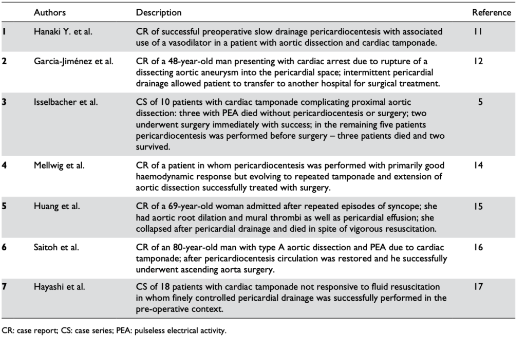
Pericardiocentesis can definitely be life-saving, restoring vital organ perfusion and buying time to get the patient to definitive surgery. Numerous case reports and case series provide evidence of its utility, even in patients in PEA cardiac arrest(1). The authors of the two largest cases series both used 8F pigtail drainage catheters(1,2).


Reports in the literature regarding the pericardiocentesis of critical cardiac tamponade complicating aortic dissection from Cruz et al


One key component of procedural success was controlled pericardial drainage, removing small volumes and reassessing the blood pressure, aiming for a SBP of 90 mmHg. The danger is overshooting, resulting in hypertension and extending the underlying aortic dissection which can be fatal (3).


Those still unconvinced by the evidence may be swayed by guidelines. The 2015 European Society of Cardiology Guidelines for the diagnosis and management of pericardial diseases (4) state:


“In the setting of aortic dissection with haemopericardium and suspicion of cardiac tamponade, emergency transthoracic echocardiography or a CT scan should be performed to confirm the diagnosis. In such a scenario, controlled pericardial drainage of very small amounts of the haemopericardium can be attempted to temporarily stabilize the patient in order to maintain blood pressure at 90 mmHg. (Class IIa, Level C)”


Intubation

Deterioration of tamponade patients following intubation is well described in the literature and the risk is well appreciated by cardiothoracic anaesthetists(5).


Once positive pressure ventilation is started, positive pleural pressure is transmitted to the pericardium, where pressures can exceed right ventricular diastolic pressure and prevent cardiac filling. The result is a fall in and possible loss of cardiac output. This is further exacerbated by the addition of PEEP(6).


One suggested approach if the patient must be intubated for airway protection but is not yet in the operating room with a surgeon ready to cut, is to consider intubation under local anaesthesia and allow the patient to breathe spontaneously (maintaining negative pleural pressure) through the tube until the surgeon is ready to open the chest(5).


Alternatively preload with fluid, use cautious doses of induction agent, and ventilate with low tidal volumes and zero PEEP. However the patient can still crash, so remember that these effects of ventilation on cardiac output in tamponade can be mitigated by the removal of a relatively small volume of pericardial fluid(6).


Cardiac Arrest

In cardiac arrest, external chest compressions are unlikely to be of benefit. In a study on baboons subjected to cardiac tamponade, closed chest massage resulted in an increase in intrapericardial pressure. There was an increase in systolic pressure, but a marked decrease in diastolic pressure, with an overall decrease in mean arterial pressure(7).
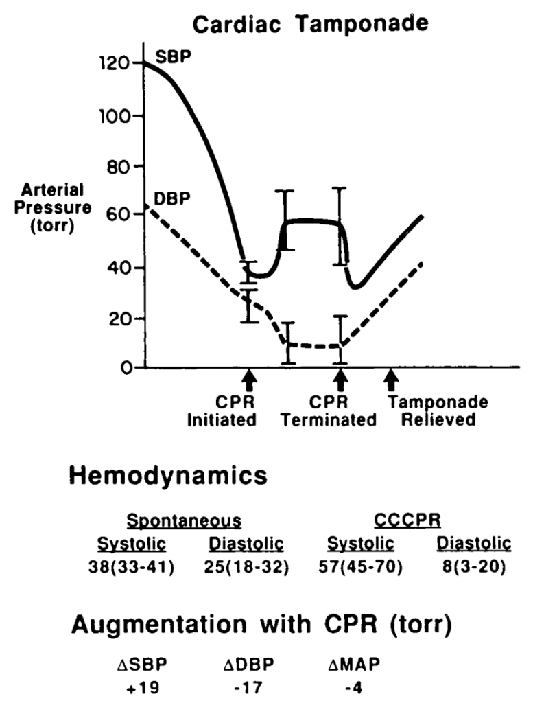
Pressure changes from CPR during tamponade in baboons. From Luna et al (7)
This would lead to impaired coronary perfusion and would be very unlikely to result in return of spontaneous circulation (ROSC). In the clinical situation described above, it is only relief of tamponade that is going to provide an arrested patient with a chance of recovery.


Transport

For patients with cardiac tamponade requiring interhospital (or intrahospital) transfer, it would seem vital therefore that the patient is accompanied by a clinician willing and capable to perform pericardiocentesis in the event of severe deterioriation or arrest en route. This simple life-saving intervention to deliver the patient alive to the operating room should be made available should the need arise.


Summary

  • Patients presenting in shock from cardiac tamponade often have treatable underlying causes and represent a situation where the planning and actions of the resuscitationist can be truly life-saving.
  • Pericardiocentesis is recommended in profound shock to buy time for definitive intervention. Controlled pericardiocentesis should be performed paying strict attention to SBP to avoid ‘overshooting’ to a hypertensive state in type A aortic dissection. In cardiac arrest, chest compressions are likely to be ineffective and pericardiocentesis is mandatory for ROSC.
  • The institution of positive pressure ventilation often results in worsened shock or cardiac arrest, and this is exacerbated by PEEP. Where possible, avoid intubation until the patient is in the operating room, or use low tidal volumes and no PEEP. Even then pericardiocentesis may be necessary to maintain or restore cardiac output.
  • Patients requiring transport who have tamponade should be accompanied by a clinician able to perform pericardiocentesis in the event of en route deterioration.


References

  1. Cruz I, Stuart B, Caldeira D, Morgado G, Gomes AC, Almeida AR, et al. Controlled pericardiocentesis in patients with cardiac tamponade complicating aortic dissection: Experience of a centre without cardiothoracic surgery. European Heart Journal: Acute Cardiovascular Care. 2015 Mar 19;4(2):124–8.
  2. Hayashi T, Tsukube T, Yamashita T, Haraguchi T, Matsukawa R, Kozawa S, et al. Impact of Controlled Pericardial Drainage on Critical Cardiac Tamponade With Acute Type A Aortic Dissection. Circulation. 2012 Sep 10;126(11_suppl_1):S97–S101.
  3. Isselbacher EM, Cigarroa JE, Eagle KA. Cardiac tamponade complicating proximal aortic dissection. Is pericardiocentesis harmful? Circulation. 1994 Nov 1;90(5):2375–8.
  4. Adler Y, Charron P, Imazio M, Badano L, Barón-Esquivias G, Bogaert J, et al. 2015 ESC Guidelines for the diagnosis and management of pericardial diseases. European Heart Journal. 2015 Nov 7;36(42):2921–64.
  5. Ho AMH, Graham CA, Ng CSH, Yeung JHH, Dion PW, Critchley LAH, et al. Timing of tracheal intubation in traumatic cardiac tamponade: A word of caution. Resuscitation. 2009 Feb;80(2):272–4.
  6. Möller CT, Schoonbee CG, Rosendorff C. Haemodynamics of cardiac tamponade during various modes of ventilation. Br J Anaesth. 1979 May;51(5):409–15.
  7. Luna GK, Pavlin EG, Kirkman T, Copass MK, Rice CL. Hemodynamic effects of external cardiac massage in trauma shock. The Journal of Trauma: Injury, Infection, and Critical Care. 1989 Oct;29(10):1430–3.
CPR in Pectus Excavatum

nussSome pectus excavatum patients have a metal ‘Nuss bar’ inserted below the sternum which can make chest compressions more difficult. In those without one, standard compression depths compress the left ventricle more than in non-pectus subjects, and might lead to myocardial injury.
This has led to a recommendation in the journal Resuscitation:
Until further studies are available, we recommend strong chest compressions, according to the current guidelines, in PE patients with a sternal Nuss bar and, to minimize the risk of myocardial injury, we suggest a reduced chest compression depth (approximately 3–4 cm) at the level of lower half of the sternum in PE patients who have not had corrective surgery.
 
Cardiopulmonary resuscitation in pectus excavatum patients: Is it time to say more?
Resuscitation. 2014 Dec 10.[Epub ahead of print]

London Cardiac Arrest Symposium 2014

The focus of the entire day is cardiac arrest and this is the second day of the London Cardiac Arrest Symposium.

Professor Niklas Nielsen kicked off with a presentation of his Targeted Temperature Management trial.  It seems that even now there is uncertainty in the interpretation of this latest study. I take heart from the knowledge that Prof Nielsen has changed the practice of his institution to reflect the findings of his study – I have certainly changed my practice. But we need to remain aware that there is more work to be done to answer the multiple questions that remain and the need for further RCTs is recognised.

The management of Cardiac arrest after avalanche is not a clinical scenario that I imagine I’ll ever find myself in. The management is well documented in the ICAR MEDCOM guidelines 2012. Dr Peter Paal reminded us that you’re not dead until you’re rewarmed and dead unless: with asystole, CPR may be terminated (or withheld) if a patient is lethally injured or completely frozen, the airway is blocked and duration of burial >35 min, serum potassium >12 mmol L(-1), risk to the rescuers is unacceptably high or a valid do-not-resuscitate order exists.

The age old question about prognostication after cardiac arrest was tackled by Prof Mauro Oddo. He covered the evidence for clinical examination, SSPE, EEG, and neurone specific enolase. Bottom line, all of these modalities are useful but none are specific enough to be used as a stand alone test so multiple modalities are required.

SAMU is leading the way with prehospital ECMO. They have mastered the art of cannulation (in the Louvre no less!) but there haven’t enough cases to demonstrate a mortality benefit. The commencement of ECMO prehospital reduces low flow time and theoretically should improve outcomes. This is begging for a RCT.

The experience of the Italians with in hospital ECMO shoes a better survival rate for in-hospital rather than out of hospital cardiac arrests, explained Dr Tomasso Mauri. They treat patients with a no flow time of <6min and low flow rate of <45min and had a 31% ICU survival rate. If you want to learn more about ED ECMO go to http://edecmo.org.

VA-ECMO

The Douglas Chamberlain lecture this year was Selective aortic arch perfusion presented by Prof James Manning. He spoke about the use of this technique in cardiac arrest and also in trauma (where it is known to you as Zone 1 REBOA).

image-1

In cardiac arrest the aim is to improve coronary perfusion, to preserve perfusion to the heart and the brain, offer a route of rapid temperature control and offer a direct route of administration of adrenaline. Coronary perfusion is seen to be supra normal after SAAP. And the suggested place for SAAP is prior to ECMO.

image-5

It’s more familiar ground talking about SAAP in trauma. This Zone 1 occlusion preserves cerebral and cardiac perfusion while blood loss is limited and rapid fluid resuscitation can occur.

image-3

You can hear Prof Manning on SAAP over at EMCrit (of course!). 

It’s been another great conference. Put the dates for next year’s London Trauma & Cardiac Arrest Conferences in your diary: 8th-10th December 2015!

Happy Holidays & Keep Well

Louisa Chan


Early femur fixation recommended

The Eastern Association for the Surgery of Trauma recommends early (<24 hours) open reduction and internal fracture fixation in trauma patients with open or closed femur fractures.
They acknowledge the strength of the evidence is low, but suggests a trend toward lower risk of infection, mortality, and venous thromboembolic disease.
They conclude: “the desirable effects of early femur fracture stabilization probably outweigh the undesirable effects in most patients
Check out the rest of the EAST Trauma Guidelines
Optimal timing of femur fracture stabilization in polytrauma patients: A practice management guideline from the Eastern Association for the Surgery of Trauma
Journal of Trauma and Acute Care Surgery 2014;77(5):787-795
[EXPAND Abstract]


BACKGROUND
Femur fractures are common among trauma patients and are typically seen in patients with multiple injuries resulting from high-energy mechanisms. Internal fixation with intramedullary nailing is the ideal method of treatment; however, there is no consensus regarding the optimal timing for internal fixation. We critically evaluated the literature regarding the benefit of early (<24 hours) versus late (>24 hours) open reduction and internal fixation of open or closed femur fractures on mortality, infection, and venous thromboembolism (VTE) in trauma patients.

METHODS
A subcommittee of the Practice Management Guideline Committee of the Eastern Association for the Surgery of Trauma conducted a systematic review and meta-analysis for the earlier question. RevMan software was used to generate forest plots. Grading of Recommendations, Assessment, Development, and Evaluations methodology was used to rate the quality of the evidence, using GRADEpro software to create evidence tables.

RESULTS
No significant reduction in mortality was associated with early stabilization, with a risk ratio (RR) of 0.74 (95% confidence interval [CI], 0.50–1.08). The quality of evidence was rated as “low.” No significant reduction in infection (RR, 0.4; 95% CI, 0.10–1.6) or VTE (RR, 0.63; 95% CI, 0.37–1.07) was associated with early stabilization. The quality of evidence was rated “low.”

CONCLUSION
In trauma patients with open or closed femur fractures, we suggest early (<24 hours) open reduction and internal fracture fixation. This recommendation is conditional because the strength of the evidence is low. Early stabilization of femur fractures shows a trend (statistically insignificant) toward lower risk of infection, mortality, and VTE. Therefore, the panel concludes the desirable effects of early femur fracture stabilization probably outweigh the undesirable effects in most patients.

[/EXPAND]

ARISE study: EGDT no better than standard care

periph-vasoactive-iconThe second of three major trials assessing early goal directed therapy (EGDT) in sepsis – the Australasian ARISE Trial – has been published.
ARISE tested the hypothesis that EGDT, as compared with usual care, would decrease 90-day all-cause mortality among patients presenting to the emergency department with early septic shock in diverse health care settings.
There was no difference in all-cause mortality at 90 days between EGDT and standard care, in keeping with the results from ProCESS.
Why are the results so different from Rivers’ original EGDT study? The authors explain:


“although our results differ from those in the original trial, they are consistent with previous studies showing that bias in small, single-center trials may lead to inflated effect sizes”

This cautions us all against making major practice changes based on one single centre study. In critical care we’ve learned this before with subjects like tight glycaemic control and Activated Protein C. However I do believe that the things we know to be of benefit – early recognition, source control, antibiotics, and fluids – are effective in making ‘standard’ care “as good as” EGDT because of heightened awareness of the condition and its treatment, and Rivers’ initial study and the subsequent Surviving Sepsis Campaign Guidelines have played a major role in raising that awareness.
The ARISE study is appraised by Wessex’s The Bottom Line and discussed on the one and only EMCrit podcast.
The ARISE Investigators and the ANZICS Clinical Trials Group.
Goal-Directed Resuscitation for Patients with Early Septic Shock
N Engl J Med. 2014 Oct;:141001063014008.Full Text
[EXPAND Abstract]


Background
Early goal-directed therapy (EGDT) has been endorsed in the guidelines of the Surviving Sepsis Campaign as a key strategy to decrease mortality among patients presenting to the emergency department with septic shock. However, its effectiveness is uncertain.

Methods In this trial conducted at 51 centers (mostly in Australia or New Zealand), we randomly assigned patients presenting to the emergency department with early septic shock to receive either EGDT or usual care. The primary outcome was all-cause mortality within 90 days after randomization.

Results Of the 1600 enrolled patients, 796 were assigned to the EGDT group and 804 to the usual-care group. Primary outcome data were available for more than 99% of the patients. Patients in the EGDT group received a larger mean (±SD) volume of intravenous fluids in the first 6 hours after randomization than did those in the usual-care group (1964±1415 ml vs. 1713±1401 ml) and were more likely to receive vasopressor infusions (66.6% vs. 57.8%), red-cell transfusions (13.6% vs. 7.0%), and dobutamine (15.4% vs. 2.6%) (P<0.001 for all comparisons). At 90 days after randomization, 147 deaths had occurred in the EGDT group and 150 had occurred in the usual-care group, for rates of death of 18.6% and 18.8%, respectively (absolute risk difference with EGDT vs. usual care, -0.3 percentage points; 95% confidence interval, -4.1 to 3.6; P=0.90). There was no significant difference in survival time, in-hospital mortality, duration of organ support, or length of hospital stay.

Conclusions In critically ill patients presenting to the emergency department with early septic shock, EGDT did not reduce all-cause mortality at 90 days.

[/EXPAND]

Non-ST-Elevation Acute Coronary Syndromes

The latest AHA/ACC guidelines on NSTEACS have been published ahead of print in Circulation.
Full text is available, and the Executive Summary is available here
Amsterdam EA, Wenger NK, Brindis RG, Casey DE, Ganiats TG, Holmes DR, et al.
2014 AHA/ACC Guideline for the Management of Patients With Non-ST-Elevation Acute Coronary Syndromes: Executive Summary: A Report of the American College of Cardiology/American Heart Association Task Force on Practice Guidelines.
Circulation. 2014 Sep 23. [Epub ahead of print]

No Benefit From Early Goal Directed Therapy

The first of three major trials assessing early goal directed therapy (EGDT) in sepsis – the American ProCESS Trial – has been published.
It showed what many of us thought – that the specific monitoring via a central line of central venous oxygen saturation – was not necessary for improved survival.
However the trial randomised 1341 patients to one of three arms:
(1) protocolised EGDT
(2) protocol-based standard therapy that did not require the placement of a central venous catheter, administration of inotropes, or blood transfusions
(3) ‘usual care’ which was not standardised.
There were no differences in any of the primary or secondary outcomes between the groups.
Interestingly, in the six hours of early care that the trial dictated, the volume of intravenous fluids administered differed significantly among the groups (2.8 litres in the protocol-based EGDT group, 3.3 litres in the protocol-based standard-therapy group, and 2.3 litres in the usual-care group).
There was also a difference in the amount of vasopressor given, with more patients in the two protocol-based groups receiving vasopressors (54.9% in the protocol-based EGDT group, 52.2% in the protocol-based standard-therapy group, 44.1% in the usual-care group).
The use of intravenous fluids, vasopressors, dobutamine, and blood transfusions between 6 and 72 hours did not differ significantly among the groups.
Overall 60 day mortality was in the region of 20% for all groups.
What are the take home points here? Firstly, overall sepsis outcomes have improved over recent years, and early recognition and antibiotic administration may be the most important components of care. In the early emergency department phase of care, protocolised fluid and vasopressor therapy may not be as important as we thought. Good clinical assessment and regular review seem to be as effective and perhaps more important than any specific monitoring modality or oxygen delivery-targeted drug and blood therapy.
We all await the ARISE and ProMISE studies which may shed more light on the most important components of early sepsis care.
A Randomized Trial of Protocol-Based Care for Early Septic Shock
NEJM Mar 18 2014 (Full Text Link)
[EXPAND Abstract]


Background: In a single-center study published more than a decade ago involving patients presenting to the emergency department with severe sepsis and septic shock, mortality was markedly lower among those who were treated according to a 6-hour protocol of early goal-directed therapy (EGDT), in which intravenous fluids, vasopressors, inotropes, and blood transfusions were adjusted to reach central hemodynamic targets, than among those receiving usual care. We conducted a trial to determine whether these findings were generalizable and whether all aspects of the protocol were necessary.

Methods: In 31 emergency departments in the United States, we randomly assigned patients with septic shock to one of three groups for 6 hours of resuscitation: protocol-based EGDT; protocol-based standard therapy that did not require the placement of a central venous catheter, administration of inotropes, or blood transfusions; or usual care. The primary end point was 60-day in-hospital mortality. We tested sequentially whether protocol-based care (EGDT and standard-therapy groups combined) was superior to usual care and whether protocol-based EGDT was superior to protocol-based standard therapy. Secondary outcomes included longer-term mortality and the need for organ support.

Results: We enrolled 1341 patients, of whom 439 were randomly assigned to protocol-based EGDT, 446 to protocol-based standard therapy, and 456 to usual care. Resuscitation strategies differed significantly with respect to the monitoring of central venous pressure and oxygen and the use of intravenous fluids, vasopressors, inotropes, and blood transfusions. By 60 days, there were 92 deaths in the protocol-based EGDT group (21.0%), 81 in the protocol-based standard-therapy group (18.2%), and 86 in the usual-care group (18.9%) (relative risk with protocol-based therapy vs. usual care, 1.04; 95% confidence interval [CI], 0.82 to 1.31; P=0.83; relative risk with protocol-based EGDT vs. protocol-based standard therapy, 1.15; 95% CI, 0.88 to 1.51; P=0.31). There were no significant differences in 90-day mortality, 1-year mortality, or the need for organ support.

Conclusions: In a multicenter trial conducted in the tertiary care setting, protocol-based resuscitation of patients in whom septic shock was diagnosed in the emergency department did not improve outcomes

[/EXPAND]

Is 4 Joules per kg enough in kids?

glash-sim-paed-face-smResearchers from the Iberian-American Paediatric Cardiac Arrest Study Network challenge the evidence base behind defibrillation shock dose recommendations in children.
In a study of in-hospital pediatric cardiac arrest due to VT or VF, clinical outcome was not related to the cause or location of arrest, type of defibrillator and waveform, energy dose per shock, number of shocks, or cumulative energy dose, although there was a trend to better survival with higher doses per shock. 50% of children required more than the recommended 4J per kg and in over a quarter three or more shocks were needed to achieve defibrillation.
 
Shockable rhythms and defibrillation during in-hospital pediatric cardiac arrest
Resuscitation. 2014 Mar;85(3):387-91
[EXPAND Abstract]


OBJECTIVE: To analyze the results of cardiopulmonary resuscitation (CPR) that included defibrillation during in-hospital cardiac arrest (IH-CA) in children.

METHODS: A prospective multicenter, international, observational study on pediatric IH-CA in 12 European and Latin American countries, during 24 months. Data from 502 children between 1 month and 18 years were collected using the Utstein template. Patients with a shockable rhythm that was treated by electric shock(s) were included. The primary endpoint was survival at hospital discharge. Univariate logistic regression analysis was performed to find outcome factors.

RESULTS: Forty events in 37 children (mean age 48 months, IQR: 7-15 months) were analyzed. An underlying disease was present in 81.1% of cases and 24.3% had a previous CA. The main cause of arrest was a cardiac disease (56.8%). In 17 episodes (42.5%) ventricular fibrillation (VF) or pulseless ventricular tachycardia (pVT) was the first documented rhythm, and in 23 (57.5%) it developed during CPR efforts. In 11 patients (27.5%) three or more shocks were needed to achieve defibrillation. Return of spontaneous circulation (ROSC) was obtained in 25 cases (62.5%), that was sustained in 20 (50.0%); however only 12 children (32.4%) survived to hospital discharge. Children with VF/pVT as first documented rhythm had better sustained ROSC (64.7% vs. 39.1%, p=0.046) and survival to hospital discharge rates (58.8% vs. 21.7%, p=0.02) than those with subsequent VF/pVT. Survival rate was inversely related to duration of CPR. Clinical outcome was not related to the cause or location of arrest, type of defibrillator and waveform, energy dose per shock, number of shocks, or cumulative energy dose, although there was a trend to better survival with higher doses per shock (25.0% with <2Jkg(-1), 43.4% with 2-4Jkg(-1) and 50.0% with >4Jkg(-1)) and worse with higher number of shocks and cumulative energy dose.

CONCLUSION: The termination of pediatric VF/pVT in the IH-CA setting is achieved in a low percentage of instances with one electrical shock at 4Jkg(-1). When VF/pVT is the first documented rhythm, the results of defibrillation are better than in the case of subsequent VF/pVT. No clear relationship between defibrillation protocol and ROSC or survival has been observed. The optimal pediatric defibrillation dose remains to be determined; therefore current resuscitation guidelines cannot be considered evidence-based, and additional research is needed.

[/EXPAND]