Tag Archives: dysrhythmia

Spot the WOBBLER in syncope!

Syncope is a common ED presentation. An ECG is a critical investigation in syncope to identify the cause, including rare conditions associated with risk of sudden cardiac death.
So we should be really grateful when we are invited to interpret an ECG while we’re in the middle of six other tasks.

The problem with syncope is that some of the important life-threatening causes have fairly obscure ECG features that might be hard to remember. Some of these disorders and their ECG features are not entirely familiar to the clinicians who first screen the ECG.

When you’re busy and cognitively stretched you can save time and reduce the risk of missing important findings by having a structured, memorable checklist. I use the acronym WOBBLER, because I don’t want these people to wobble and kiss the dirt again.

The nice thing about WOBBLER is that it uses the sequence that you follow when you look at an ECG, ie from left to right, or from P wave to T wave.

The key is that this is for ECGs without obvious ischaemia or dysrhythmia. If you see something like this (STEMI):

or this (VT):

you don’t need WOBBLER, you need to be treating that patient.

So we apply WOBBLER to screen the ECG of well-looking syncope patients without obvious ischaemia or dysrhythmia.

Here goes…

W is Wolff-Parkinson-White syndrome – look for a short PR interval or delta wave:

O is obstructed AV pathway – look for 2nd or 3rd degree block:

or axis deviation:

…which is the first step in looking for B bifascicular block, or the combination of axis deviation and right bundle branch block:

the second B is Brugada, looking for characteristic morphology of the ST segment, so called coved ST elevation:

Now syncope, especially exertional syncope, can be caused by left ventricular outflow tract obstruction. Two conditions not to be missed associated with LVOTO (and exertional syncope) are hypertrophic cardiomyopathy and aortic stenosis. These both characteristically cause Lleft ventricular hypertrophy:

This example of Hypertrophic Cardiomyopathy has voltage criteria for LVH in the precordial leads as well as narrow, “dagger-like” Q waves in lateral and inferior leads

E– stands for epsilon wave, a feature of arrythmogenic right ventricular dysplasia, a rare disorder associated with sudden cardiac death. The epsilon wave looks a bit like the J wave of hypothermia and may be associated with other T wave abnormalities in V1-V3:

Epsilon wave in more detail

Finally, R stands for Repolarisation abnormality, particularly delayed Repolarisation as found in long QT syndrome:

but remember there is also a short QT syndrome too:

So WOBBLER may help you find the important and rare abnormalities not to be missed in the syncope patient, going from left to right from P wave through to T wave, in the patient that does not have obvious dysrhythmia or ischaemia. I hope you find it a helpful cognitive forcing tool.

All ECGs reproduced with kind permission of Life in the Fast Lane

Pre-SMACC mini RAGE

Currently the RAGE Podcast site is recovering from a cold, so here are the show notes for the pre-SMACC mini RAGE episode released June 2015.
Here is the podcast
And here are the references:
SMACC Conference
It’s a knockout

GoodSAM
GoodSAM App
Oxygen therapy
AVOIDthis2
AVOID: Air Versus Oxygen in ST-Segment Elevation Myocardial Infarction.
HOT or NOT trial: HyperOxic Therapy OR NormOxic Therapy after out-of-hospital cardiac arrest (HOT OR NOT): a randomised controlled feasibility trial.
Helicopter Emergency Medical Services
Survival benefit of a physician-staffed HEMS assistance for severely injured patients
Willingness to pay for lives saved by HEMS
Intraosseous access
Intraosseous infusion rates under high pressure: a cadaveric comparison of anatomic sites.
Intraosseous hypertonic saline: myonecrosis in swine
Intraosseous hypertonic saline: safe in swine
Discussion post about intraosseous hypertonic saline at Sydney HEMS
Handstands
Handstands: a treatment for supraventricular tachycardia?
Impact of a modified Valsalva manoeuvre in the termination of paroxysmal supraventricular tachycardia
Handstands keep you awake
 
The next RAGE Podcast will air late August / early September

Esmolol for refractory VF

Already well publicised on social media, the team at Hennepin County published a retrospective comparison between patients with refractory VF who received esmolol with those who did not(1). The results are impressive and I look forward to further studies on this.
I work in an ED in a hospital with no cath lab and no access to extracorporeal life support, limiting our options for patients who remain in shockable rhythms despite ACLS interventions. We now have esmolol available in our resus room. You might want to keep it in your list of options for ACLS-refractory VF, which might also include double sequential external defibrillation(2) and even stellate ganglion block.
The dose of esmolol used was: loading dose 500 mcg/kg, followed by infusions of 0, 50, or 100 mcg/kg/min
An important point to note in the esmolol study is that almost all patients received high-quality mechanical CPR with the combined use of an impedence threshold device to augment venous return and cardiac output. The authors “speculate that this additional hemodynamic support may be essential given the hypotensive effects of esmolol.”
1. Use of esmolol after failure of standard cardiopulmonary resuscitation to treat patients with refractory ventricular fibrillation
Resuscitation. 2014 Oct;85(10):1337-41
[EXPAND Abstract]


INTRODUCTION: We compare the outcomes for patients who received esmolol to those who did not receive esmolol during refractory ventricular fibrillation (RVF) in the emergency department (ED).

METHODS: A retrospective investigation in an urban academic ED of patients between January 2011 and January 2014 of patients with out-of-hospital or ED cardiac arrest (CA) with an initial rhythm of ventricular fibrillation (VF) or ventricular tachycardia (VT) who received at least three defibrillation attempts, 300mg of amiodarone, and 3mg of adrenaline, and who remained in CA upon ED arrival. Patients who received esmolol during CA were compared to those who did not.

RESULTS: 90 patients had CA with an initial rhythm of VF or VT; 65 patients were excluded, leaving 25 for analysis. Six patients received esmolol during cardiac arrest, and nineteen did not. All patients had ventricular dysrhythmias refractory to many defibrillation attempts, including defibrillation after administration of standard ACLS medications. Most received high doses of adrenaline, amiodarone, and sodium bicarbonate. Comparing the patients that received esmolol to those that did not: 67% and 42% had temporary return of spontaneous circulation (ROSC); 67% and 32% had sustained ROSC; 66% and 32% survived to intensive care unit admission; 50% and 16% survived to hospital discharge; and 50% and 11% survived to discharge with a favorable neurologic outcome, respectively.

CONCLUSION: Beta-blockade should be considered in patients with RVF in the ED prior to cessation of resuscitative efforts.

[/EXPAND]
2. Double Sequential External Defibrillation in Out-of-Hospital Refractory Ventricular Fibrillation: A Report of Ten Cases.
Prehosp Emerg Care. 2015 January-March;19(1):126-130
[EXPAND Abstract]


Background. Ventricular fibrillation (VF) is considered the out-of-hospital cardiac arrest (OOHCA) rhythm with the highest likelihood of neurologically intact survival. Unfortunately, there are occasions when VF does not respond to standard defibrillatory shocks. Current American Heart Association (AHA) guidelines acknowledge that the data are insufficient in determining the optimal pad placement, waveform, or energy level that produce the best conversion rates from OOHCA with VF.

Objective. To describe a technique of double sequential external defibrillation (DSED) for cases of refractory VF (RVF) during OOHCA resuscitation.

Methods. A retrospective case series was performed in an urban/suburban emergency medical services (EMS) system with advanced life support care and a population of 900,000. Included were all adult OOHCAs having RVF during resuscitation efforts by EMS providers. RVF was defined as persistent VF following at least 5 unsuccessful single shocks, epinephrine administration, and a dose of antiarrhythmic medication. Once the patient was in RVF, EMS personnel applied a second set of pads and utilized a second defibrillator for single defibrillation with the new monitor/pad placement. If VF continued, EMS personnel then utilized the original and second monitor/defibrillator charged to maximum energy, and shocks were delivered from both machines simultaneously. Data were collected from electronic dispatch and patient care reports for descriptive analysis.

Results. From 01/07/2008 to 12/31/2010, a total of 10 patients were treated with DSED. The median age was 76.5 (IQR: 65-82), with median resuscitation time of 51minutes (IQR: 45-62). The median number of single shocks was 6.5 (IQR: 6-11), with a median of 2 (IQR: 1-3) DSED shocks delivered. VF broke after DSED in 7 cases (70%). Only 3 patients (30%) had ROSC in the field, and none survived to discharge.

Conclusion. This case series demonstrates that DSED may be a feasible technique as part of an aggressive treatment plan for RVF in the out-of-hospital setting. In this series, RVF was terminated 70% of the time, but no patient survived to discharge. Further research is needed to better understand the characteristics of and treatment strategies for RVF.

[/EXPAND]

The 'Magic Eye®' method of rhythm assessment

Are you someone who tries to determine whether an ECG trace is ‘irregularly irregular’ by drawing little dots on a piece of paper level with the R waves to see if they are evenly spaced? I’d done that for years until I read this fantastic suggestion, which I’ve been following for over a year now.
In the 1990s there was a popular series of posters and books called ‘Magic Eye‘. These contained a ‘random dot autostereogram‘ which appeared as a mish-mash of coloured dots, but when you stared at it for a while the illusion of a 3D image would emerge. They looked a bit like this (although this one won’t work at such reduced resolution):

Image Credit: Wikimedia Commons

Dr Broughton and colleagues from Cambridge, UK, discovered that this technique, which involves forcing a divergent gaze to get repeating patterns to appear to overlap, can be applied to an ECG trace.
Stereoviewing an ECG trace causes successive QRS complexes to visually overlap and produce a new image. As Broughton and colleagues point out:
When achieved, this will lead to one of three outcomes. Entirely regular rhythms will ‘click’ into place as a new image at fixed depth. Rhythms with only mild irregularity may be stereoviewable, and if so, will appear to show successive QRS complexes at subtly varying depths. Rhythms with marked irregularity will not be stereoviewable, instead (in our experience) merely giving the viewer sore eyes after several failed viewing attempts.”
The authors assert that this can be applied to continuous ECG monitors, although unless you are really good at stereoviewing while moving your head/eyes horizontally, you should really freeze the trace on the screen first.
The ‘Magic Eye®’ method of rhythm assessment
Anaesthesia. 2012 Oct;67(10):1170-1

Caution with intraosseous adenosine

Two cases of failed cardioversion of SVT after tibial intraosseous administration of adenosine in infants are described in this month’s Pediatric Emergency Care. Both cases were subsequently cardioverted by intravenous adenosine. The maximum intraosseous dose given was 0.25 mg/kg. The successful IV doses were not higher than the IO doses.
It has been noted before that infants may require relatively higher doses of adenosine than children and that 0.2 mg/kg might even be considered a starting dose in infancy. I wonder if a bigger IO dose would have been effective, or whether a proximal humeral insertion site would make a difference. IO adenosine has been successfully used in infants and piglets.
This interesting case series provides a helpful caution in the management of paediatric SVT.

ABSTRACT: Supraventricular tachycardia (SVT) is a common tachyarrhythmia in the pediatric population that can necessitate immediate treatment. Adenosine has been well studied as a mainstay treatment, but the methods of adenosine administration have not been very well delineated. The intraosseous technique has presented itself as a possible method of administration. We describe 2 cases in which adenosine was administered through bone marrow infusion to convert SVT without success. The cases we describe show that intraosseous is not a reliable method of administering adenosine to stop SVT. Both patients presented with SVT refractory to vagal maneuvers and difficult intravenous placement. Intraosseous access was achieved, but administration of adenosine at increasing doses was unable to successfully convert the arrhythmia.

Intraosseous Infusion Is Unreliable for Adenosine Delivery in the Treatment of Supraventricular Tachycardia
Pediatr Emerg Care. 2012 Jan;28(1):47-8

AF in sepsis and risk of stroke

Atrial fibrillation can occur in the setting of severe sepsis, and often presents a therapeutic conundrum for critical care physicians, in that it can be relatively resistant to treatment until the sepsis has resolved, and its prognostic significance is unclear. A new study on a massive dataset shows atrial fibrillation in the setting of severe sepsis is associated with an increased risk of stroke and increased hospital mortality. Patients with severe sepsis who developed new-onset AF had a greater risk of in-hospital stroke than patients with preexisting AF and individuals without a history of AF.


Context New-onset atrial fibrillation (AF) has been reported in 6% to 20% of patients with severe sepsis. Chronic AF is a known risk factor for stroke and death, but the clinical significance of new-onset AF in the setting of severe sepsis is uncertain.

Objective To determine the in-hospital stroke and in-hospital mortality risks associated with new-onset AF in patients with severe sepsis.

Design and Setting Retrospective population-based cohort of California State Inpatient Database administrative claims data from nonfederal acute care hospitals for January 1 through December 31, 2007.

Patients Data were available for 3 144 787 hospitalized adults. Severe sepsis (n = 49 082 [1.56%]) was defined by validated International Classification of Diseases, Ninth Revision, Clinical Modification (ICD-9-CM) code 995.92. New-onset AF was defined as AF that occurred during the hospital stay, after excluding AF cases present at admission.

Main Outcome Measures A priori outcome measures were in-hospital ischemic stroke (ICD-9-CM codes 433, 434, or 436) and mortality.

Results Patients with severe sepsis were a mean age of 69 (SD, 16) years and 48% were women. New-onset AF occurred in 5.9% of patients with severe sepsis vs 0.65% of patients without severe sepsis (multivariable-adjusted odds ratio [OR], 6.82; 95% CI, 6.54-7.11; P < .001). Severe sepsis was present in 14% of all new-onset AF in hospitalized adults. Compared with severe sepsis patients without new-onset AF, patients with new-onset AF during severe sepsis had greater risks of in-hospital stroke (75/2896 [2.6%] vs 306/46 186 [0.6%] strokes; adjusted OR, 2.70; 95% CI, 2.05-3.57; P < .001) and in-hospital mortality (1629 [56%] vs 18 027 [39%] deaths; adjusted relative risk, 1.07; 95% CI, 1.04-1.11; P < .001). Findings were robust across 2 definitions of severe sepsis, multiple methods of addressing confounding, and multiple sensitivity analyses.
Conclusion Among patients with severe sepsis, patients with new-onset AF were at increased risk of in-hospital stroke and death compared with patients with no AF and patients with preexisting AF.

Incident Stroke and Mortality Associated With New-Onset Atrial Fibrillation in Patients Hospitalized With Severe Sepsis
JAMA. 2011 Nov 13. [Epub ahead of print]

Cardiac arrest caused by subarachnoid haemorrhage

We know that subarachnoid haemorrhage (SAH) can cause cardiac arrest. Some questions we may have about this are:

Questions

  • What proportion of out-of-hospital cardiac arrests (OOHCA) who achieve return of spontaneous circulation (ROSC) are caused by SAH?
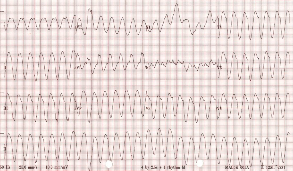
  • What is the usual presenting arrest rhythm – VT/VF or non-shockable rhythms?
  • What is the outcome of these patients – do any survive?
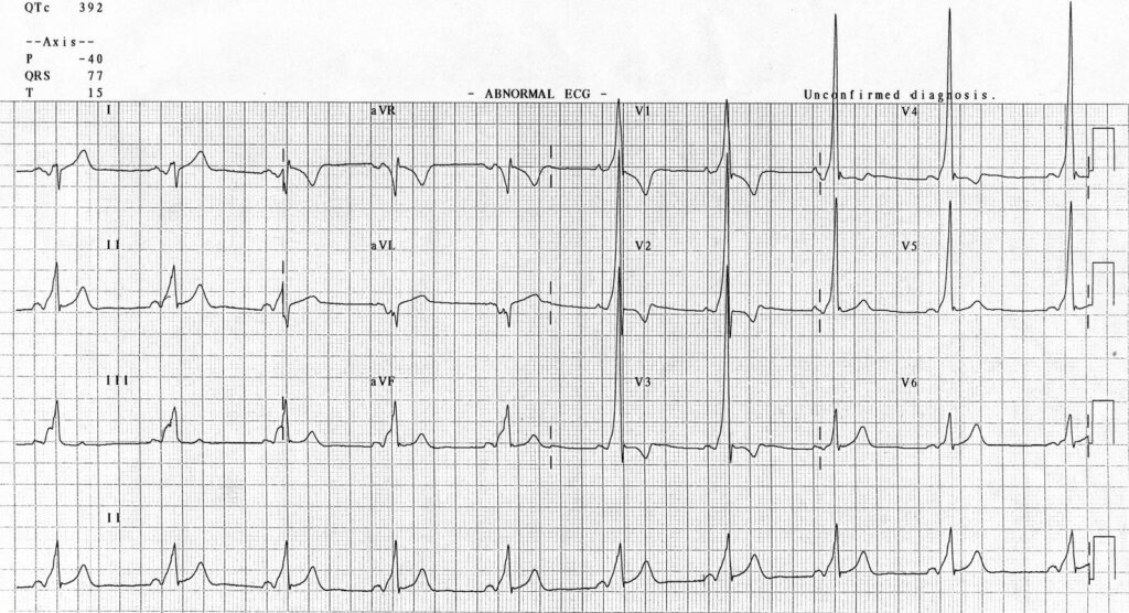
  • Do they have other characteristic cardiac features, such as ECG or echo abnormalities?
  • Should we do a head CT on all survivors of out-of-hospital cardiac arrest of uncertain aetiology?

A recent Japanese article in Resuscitation1 is the third from that country to be published on the topic in three years, the other two2,3 coming from different centres and all demonstrating some consistent answers, as do papers published in recent years from Europe4 and North America5:

Answers

  • Rates of SAH in OOHCA patients who achieve ROSC and make it to CT range from 4-16% (even higher if other sources of intracranial haemorrhage are included).
  • Studies consistently demonstrate VT/VF to be very rare – PEA and asystole are by far the commonest presenting arrest rhythms.
  • Almost no patients with this presentation due to SAH survive to hospital discharge.
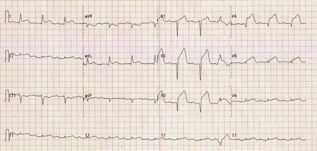
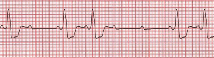
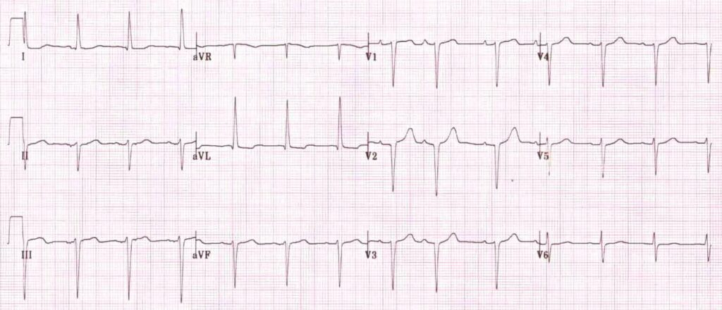
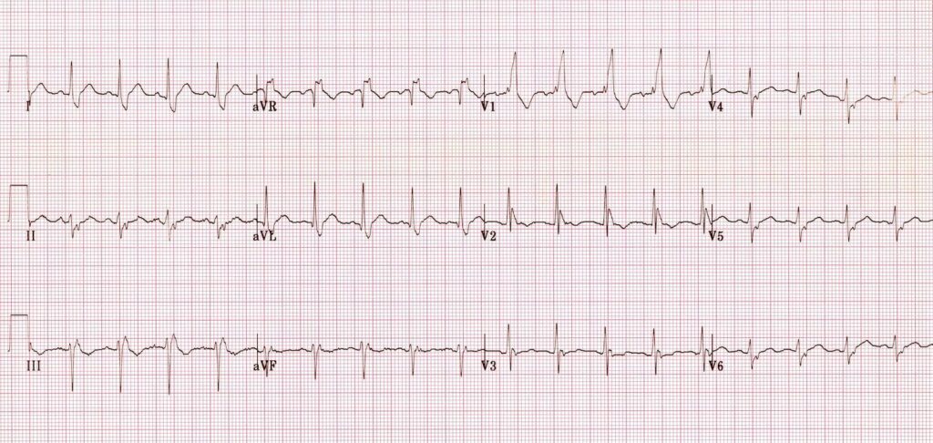
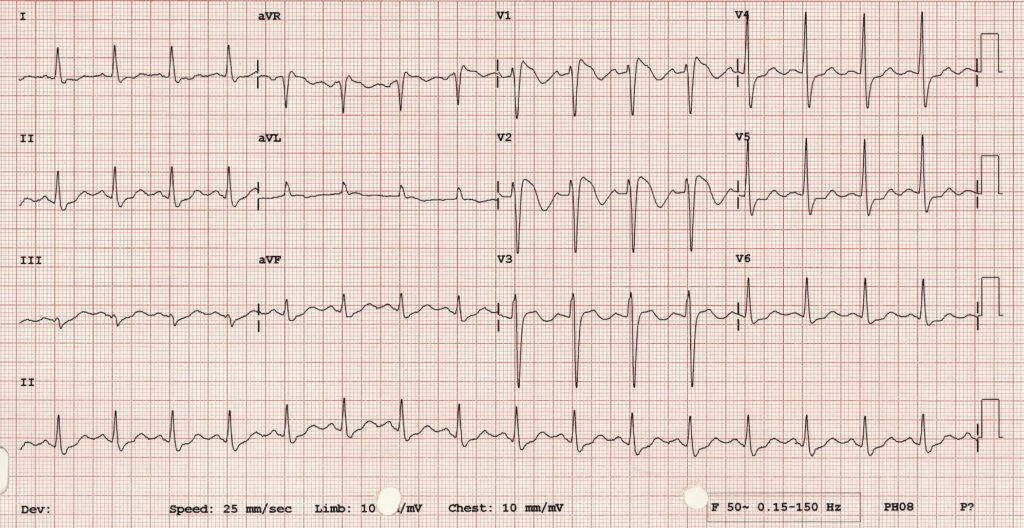
  • In the most recent study, all patients who survived long enough to get a 12 lead showed ST-T abnormalities and/or QT prolongation, although echocardiograms were mostly normal.
  • Rates of SAH in OOHCA patients who achieve ROSC seem to be sufficiently high to seriously consider head CT in these patients if there is no obvious alternate explanation for the arrest.

1. Clinical and cardiac features of patients with subarachnoid haemorrhage presenting with out-of-hospital cardiac arrest
Resuscitation. 2011 Oct;82(10):1294-7
[EXPAND Abstract]


Background Subarachnoid haemorrhage (SAH) is known as one of the aetiologies of out-of-hospital cardiac arrest (OHCA). However, the mechanisms of circulatory collapse in these patients have remained unclear.

Methods and results We examined 244 consecutive OHCA patients transferred to our emergency department. Head computed tomography was performed on all patients and revealed the existence of SAH in 14 patients (5.9%, 10 females). Among these, sudden collapse was witnessed in 7 patients (50%). On their initial cardiac rhythm, all 14 patients showed asystole or pulseless electrical activity, but no ventricular fibrillation (VF). Return of spontaneous circulation (ROSC) was obtained in 10 of the 14 patients (14.9% of all ROSC patients) although all resuscitated patients died later. The ROSC rate in patients with SAH (71%) was significantly higher than that of patients with either other types of intracranial haemorrhage (25%, n = 2/8) or presumed cardiovascular aetiologies (22%, n = 23/101) (p < 0.01). On electrocardiograms, ST-T abnormalities and/or QT prolongation were found in all 10 resuscitated patients. Despite their electrocardiographic abnormalities, only 3 patients showed echocardiographic abnormalities.

Conclusions The frequency of SAH in patients with all causes of OHCA was about 6%, and in resuscitated patients was about 15%. The initial cardiac rhythm revealed no VF even though half had a witnessed arrest. A high ROSC rate was observed in patients with SAH, although none survived to hospital discharge.

[/EXPAND]
2. Assessing outcome of out-of-hospital cardiac arrest due to subarachnoid hemorrhage using brain CT during or immediately after resuscitation
Signa Vitae 2010; 5(2): 21 – 24 Full Text
[EXPAND Abstract]


Objectives. The clinical course and outcome of out-of-hospital cardiopulmonary arrest (OHCPA) due to subarachnoid hemorrhage (SAH) is unclear. The objective of this study is to clarify them.

Study design. Single- center, observational study. Setting. We usually perform a brain computed tomography (CT) in OHCPA patients who present without a clear etiology (42% of all OHCPA), such as trauma, to determine the cause of OHCPA and to guide treatment.

Patients. The study included OHCPA patients without a clear etiology, who were transferred to our center and who underwent a brain CT during resuscitation.

Methods of measurement. Patients’ records were reviewed; initial cardiac rhythm, existence of a witness and bystander cardiopulmonary resuscitation efforts (CPR) were compared with patients’ outcomes.

Results. Sixty-six patients were enrolled. 72.7% achieved return of spontaneous circulation (ROSC), 71.2% were admitted, 30.3% survived more than 7 days, and 9.1. survived-to-discharge. In 41 witnessed OHCPA, 87.8% obtained ROSC, 85.4% were admitted, and 14.6% survived-to-discharge. All survivors were witnessed. In 25 non-witnessed OHCPA, 48% obtained ROSC and were admitted, and no patients were discharged. Initial cardiac rhythm was ventricular fibrillation (VF), pulseless electrical activity (PEA) and asystole in 3.0%, 39.4%, and 47.0%. In 2 VF patients 50.0% survived-to- discharge, and there was no survivor with PEA or asystole.

Conclusion. This study shows a high rate of ROSC and admission in OHCPA patients with a SAH, and also reveals their very poor neurological outcome. We conclude that the detection of a SAH in OHCPA patients is important to determine the accurate frequency of SAH in this patient group and to guide appropriate treatment of all OHCPA patients.

[/EXPAND]
3. Subarachnoid haemorrhage as a cause of out-of-hospital cardiac arrest: A prospective computed tomography study
Resuscitation. 2009 Sep;80(9):977-80
[EXPAND Abstract]


Aim Aneurysmal subarachnoid haemorrhage (SAH) is a relatively common cause of out-of-hospital cardiac arrest (OHCA). Early identification of SAH-induced OHCA with the use of brain computed tomography (CT) scan obtained immediately after resuscitation may help emergency physicians make therapeutic decision as quickly as they can.

Methods During the 4-year observation period, brain CT scan was obtained prospectively in 142 witnessed non-traumatic OHCA survivors who remained haemodynamically stable after resuscitation. Demographics and clinical characteristics of SAH-induced OHCA survivors were compared with those with “negative” CT finding.

Results Brain CT scan was feasible with an average door-to-CT time of 40.0min. SAH was found in 16.2% of the 142 OHCA survivors. Compared with 116 survivors who were negative for SAH, SAH-induced OHCA survivors were significantly more likely to be female, to have experienced a sudden headache, and trended to have achieved return of spontaneous circulation (ROSC) prior to arrival in the emergency department less frequently. Ventricular fibrillation (VF) was significantly less likely to be seen in SAH-induced than SAH-negative OHCA (OR, 0.06; 95% CI, 0.01–0.46). Similarly, Cardiac Trop-T assay was significantly less likely to be positive in SAH-induced OHCA (OR, 0.08; 95% CI, 0.01–0.61).

Conclusion Aneurysmal SAH causes OHCA more frequently than had been believed. Immediate brain CT scan may particularly be useful in excluding SAH-induced OHCA from thrombolytic trial enrollment, for whom the use of thrombolytics is contraindicated. The low VF incidence suggests that VF by itself may not be a common cause of SAH-induced OHCA.

[/EXPAND]
4. Spontaneous subarachnoid haemorrhage as a cause of out-of-hospital cardiac arrest
Resuscitation. 2001 Oct;51(1):27-32
[EXPAND Abstract]


Objective: Spontaneous subarachnoid haemorrhage as a cause of out-of-hospital cardiac arrest is poorly evaluated. We analyse disease-specific and emergency care data in order to improve the recognition of subarachnoid haemorrhage as a cause of cardiac arrest.

Design: We searched a registry of cardiac arrest patients admitted after primarily successful resuscitation to an emergency department retrospectively and analysed the records of subarachnoid haemorrhage patients for predictive features.

Results: Over 8.5 years, spontaneous subarachnoidal haemorrhage was identified as the immediate cause in 27 (4%) of 765 out-of-hospital cardiac arrests. Of these 27 patients, 24 (89%) presented with at least three or more of the following common features: female gender (63%), age under 40 years (44%), lack of co-morbidity (70%), headache prior to cardiac arrest (39%), asystole or pulseless electric activity as the initial cardiac rhythm (93%), and no recovery of brain stem reflexes (89%). In six patients (22%), an intraventricular drain was placed, one of them (4%) survived to hospital discharge with a favourable outcome.

Conclusions: Subarachnoid haemorrhage complicated by cardiac arrest is almost always fatal even when a spontaneous circulation can be restored initially. This is due to the severity of brain damage. Subarachnoid haemorrhage may present in young patients without any previous medical history with cardiac arrest masking the diagnosis initially.

[/EXPAND]
5. Cranial computed tomography in the resuscitated patient with cardiac arrest
Am J Emerg Med. 2009 Jan;27(1):63-7
[EXPAND Abstract]


Introduction The incidence of out-of-hospital and in-hospital cardiorespiratory arrest from all causes in the United States occurs not infrequently. Postresuscitation care should include the identification of the inciting arrest event as well as therapy tailored to support the patient and treat the primary cause of the decompensation. The application of one particular testing modality, cranial computed tomography (CT) of the head, has not yet been determined. We undertook an evaluation of the use of head CT in patients who were resuscitated from cardiac arrest.

Methods Prehospital (emergency medical services), ED, and hospital records were reviewed for patients of all ages with cardiorespiratory arrest over a 4-year period (July 1996-June 2000). Information regarding diagnosis, management, and outcome was recorded. The results of cranial CT, if performed, and any apparent resulting therapeutic changes were recorded. Patients with a known traumatic mechanism for the cardiorespiratory arrest were excluded.

Results A total of 454 patients (mean age 58.3 years with 60% male) with cardiorespiratory arrest were entered in the study with 98 (22%) individuals (mean age 58.5 years with 53% male) undergoing cranial CT. Arrest location was as follows: emergency medical services, 41 (42%); ED, 11 (11%); and hospital, 46 (47%). Seventy-eight (79%) patients demonstrated 111 CT abnormalities: edema, 35 (32%); atrophy, 24 (22%); extra-axial hemorrhage, 14 (13%); old infarct, 12 (11%); new infarct, 11 (10%); intraparenchymal hemorrhage, 6 (5%); skull fracture, 5 (4%); mass, 3 (2%); and foreign body, 1 (1%). Therapeutic and diagnostic alterations in care were made in 38 (39%) patients—35 abnormal and 3 normal CTs. The following alterations occurred: medication administration, 26; withdrawal of life support, 7; additional diagnostic study, 6; neurologic consultation, 6; and intracranial pressure monitoring. 4. No patient survived to discharge.

Conclusion In this subset of resuscitated patients with cardiac arrest, abnormalities on the head CT were not uncommon. Alterations in management did occur in those patients with abnormalities. The indications and impact of head CT in the population of resuscitated patients with cardiac arrest remain unknown, warranting further investigation.

[/EXPAND]

Verapamil vs adenosine for SVT

Most people reach for the adenosine once vagotonic manouevres have failed in SVT, but some patients find the side effects – albeit short-lived – pretty unpleasant. For this reason I’ve heard Jerry Hoffman espouse the relative benefits of verapamil in patients without contra-indications. A recent meta-analysis suggests both verapamil and adenosine have about a 90% success rate. The study did not look at recurrence rates of SVT, which one might expect to be higher with the shorter-acting adenosine.
The authors conclude:
The choice between the agents should be made on a case by case basis with awareness of the respective adverse effect profiles, and should involve informed discussion with the patient where appropriate.

OBJECTIVE: Verapamil and adenosine are the most common agents used to treat paroxysmal supraventricular tachycardia (PSVT). We performed a systematic review and meta-analysis to determine the relative effectiveness of these drugs and to examine their respective adverse effect profiles.
METHODS: We searched MEDLINE, EMBASE, CINAHL, the Cochrane database, and international clinical trial registers for randomized controlled trials comparing adenosine (or adenosine compounds) with verapamil for the treatment of PSVT in stable adult patients. The primary outcome was rate of reversion to sinus rhythm. Secondary outcome was occurrence of pooled adverse events. Odds ratios and 95% confidence intervals (CIs) were calculated using a random effects model (RevMan v5).
RESULTS: Eight trials were appropriate and had the available data. The reversion rate for adenosine was 90.8% (95% CI: 87.3-93.4%) compared with 89.9% for verapamil (95% CI: 86.0-92.9%). The pooled odds ratio for successful reversion was 1.27 (95% CI: 0.63-2.57) favouring adenosine. This was not statistically significant. There was a higher rate of minor adverse effects described with adenosine (16.7-76%) compared with verapamil (0-9.9%). The rate of hypotension was lower with adenosine [0.6% (95% CI: 0.1-2.4%)] compared with verapamil [3.7% (95% CI: 1.9-6.9%)].
CONCLUSION: Adenosine and verapamil have similar efficacy in treating PSVT. Adenosine has a higher rate of minor adverse effects, and of overall adverse effects, whereas verapamil has a higher rate of causing hypotension. A decision between the two agents should be made on a case-by-case basis and ideally involve informed discussion with the patient where appropriate.

The relative efficacy of adenosine versus verapamil for the treatment of stable paroxysmal supraventricular tachycardia in adults: a meta-analysis
Eur J Emerg Med. 2011 Jun;18(3):148-52

Drugs in cardiac arrest – guess what works?

Just like epinephrine (adrenaline), amiodarone does not increase survival to hospital discharge in cardiac arrest patients. I doubt his will deter the people in the resuscitation room with their stopwatches from handing me these drugs and telling me I ought to be giving them though.

Amiodarone - a load of balls


AIMS: In adult cardiac arrest, antiarrhythmic drugs are frequently utilized in acute management and legions of medical providers have memorized the dosage and timing of administration. However, data supporting their use is limited and is the focus of this comprehensive review.

METHODS: Databases including PubMed, Cochrane Library (including Cochrane database for systematic reviews and Cochrane Central Register of Controlled Trials), Embase, and AHA EndNote Master Library were systematically searched. Further references were gathered from cross-references from articles and reviews as well as forward search using SCOPUS and Google scholar. The inclusion criteria for this review included human studies of adult cardiac arrest and anti-arrhythmic agents, peer-review. Excluded were review articles, case series and case reports.

RESULTS: Of 185 articles found, only 25 studies met the inclusion criteria for further review. Of these, 9 were randomised controlled trials. Nearly all trials solely evaluated Ventricular Tachycardia (VT) and Ventricular Fibrillation (VF), and excluded Pulseless Electrical Activity (PEA) and asystole. In VT/VF patients, amiodarone improved survival to hospital admission, but not to hospital discharge when compared to lidocaine in two randomized controlled trials.

CONCLUSION: Amiodarone may be considered for those who have refractory VT/VF, defined as VT/VF not terminated by defibrillation, or VT/VF recurrence in out of hospital cardiac arrest or in-hospital cardiac arrest. There is inadequate evidence to support or refute the use of lidocaine and other antiarrythmic agents in the same settings.

The use of antiarrhythmic drugs for adult cardiac arrest: A systematic review
Resuscitation. 2011 Jun;82(6):665-70