Tag Archives: investigation

Spot the WOBBLER in syncope!

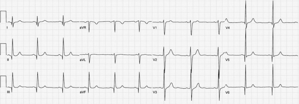
Syncope is a common ED presentation. An ECG is a critical investigation in syncope to identify the cause, including rare conditions associated with risk of sudden cardiac death.
So we should be really grateful when we are invited to interpret an ECG while we’re in the middle of six other tasks.
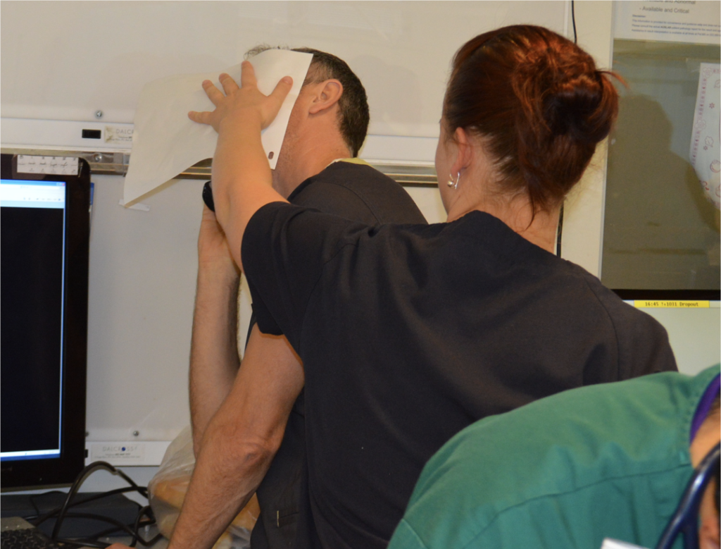
The problem with syncope is that some of the important life-threatening causes have fairly obscure ECG features that might be hard to remember. Some of these disorders and their ECG features are not entirely familiar to the clinicians who first screen the ECG.

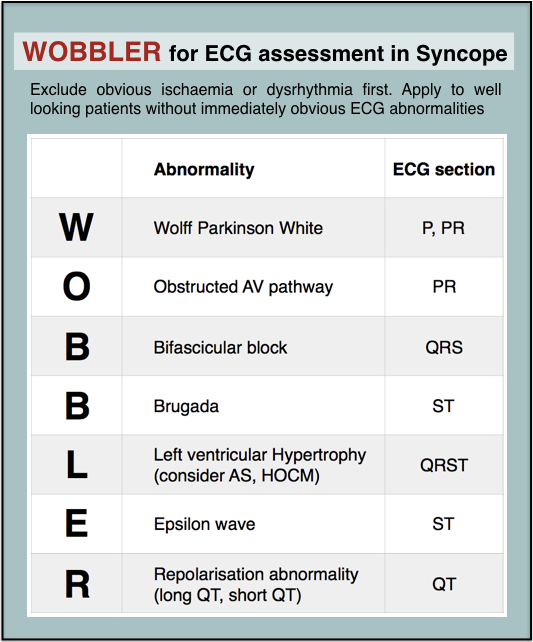
When you’re busy and cognitively stretched you can save time and reduce the risk of missing important findings by having a structured, memorable checklist. I use the acronym WOBBLER, because I don’t want these people to wobble and kiss the dirt again.

The nice thing about WOBBLER is that it uses the sequence that you follow when you look at an ECG, ie from left to right, or from P wave to T wave.

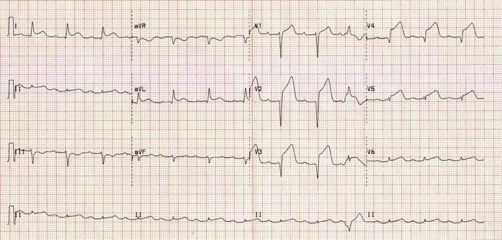
The key is that this is for ECGs without obvious ischaemia or dysrhythmia. If you see something like this (STEMI):
or this (VT):
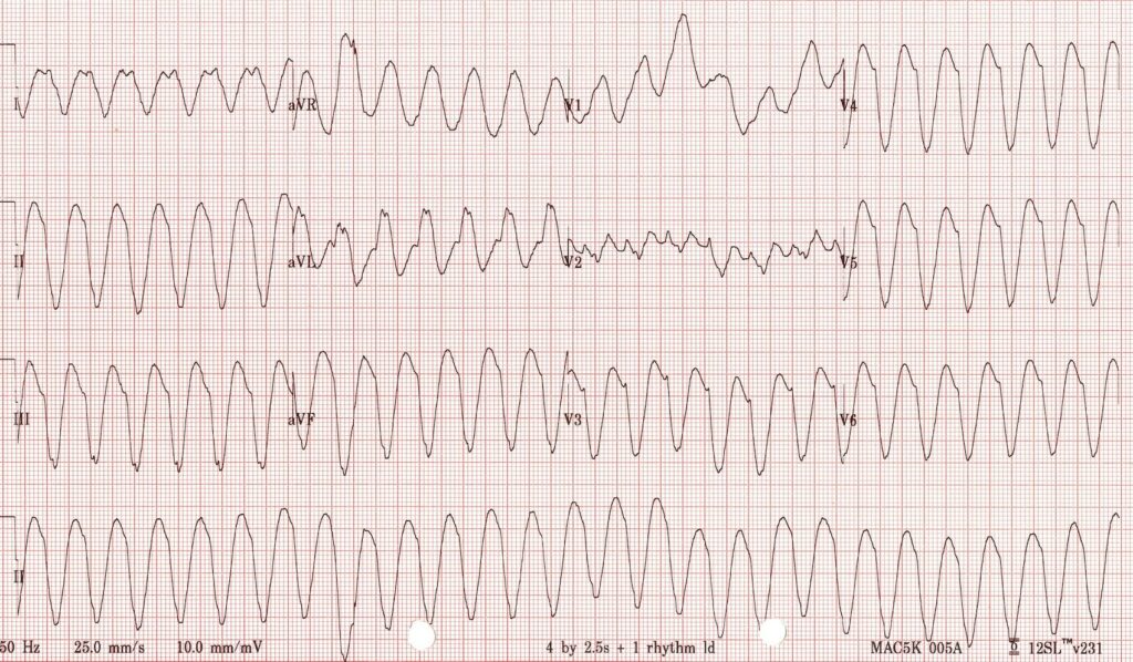
you don’t need WOBBLER, you need to be treating that patient.

So we apply WOBBLER to screen the ECG of well-looking syncope patients without obvious ischaemia or dysrhythmia.

Here goes…

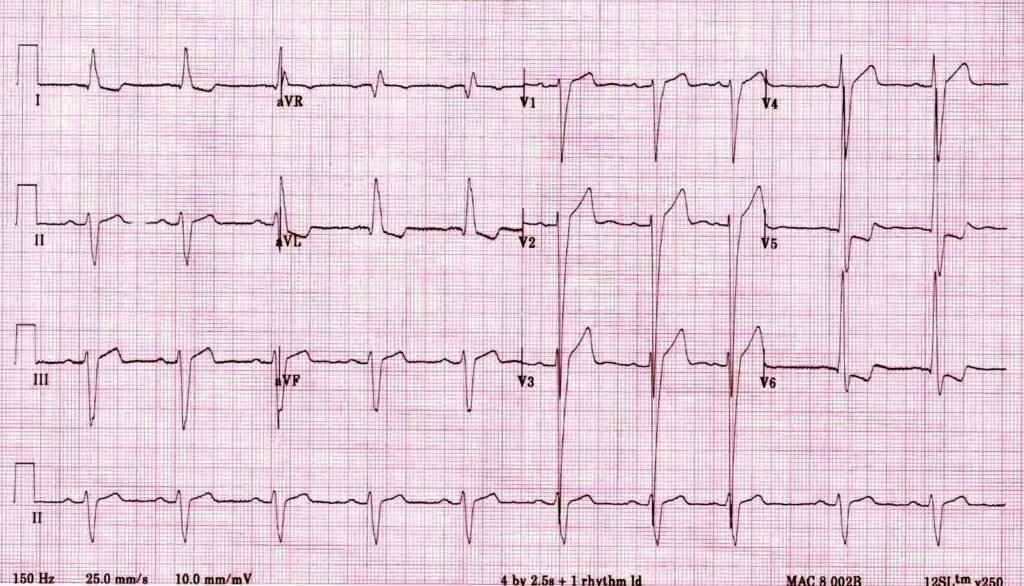
W is Wolff-Parkinson-White syndrome – look for a short PR interval or delta wave:
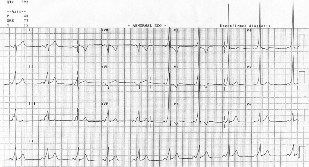
O is obstructed AV pathway – look for 2nd or 3rd degree block:

or axis deviation:
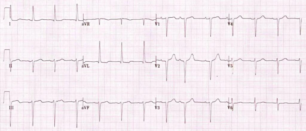
…which is the first step in looking for B bifascicular block, or the combination of axis deviation and right bundle branch block:
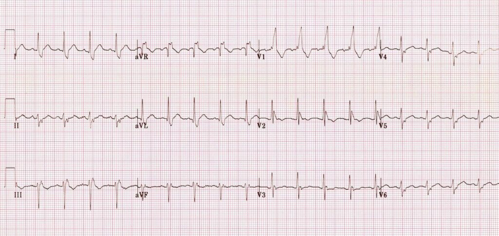
the second B is Brugada, looking for characteristic morphology of the ST segment, so called coved ST elevation:
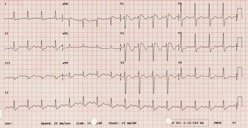
Now syncope, especially exertional syncope, can be caused by left ventricular outflow tract obstruction. Two conditions not to be missed associated with LVOTO (and exertional syncope) are hypertrophic cardiomyopathy and aortic stenosis. These both characteristically cause Lleft ventricular hypertrophy:

This example of Hypertrophic Cardiomyopathy has voltage criteria for LVH in the precordial leads as well as narrow, “dagger-like” Q waves in lateral and inferior leads

E– stands for epsilon wave, a feature of arrythmogenic right ventricular dysplasia, a rare disorder associated with sudden cardiac death. The epsilon wave looks a bit like the J wave of hypothermia and may be associated with other T wave abnormalities in V1-V3:
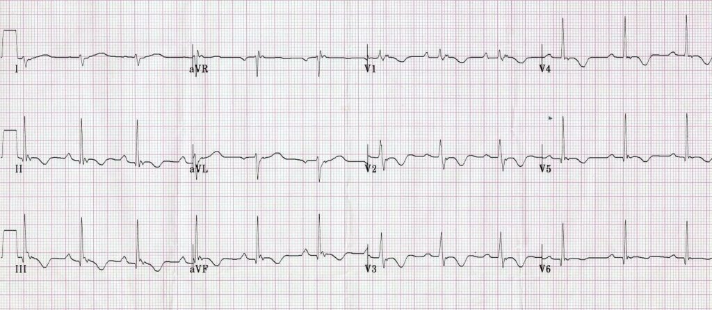
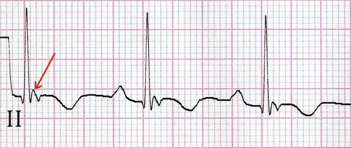
Epsilon wave in more detail

Finally, R stands for Repolarisation abnormality, particularly delayed Repolarisation as found in long QT syndrome:
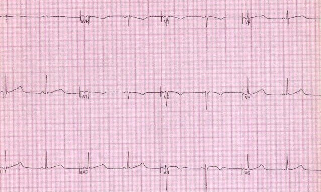
but remember there is also a short QT syndrome too:
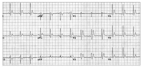
So WOBBLER may help you find the important and rare abnormalities not to be missed in the syncope patient, going from left to right from P wave through to T wave, in the patient that does not have obvious dysrhythmia or ischaemia. I hope you find it a helpful cognitive forcing tool.
All ECGs reproduced with kind permission of Life in the Fast Lane

Swallow a camera in GI bleed

Two recent studies evaluate the use of a novel ingestable camera to diagnose upper gastrointestinal bleeding in emergency department patients.
The potential advantages of video capsule endoscopy over traditional endoscopy could include immediate availability, avoidance of sedation, patient tolerance, and the ability to rule out active bleeding in the emergency department.
The device used was the PillCam ESO2 – shown here in this animation:

Further research is needed. These small interesting studies demonstrate the potential for this imaging technology to be used in stable patients presenting to emergency departments. Since it can only diagnose rather than treat, it would not appear to have any role in unstable patients.
Video capsule endoscopy in the emergency department: a prospective study of acute upper gastrointestinal hemorrhage.
Ann Emerg Med. 2013 Apr;61(4):438-443
[EXPAND Abstract]


STUDY OBJECTIVE: Video capsule endoscopy has been used to diagnose gastrointestinal hemorrhage and other small bowel diseases but has not been tested in an emergency department (ED) setting. The objectives in this pilot study are to demonstrate the ability of emergency physicians to detect blood in the upper gastrointestinal tract with capsule endoscopy after a short training period, measure ED patient acceptance of capsule endoscopy, and estimate the test characteristics of capsule endoscopy to detect acute upper gastrointestinal hemorrhage.

METHODS: During a 6-month period at a single academic hospital, eligible patients underwent video capsule endoscopy (Pillcam Eso2; Given Imaging) in the ED. Video images were reviewed by 4 blinded physicians (2 emergency physicians with brief training in capsule endoscopy interpretation and 2 gastroenterologists with capsule endoscopy experience).

RESULTS: A total of 25 subjects with acute upper gastrointestinal hemorrhage were enrolled. There was excellent agreement between gastroenterologists and emergency physicians for the presence of fresh or coffee-ground blood (0.96 overall agreement; κ=0.90). Capsule endoscopy was well tolerated by 96% of patients and showed an 88% sensitivity (95% confidence interval 65% to 100%) and 64% specificity (95% confidence interval 35% to 92%) for the detection of fresh blood. Capsule endoscopy missed 1 bleeding lesion located in the postpyloric region, which was not imaged because of expired battery life.

CONCLUSION: Video capsule endoscopy is a sensitive way to identify upper gastrointestinal hemorrhage in the ED. It is well tolerated and there is excellent agreement in interpretation between gastroenterologists and emergency physicians.

[/EXPAND]
Capsule endoscopy in acute upper gastrointestinal hemorrhage: a prospective cohort study
Endoscopy. 2013 Jan;45(1):12-9
[EXPAND Abstract]


BACKGROUND AND STUDY AIMS: Capsule endoscopy may play a role in the evaluation of patients presenting with acute upper gastrointestinal hemorrhage in the emergency department.

METHODS: We evaluated adults with acute upper gastrointestinal hemorrhage presenting to the emergency departments of two academic centers. Patients ingested a wireless video capsule, which was followed immediately by a nasogastric tube aspiration and later by esophagogastroduodenoscopy (EGD). We compared capsule endoscopy with nasogastric tube aspiration for determination of the presence of blood, and with EGD for discrimination of the source of bleeding, identification of peptic/inflammatory lesions, safety, and patient satisfaction.

RESULTS:The study enrolled 49 patients (32 men, 17 women; mean age 58.3 ± 19 years), but three patients did not complete the capsule endoscopy and five were intolerant of the nasogastric tube. Blood was detected in the upper gastrointestinal tract significantly more often by capsule endoscopy (15 /18 [83.3 %]) than by nasogastric tube aspiration (6 /18 [33.3 %]; P = 0.035). There was no significant difference in the identification of peptic/inflammatory lesions between capsule endoscopy (27 /40 [67.5 %]) and EGD (35 /40 [87.5 %]; P = 0.10, OR 0.39 95 %CI 0.11 - 1.15). Capsule endoscopy reached the duodenum in 45 /46 patients (98 %). One patient (2.2 %) had self-limited shortness of breath and one (2.2 %) had coughing on capsule ingestion.

CONCLUSION:In an emergency department setting, capsule endoscopy appears feasible and safe in people presenting with acute upper gastrointestinal hemorrhage. Capsule endoscopy identifies gross blood in the upper gastrointestinal tract, including the duodenum, significantly more often than nasogastric tube aspiration and identifies inflammatory lesions, as well as EGD. Capsule endoscopy may facilitate patient triage and earlier endoscopy, but should not be considered a substitute for EGD.

[/EXPAND]

Potassium levels and AMI death

An association is demonstrated between abnormal (both high and low) serum potassium levels and in-hospital mortality in patients with acute myocardial infarction. These findings do not necessarily imply a causal relationship, since abnormal potassium levels might be a marker of increased risk of death due to other illness factors rather than a risk of death per se.
Acknowledging that a randomised trial of potassium replacement is unlikely to happen, the authors pragmatically advise:
Our data suggest that the optimal range of serum potassium levels in AMI patients may be between 3.5 and 4.5 mEq/L and that potassium levels of greater than 4.5 mEq/L are associated with increased mortality and should probably be avoided.


Context Clinical practice guidelines recommend maintaining serum potassium levels between 4.0 and 5.0 mEq/L in patients with acute myocardial infarction (AMI). These guidelines are based on small studies that associated low potassium levels with ventricular arrhythmias in the pre−β-blocker and prereperfusion era. Current studies examining the relationship between potassium levels and mortality in AMI patients are lacking.

Objective To determine the relationship between serum potassium levels and in-hospital mortality in AMI patients in the era of β-blocker and reperfusion therapy.

Design, Setting, and Patients Retrospective cohort study using the Cerner Health Facts database, which included 38 689 patients with biomarker-confirmed AMI, admitted to 67 US hospitals between January 1, 2000, and December 31, 2008. All patients had in-hospital serum potassium measurements and were categorized by mean postadmission serum potassium level (<3.0, 3.0-<3.5, 3.5-<4.0, 4.0-<4.5, 4.5-<5.0, 5.0-<5.5, and ≥5.5 mEq/L). Hierarchical logistic regression was used to determine the association between potassium levels and outcomes after adjusting for patient- and hospital-level factors.
Main Outcome Measures All-cause in-hospital mortality and the composite of ventricular fibrillation or cardiac arrest.

Results There was a U-shaped relationship between mean postadmission serum potassium level and in-hospital mortality that persisted after multivariable adjustment. Compared with the reference group of 3.5 to less than 4.0 mEq/L (mortality rate, 4.8%; 95% CI, 4.4%-5.2%), mortality was comparable for mean postadmission potassium of 4.0 to less than 4.5 mEq/L (5.0%; 95% CI, 4.7%-5.3%), multivariable-adjusted odds ratio (OR), 1.19 (95% CI, 1.04-1.36). Mortality was twice as great for potassium of 4.5 to less than 5.0 mEq/L (10.0%; 95% CI, 9.1%-10.9%; multivariable-adjusted OR, 1.99; 95% CI, 1.68-2.36), and even greater for higher potassium strata. Similarly, mortality rates were higher for potassium levels of less than 3.5 mEq/L. In contrast, rates of ventricular fibrillation or cardiac arrest were higher only among patients with potassium levels of less than 3.0 mEq/L and at levels of 5.0 mEq/L or greater.

Conclusion Among inpatients with AMI, the lowest mortality was observed in those with postadmission serum potassium levels between 3.5 and <4.5 mEq/L compared with those who had higher or lower potassium levels.

Serum Potassium Levels and Mortality in Acute Myocardial Infarction
JAMA Jan 11 2012,307(2):115-213

What do I do with a high sensitivity troponin?

Newer high-sensitivity troponin tests can be positive in patients who would have negative tests with the ‘traditional’ assay, which can result in confusion about what to do with the patient, particularly those patients without an obvious cardiac presentation. A recent study1 shows that the majority of patients that fall into this group had non-cardiac discharge diagnoses.


Background: High sensitivity troponin T (hsTnT) detects lower levels of troponin T with greater precision than the 4th generation (cTnT) assay. However, the clinical implications of this are uncertain.

Objectives: Primary: Describe the proportion of patients who test ‘positive’ with hsTnT but negative with cTnT. Secondary: Determine proportion in each group with an adverse event (representation, AMI or died) within 90 days of the index test.

Method: 161 patients samples were tested with cTnT and hsTNT assays. McNemar’s test was used to compare paired samples. Electronic medical records were reviewed, with discharge diagnosis and 90 day outcomes determined blind to hsTnT results. Patients were then classified as ‘TnT negative’ (hsTnT was <0.014 mcg/mL), 'new positive' (hsTnT was ≥0.014 mcg/mL and cTnT <0.03 mcg/mL) and 'TnT positive' (cTNT was ≥0.03 mcg/mL)
Results: Positive results more than doubled with the hsTnT assay (50% vs 22%, P < 0.001). 81 patients were ‘TnT negative’, 44 were ‘new positive’ and 36 ‘cTnT positive’. The discharge diagnosis for ‘new positives’ was AMI in 4 (9%), other cardiac in 13 (30%) and non-cardiac in 27 (61%). At 90 days adverse events occurred in 30%, 54% and 50% of the groups respectively. There were no late cases of AMI or cardiovascular death in ‘new positive’ patients.

Conclusion: Many patients with diagnoses other than AMI will have hsTNT above the reference level. Indiscriminate testing with hsTnT might lead to more patients requiring serial troponin testing and/or invasive further tests, which will have process and resource implications for EDs and health services.

An accompanying editorial2 highlights that:

Elevations are seen in pathological conditions, including structural heart disease, renal impairment and pulmonary embolism, but might also be seen in extreme exertion, such as marathon runners. It is now clear that when using a highly sensitive assay, circulating levels of troponin will be detected in many normal people.

The editorial makes the interesting observation that the duration of rise may help elucidate the cause; ischaemic elevation of troponin falls rapidly, since the rise might be due to the release of small amounts of troponin that exist free within the cytoplasm, in contrast to the more persistent elevation seen with myocardial necrosis. The editorialist provides the following caution:


Overall, our practice for ordering troponin will need to be urgently reviewed. Single troponin values will continue to be of little to no use in defining disease states in the ED. Identifying a chronic versus an acute elevation will only be revealed by serial troponin testing. The time interval between testing is currently contentious.

High sensitivity troponins are referred to in the newly published 2011 Addendum to the National Heart Foundation of Australia/Cardiac Society of Australia and New Zealand Guidelines for the Management of Acute Coronary Syndromes (full text link below)3:

RECOMMENDED PROTOCOL FOR TROPONIN TESTING USING HIGH SENSITIVITY ASSAYS IN “RULING-OUT” ACS

  • All patients with a suspected ACS should undergo troponin testing on arrival at ED to ‘rule in’ ACS within the differential diagnosis
  • For a patient with a positive troponin result or a change in troponin levels over time, neither ACS nor other significant pathology (e.g. pulmonary embolus, aortic dissection, and sepsis) can be excluded. These patients are at higher risk of subsequent events. A positive result should be considered within the entire clinical context (history, examination, ECG findings and other investigations). Further investigations directed at all plausible clinical diagnoses should be considered and, if ACS is thought to be the likely cause, these patients may require cardiology assessment.
  • All patients with a negative result should undergo repeat testing 3–4 hours later.
  • The testing interval to ‘rule out’ MI may be reduced to 3 hours, provided that one sample is taken at least 6 hours after symptom onset:
  • Patients with a negative result at 3 hours after presentation and at least 6 hours after the onset of pain should be considered for early assessment by non-invasive anatomic or functional testing, as determined by local availability.
  • For patients presenting more than 6 hours after pain onset, a single high sensitivity troponin assay is sufficient to rule out myocardial infarction in the absence of ongoing chest pain.

High sensitivity troponin assays have an increased sensitivity for the detection of “myonecrosis”, but a reduced specificity for the diagnosis of “MI”. A positive result (≥99th centile for reference population OR where there is a change of ≥50% above an initial baseline level) should be interpreted in the context of the entire clinical presentation and does not necessarily represent an indication for coronary angiography. The management MI secondary to other conditions (e.g. anaemia, thyrotoxicosis, and sepsis) should be primarily directed at those conditions.
The finding of troponin concentrations that remain stable over time suggests that the presence of troponin is due to chronic disease. Acute exacerbations of chronic disease that result in elevated troponin levels can mimic an MI release pattern.

1. Clinical diagnosis and outcomes for Troponin T ‘positive’ patients assessed by a high sensitivity compared with a 4th generation assay
Emerg Med Australas. 2011 Aug;23(4):490-501
2. Troponin: A risk-defining biomarker for emergency department physicians
Emerg Med Australas. 2011 Aug;23(4):391-4
3. 2011 Addendum to the National Heart Foundation of Australia/Cardiac Society of Australia and New Zealand Guidelines for the Management of Acute Coronary Syndromes
Heart, Lung and Circulation 2011 Aug;28(8):487-502 Free Full Text

IVC collapse depends on breathing pattern

A high degree of sonographically-visualised collapse of the inferior vena cava (IVC) during inspiration suggests a volume-responsive cardiac output. This inspiratory collapse is said to be due to a fall in intra-thoracic pressure. However, the IVC traverses the abdominal compartment and is therefore under the influences of hepatic weight, intra-abdominal pressure, and venous return of pooled splanchnic and lower extremity blood.
Diaphragmatic descent, which increases intra-abdominal pressure, may contribute to the respiratory change in IVC diameter. This was borne out in a volunteer study in which diaphragmatic breathing was compared with chest wall breathing. With diaphragmatic breathing there was a trend for a larger IVC collapse index (median 0.80, range 0.48–1.00 vs. 0.57, range 0.13–1.00, P = 0.053). The authors state:
These findings suggest that during inspiration the IVC, in addition to responding to falling intra-thoracic pressure, may also be compressed with diaphragmatic descent and have implications regarding the use of IVC diameters to estimate the central venous pressure without knowing the manner of breathing, intra-abdominal pressure, or magnitude of diaphragmatic excursion.”
The take home message for me is that there is probably a more complex mechanism of IVC behaviour during respiration than is often taught, and that breathing pattern and abdominal issues may influence the IVC diameter and degree of collapse seen on ultrasound. This might not however negate the correlation between a high degree of collapse and fluid-responsiveness, which is what I’m looking for in my patients with shock or hypotension.
Incidentally the first author of this study is Bruce Kimura, a pioneer of focused echo in the emergency setting and author of a fantastic little book all about the parasternal long axis approach, which seems to be impossible to source on the web at the moment.

AIMS: Although the inspiratory ‘collapse’ of the inferior vena cava (IVC) has been used to signify normal central venous pressure, the effect of the manner of breathing IVC size is incompletely understood. As intra-abdominal pressure rises during descent of the diaphragm, we hypothesized that inspiration through diaphragmatic excursion may have a compressive effect on the IVC.
METHODS AND RESULTS: We measured minimal and maximal intrahepatic IVC diameter on echocardiography and popliteal venous return by spectral Doppler during isovolemic inspiratory efforts in 19 healthy non-obese volunteers who were instructed to inhale using either diaphragmatic or chest wall expansion. During inspiration, the maximal diaphragmatic excursion and popliteal vein flow were compared between breathing methods. The IVC ‘collapsibility index,’ IVCCI, was calculated as (IVC(max)-IVC(min))/IVC(max). The difference in diaphragmatic excursion between diaphragmatic and chest wall breaths in each subject was correlated with the corresponding change in IVCCI. Diaphragmatic breathing resulted in more diaphragmatic excursion than chest wall breathing (median 3.4 cm, range 1.7-5.8 vs. 2.2 cm, range 1.0-5.2, P= 0.0003), and was universally associated with decreased popliteal venous return (19/19 vs. 9/19 subjects, P< 0.004). The difference in diaphragmatic excursion correlated with the difference in IVCCI (Spearman’s rho = 0.53, P= 0.024).
CONCLUSION: During inspiration of equivalent tidal volumes, the reduction in IVC diameter and lower extremity venous return was related to diaphragmatic excursion, suggesting that the IVC may be compressed through descent of the diaphragm.

The effect of breathing manner on inferior vena caval diameter
Eur J Echocardiogr. 2011 Feb;12(2):120-3

TIA workup renders ABCD2 unhelpful

ABCD2 is recommended to stratify the risk of stroke in patients presenting to the ED with TIA symptoms. In some centres this is used to differentiate those that need to be admitted for further evaluation and treatment from those that can be followed up in the outpatient setting. A recent study showed that if a detailed work up was done in the ED on all TIA patients (followed by appropriate intervention), the ABCD2 score did not predict adverse outcomes, which were lower in this cohort than in the original ABCD2 cohort.

STUDY OBJECTIVE: We study the incremental value of the ABCD2 score in predicting short-term risk of ischemic stroke after thorough emergency department (ED) evaluation of transient ischemic attack.
METHODS: This was a prospective observational study of consecutive patients presenting to the ED with a transient ischemic attack. Patients underwent a full ED evaluation, including central nervous system and carotid artery imaging, after which ABCD2 scores and risk category were assigned. We evaluated correlations between risk categories and occurrence of subsequent ischemic stroke at 7 and 90 days.
RESULTS: The cohort consisted of 637 patients (47% women; mean age 73 years; SD 13 years). There were 15 strokes within 90 days after the index transient ischemic attack. At 7 days, the rate of stroke according to ABCD2 category in our cohort was 1.1% in the low-risk group, 0.3% in the intermediate-risk group, and 2.7% in the high-risk group. At 90 days, the rate of stroke in our ED cohort was 2.1% in the low-risk group, 2.1% in the intermediate-risk group, and 3.6% in the high-risk group. There was no relationship between ABCD2 score at presentation and subsequent stroke after transient ischemic attack at 7 or 90 days.
CONCLUSION: The ABCD2 score did not add incremental value beyond an ED evaluation that includes central nervous system and carotid artery imaging in the ability to risk-stratify patients with transient ischemic attack in our cohort. Practice approaches that include brain and carotid artery imaging do not benefit by the incremental addition of the ABCD2 score. In this population of transient ischemic attack patients, selected by emergency physicians for a rapid ED-based outpatient protocol that included early carotid imaging and treatment when appropriate, the rate of stroke was independent of ABCD2 stratification.

An Assessment of the Incremental Value of the ABCD2 Score in the Emergency Department Evaluation of Transient Ischemic Attack
Ann Emerg Med. 2011 Jan;57(1):46-51

Algorithm for Body Packers

‘Mules’ or body packers are people who transport illegal drugs by packet ingestion into the gastrointestinal tract. A large study of body packers apprehended by United State Customs officials at JFK International Airport, New York describes experience with body packers and an algorithm for conservative and surgical management.

Of 56 patients requiring admission out of a total of 1250 subjects confirmed to be body packers, 25 patients (45%) required surgical intervention, whereas 31 patients (55%) were successfully managed conservatively.
Diagnosis:

  • Plain abdominal x-ray was diagnostic in 49 patients (88% of all hospitalised patients).
  • Non-contrast CT of the abdomen and pelvis is required if AXR is negative
  • Forty-eight per cent of body packers had positive urine toxicology for illicit substances.


Management:

  • Indications for intervention included:
  • bowel obstruction
  • packet rupture/toxicity
  • delayed progression of packet transit on conservative management.
  • Patients with packets found predominantly in the proximal gastrointestinal tract failed conservative management more frequently than those with packets found in the distal gastrointestinal tract.

Multiple intraoperative manoeuvres were used to remove the foreign bodies:

  • gastrotomy
  • enterotomy
  • colotomy.

Wound infection was the most common complication and is associated with distal enterotomy and colotomy.
The authors recommend a confirmatory radiological study to demonstrate complete clearance of packets
Establishment of a definitive protocol for the diagnosis and management of body packers (drug mules).
Emerg Med J 2011;28:98-10

Venous gas in COPD exacerbation

Prolific emergency medicine researcher Anne-Maree Kelly and colleague Dr Lim from Tan Tock Seng Hospital in Singapore have published a systematic review of articles assessing the utility of peripheral venous blood gases (pVBG) in exacerbations of COPD1. Their conclusion:
Available evidence suggests that there is good agreement for pH and HCO3 values between arterial and pVBG results in patients with COPD, but not for pO2 or pCO2. Widespread clinical use is limited because of the lack of validation studies on clinical outcomes
pVBG may however be useful as a screening test for significant arterial hypercarbia; Kelly et al. previously reported2 a cutoff value of 45 mmHg (5.9 kPa).
1. A meta-analysis on the utility of peripheral venous blood gas analyses in exacerbations of chronic obstructive pulmonary disease in the emergency department
Eur J Emerg Med. 2010 Oct;17(5):246-8
2. Kelly AM, Kerr D, Middleton P. Validation of venous pCO2 to screen for arterial hypercarbia in patients with chronic obstructive airways disease.
J Emerg Med 2005; 28:377–379

Higher D Dimer cut-off in older patients

D-dimer levels below the conventional cut-off point of 500 µg/l combined with a “low/intermediate” or “unlikely” clinical probability can safely rule out the diagnosis in about 30% of patients with suspected pulmonary embolism.
However, the D-dimer concentration increases with age and its specificity for embolism decreases, which makes the test less useful to exclude pulmonary embolism in older patients; the test is able to rule out pulmonary embolism in 60% of patients aged <40 years, but in only 5% of patients aged >80.
A new, age dependent cut-off value was derived and then validated in two independent retrospective datasets from Belgium, France, the Netherlands, and Switzerland. They studied over 5000 patients aged >50 years.
The new D-dimer cut-off value was defined as (patient’s age x 10) µg/l in patients aged >50.
In 1331 patients in the derivation set with an “unlikely” score from clinical probability assessment, pulmonary embolism could be excluded in 42% with the new cut-off value versus 36% with the old cut-off value (<500 µg/l). In the two validation sets, the increase in the proportion of patients with a D-dimer below the new cut-off value compared with the old value was 5% and 6%. This absolute increase was largest among patients aged >70 years, ranging from 13% to 16% in the three datasets. The failure rates (all ages) were 0.2% (95% CI 0% to 1.0%) in the derivation set and 0.6% (0.3% to 1.3%) and 0.3% (0.1% to 1.1%) in the two validation sets.
Potential of an age adjusted D-dimer cut-off value to improve the exclusion of pulmonary embolism in older patients: a retrospective analysis of three large cohorts.

BMJ. 2010 Mar 30;340:c1475

Extreme white cell counts

Febrile children aged three months to three years with a white cell count over 25000/mm3 and fever were compared with controls whose leucoytosis was less extreme (15000-24999). The ‘extreme’ group had serious bacterial infection (SBI) in 39% compared with 15.4% controls. Pneumonia was the commonest SBI.
The authors conclude that in febrile children aged 3–36 months, the presence of extreme leucocytosis is associated with a 39% risk of having SBIs. The increased risk for SBI is mainly due to a higher risk for pneumonia. I conclude that leucocytosis is like fever: the cause may be benign, but the higher the number the less likely that is, even though the majority still won’t have SBI.
Extreme leucocytosis and the risk of serious bacterial infections in febrile children
Arch Dis Child. 2010 Mar;95(3):209-12