Tag Archives: VTE

Does RV enlargement on echo predict PE?

A nice paper from Annals of Emergency Medicine showing the test characteristcs of some of the common signs we look for on basic 2D echo that suggest the presence of (sub)massive pulmonary embolism:
Right Ventricular Dilatation on Bedside Echocardiography Performed by Emergency Physicians Aids in the Diagnosis of Pulmonary Embolism
Ann Emerg Med. 2014 Jan;63(1):16-24
[EXPAND Abstract]


STUDY OBJECTIVE: The objective of this study was to determine the diagnostic performance of right ventricular dilatation identified by emergency physicians on bedside echocardiography in patients with a suspected or confirmed pulmonary embolism. The secondary objective included an exploratory analysis of the predictive value of a subgroup of findings associated with advanced right ventricular dysfunction (right ventricular hypokinesis, paradoxical septal motion, McConnell’s sign).

METHODS: This was a prospective observational study using a convenience sample of patients with suspected (moderate to high pretest probability) or confirmed pulmonary embolism. Participants had bedside echocardiography evaluating for right ventricular dilatation (defined as right ventricular to left ventricular ratio greater than 1:1) and right ventricular dysfunction (right ventricular hypokinesis, paradoxical septal motion, or McConnell’s sign). The patient’s medical records were reviewed for the final reading on all imaging, disposition, hospital length of stay, 30-day inhospital mortality, and discharge diagnosis.

RESULTS: Thirty of 146 patients had a pulmonary embolism. Right ventricular dilatation on echocardiography had a sensitivity of 50% (95% confidence interval [CI] 32% to 68%), a specificity of 98% (95% CI 95% to 100%), a positive predictive value of 88% (95% CI 66% to 100%), and a negative predictive value of 88% (95% CI 83% to 94%). Positive and negative likelihood ratios were determined to be 29 (95% CI 6.1% to 64%) and 0.51 (95% CI 0.4% to 0.7%), respectively. Ten of 11 patients with right ventricular hypokinesis had a pulmonary embolism. All 6 patients with McConnell’s sign and all 8 patients with paradoxical septal motion had a diagnosis of pulmonary embolism. There was a 96% observed agreement between coinvestigators and principal investigator interpretation of images obtained and recorded.

CONCLUSION: Right ventricular dilatation and right ventricular dysfunction identified on emergency physician performed echocardiography were found to be highly specific for pulmonary embolism but had poor sensitivity. Bedside echocardiography is a useful tool that can be incorporated into the algorithm of patients with a moderate to high pretest probability of pulmonary embolism.

[/EXPAND]

Age adjusted D-dimer cut-off values

Age adjusted D-dimer cut-off values (age×10 µg/L) improve specificity without losing sensitivity for venous thromboembolism. This could spare many elderly patients unnecessary imaging. Full text is available free from the BMJ.
Diagnostic accuracy of conventional or age adjusted D-dimer cut-off values in older patients with suspected venous thromboembolism: systematic review and meta-analysis>
BMJ. 2013 May 3;346:f2492
[EXPAND Abstract]


OBJECTIVE: To review the diagnostic accuracy of D-dimer testing in older patients (>50 years) with suspected venous thromboembolism, using conventional or age adjusted D-dimer cut-off values.

DESIGN Systematic review and bivariate random effects meta-analysis.

DATA SOURCES: We searched Medline and Embase for studies published before 21 June 2012 and we contacted the authors of primary studies.

STUDY SELECTION: Primary studies that enrolled older patients with suspected venous thromboembolism in whom D-dimer testing, using both conventional (500 µg/L) and age adjusted (age × 10 µg/L) cut-off values, and reference testing were performed. For patients with a non-high clinical probability, 2 × 2 tables were reconstructed and stratified by age category and applied D-dimer cut-off level.

RESULTS: 13 cohorts including 12,497 patients with a non-high clinical probability were included in the meta-analysis. The specificity of the conventional cut-off value decreased with increasing age, from 57.6% (95% confidence interval 51.4% to 63.6%) in patients aged 51-60 years to 39.4% (33.5% to 45.6%) in those aged 61-70, 24.5% (20.0% to 29.7% in those aged 71-80, and 14.7% (11.3% to 18.6%) in those aged >80. Age adjusted cut-off values revealed higher specificities over all age categories: 62.3% (56.2% to 68.0%), 49.5% (43.2% to 55.8%), 44.2% (38.0% to 50.5%), and 35.2% (29.4% to 41.5%), respectively. Sensitivities of the age adjusted cut-off remained above 97% in all age categories.

CONCLUSIONS: The application of age adjusted cut-off values for D-dimer tests substantially increases specificity without modifying sensitivity, thereby improving the clinical utility of D-dimer testing in patients aged 50 or more with a non-high clinical probability.

[/EXPAND]

Surgical treatment for acute massive pulmonary embolism

A recent paper reminds us that surgery is an option in the management of massive pulmonary embolism(1), to be considered in the patient for whom thrombolysis has failed or is contraindicated. Good outcomes were produced when surgery was performed in a centre capable of cardiopulmonary bypass (6% 30-day postoperative mortality), but is surgery an option when these facilities are unavailable?

The “venous inflow occlusion” technique involves clamping the venae cavae prior to removing clot directly from the pulmonary artery and its branches after median sternotomy, and can be performed in any hospital with surgical facilities. Under normothermic conditions, speed is of the essence once cardiac arrest occurs, since the irreversible anoxic cerebral injury will occur after just a few minutes.
Clarke and Abrams wrote in the Lancet in 1972(2):

Our use of venous inflow-occlusion has given results which compare well with those obtained with extracorporeal circulation. 50% of our patients survived. All patients who had emboli removed without an episode of ventricular asystole survived surgery. Late deaths in 3 patients were from causes unrelated to pulmonary embolism, and from a further massive pulmonary embolus a week later. The technique has been applied with equal success in a major hospital fully equipped for cardiac surgery and in hospitals where resident and nursing staff had no experience of either thoracic or cardiac surgery. The simplicity and speed of the method has enabled the obstructed right ventricle to be relieved within thirty minutes of the onset of symptoms. The interval between induction of anaesthesia and the skin incision should be kept as short as possible, and drugs to maintain the blood pressure should be given. The period between skin incision and the restoration of the circulation has, with practice, been reduced to ten minutes.

But this clearly still requires surgical expertise and facilities. Emergency physicians can open the chest to deal with penetrating trauma. Could an ED thoracotomy facilitate clot removal from the pulmonary artery?

In 1969, a lady in her 50s arrested on the ward after an operation to remove a mass via a left lateral thoractomy. Pulmonary embolism was suspected and her thoracotomy wound was re-opened and the pulmonary artery incised, resulting in the removal of large amounts of clot. Return of spontaneous circulation resulted after a brief period of internal cardiac massage. Her case was written up decades later, in 1998(3):

The patient recovered rapidly and left the hospital on the 21st day without signs of cerebral damage. This patient is now 86 years old, mentally normal, living alone, and doing her own housekeeping. She remembers the hospital stay and the past years as worth living

Some patients may be considered too high risk for surgery and in some centres Extracorporeal Membrane Oxygenation (ECMO) is an option. It has been used both as life support pending surgery(4), or as an alternative to surgery to allow heparinisation to be used(5,6).

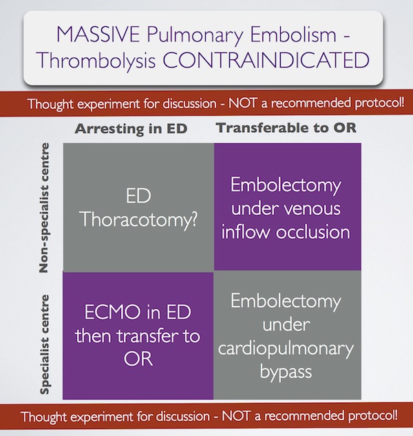
In summary, some patients with massive pulmonary embolism may benefit from surgery (contraindication to ‘lysis or failed ‘lysis). Getting them to surgery alive, or operating on them during cardiac arrest, is a challenge. Ideally they would undergo embolectomy under cardopulmonary bypass in the operating room, or could be placed on ECMO in the ED prior to going to the OR. If they present to a centre without these facilities, then the venous inflow occlusion technique could be used in the OR without bypass. Just rarely a patient may present in extremis with PE to an ED without these options. If that patient has major contraindications to thrombolysis, would an ED thoracotomy be something you would entertain?

I have done several thoracotomies for penetrating trauma but never for PE. I do not pretend to know how, and cannot find a case report of ED thoracotomy for pulmonary embolism in the literature. I’m therefore NOT recommending it. However, I would love to know people’s views on its feasibility. A possible approach could be summarised as:
Massive pulmonary embolism fascinates me, because it’s seen in the ‘talk and die’ patient. It is a single, treatable pathology that if diagnosed and treated appropriately truly makes the difference between life and death. When medicine presents us with an opportunity ‘on a plate’ like that to save a life, we need to be prepared. I have had great saves with this diagnosis and sadly have seen disastrous failures to act. When the time comes, we need to ask: ‘have we explored all options?’.

1. Surgical treatment of acute pulmonary embolism–a 12-year retrospective analysis.
Scand Cardiovasc J. 2012 Jun;46(3):172-6. Epub 2012 Mar 27.

OBJECTIVES: Surgical embolectomy for acute pulmonary embolism (PE) is considered to be a high risk procedure and therefore a last treatment option. We wanted to evaluate the procedures role in modern treatment of acute PE.
DESIGN: All data on patients treated with surgical embolectomy for acute PE were retrieved from our clinical database. The mortality was extracted from the Danish mortality register.
RESULTS: From October 1998 to July 2010, 33 patients underwent surgical embolectomy. All procedures were done through a median sternotomy and extracorporeal circulation. Twenty-six patients were diagnosed with a high risk PE and 7 with an intermediate risk PE and intracardial pathology. Six patients had been insufficiently treated with thrombolysis. Thirteen patients had contraindication for thrombolysis. Six patients were brought to the operating theatre in cardiogenic shock, 8 needed ventilator support, and 1 was in cardiac arrest. The postoperative 30-day mortality was 6% and during the 12-year follow-up the cumulative survival was 80% with 4 late deaths.
CONCLUSION: Surgical pulmonary embolectomy can be performed with low mortality although the treated patients belong to the most compromised part of the PE population. The results support surgical embolectomy as a vital part of the treatment algorithm for acute PE.

2. Pulmonary embolectomy with venous inflow-occlusion.
The Lancet 1972;1(7754):767–769

Massive pulmonary emboli have been removed surgically from 26 patients. The technique of normothermic circulatory arrest by venous inflow-occlusion was used in 25 patients. 13 patients survived. There were 10 operative deaths and 3 hospital deaths. Diagnosis was based upon clinical findings supplemented by electrocardiography and a plain radiograph of the chest. Surgery was offered to patients having a pulmonary embolus sufficiently massive to produce sustained hypotension. All patients whose hearts stopped beating before the embolectomy died. 6 successful operations were performed in hospitals without facilities for cardiac surgery. The method is recommended for its simplicity.

3. Left Anterior Thoracotomy for Pulmonary Embolectomy With 29-Year Follow-up
The Annals of Thoracic Surgery 1998, 66(4):1420-1421

Pulmonary embolectomy is usually performed in cardiopulmonary bypass. In acute situations too much time can be lost in setting up and connecting the pump oxygenator; this delay can cause cerebral damage in a patient with circulatory arrest. In such a situation left anterior thoracotomy can provide an ideal approach. An emergency thoracotomy can be performed in a few seconds. The lung automatically retracts. The phrenic nerve, pulmonary artery, and pericardium are clearly seen, and they outline the area for embolectomy. A case in which such an approach was successfully used is described.

4.ECMO treatment saved life of a young woman with acute pulmonary embolism
Lakartidningen 2004, 101(44):3420-3421

A 42-year old obese female using contraceptive medication was admitted to the emergency room because of sudden onset of dyspnoea and hypoxia. Computed tomography showed massive pulmonary emboli. Despite initial treatment with thrombolysis her condition deteriorated further and she was referred for acute surgery to our clinic. Before putting the patient to sleep extracorporeal circulation was instituted with access from the groin. After anaesthesia a median sternotomy was performed. With the heart beating, the main pulmonary artery was incised and a 9 cm long thrombus was removed. Immediate weaning from the heart-lung machine was not possible, mainly because of bleeding to the airways. The right atrium and the aorta was therefore cannulated and an extracorporeal circulation membrane oxygenator (ECMO) was used for three days. The patient required several re-entries for bleeding and a tracheotomy during the postoperative course. She was fully recovered three months after the operation.

5. Extracorporeal membrane oxygenator for pulmonary embolism.
The Annals of Thoracic Surgery 1997, 64(3):883-884 Free full text

6. Peripheral Extracorporeal Membrane Oxygenation: Comprehensive Therapy for High-Risk Massive Pulmonary Embolism
Ann Thorac Surg 2012;94:104–8

Background: Although commonly reserved as a last line of defense, experienced centers have reported excellent results with pulmonary embolectomy for massive and submassive pulmonary embolism (PE). We present a contemporary surgical series for PE that demonstrates the utility of peripheral extracorporeal membrane oxygenation (pECMO) for high-risk surgical candidates.
Methods: Between June 2005 and April 2011, 29 patients were treated for massive or submassive pulmonary embolism, with surgical embolectomy performed in 26. Four high-risk patients were placed on pECMO, established by percutaneously cannulating the right atrium through a femoral vein and perfusing by a Dacron graft anastomosed to the axillary artery. A small, extracorporeal, rotary assist device was used, interposing a compact oxygenator in the circuit, and maintaining anticoagulation with heparin.
Results: Extracorporeal membrane oxygenation was weaned in 3 of 4 patients after 5.3 days (5, 5, and 6), with normalization of right ventricular dysfunction and pulmonary artery pressure (44.0 ± 2.0 to 24.5 ± 5.5 mm Hg) by ECHO. Follow-up computed tomographies showed several peripheral, nearly resorbed emboli in 1 case and complete resolution in 2 others. The fourth patient, not improving after 10 days, underwent surgery where an embolic liposarcoma was extracted. For all 29 cases, hospital and 30-day mortality was 0% and all patients were discharged, with average postoperative length of stay of 15 days for embolectomy and 17 days for pECMO.
Conclusions: Heparin therapy with pECMO support is a rapid, effective option for patients who might benefit from pulmonary embolectomy but are at high risk for surgery.

Thrombolytic Therapy in Unstable Patients with PE

Most of us would give strong consideration to giving thrombolytics to patients with massive pulmonary embolism (PE), which is in keeping with many guidelines. Some physicians remain reluctant to do so, often citing the lack of good evidence. It is true that large scale RCTs have not been done in this population. The authors of this recent retrospective study state:


There are no definitive trials that prove the value of thrombolytic therapy in unstable patients with pulmonary embolism. It is extremely remote that a randomized controlled trial will be performed in the future. We therefore analyzed the database of the Nationwide Inpatient Sample to test the hypothesis that thrombolytic therapy reduces case fatality rate in unstable patients with acute pulmonary embolism.

They demonstrate a striking difference in mortality when thrombolysis is given to unstable patients with PE, which is further reduced with the addition of a vena cava filter. ‘Unstable’ was defined as having a listed code for shock or ventilator dependence.

Associated comorbid conditions were more often present in those who did not receive thrombolytic therapy than in those who did. However in their discussion the authors add:


Although unstable patients who received thrombolytic therapy had fewer comorbid conditions than those who did not, this would not explain the difference in case fatality rate because unstable patients with a primary diagnosis of pulmonary embolism and none of the comorbid conditions…also showed a lower case fatality rate with thrombolytic therapy. Therefore, differences in comorbid conditions in this group were eliminated as a possible cause of the lower case fatality rate in unstable patients who received thrombolytic therapy.

They round off their conclusion with:


Despite the marked reduction of case fatality rate with thrombolytic therapy in unstable patients, only 30% of unstable patients received it, and the proportion receiving thrombolytic therapy is diminishing. On the basis of these data, thrombolytic therapy in combination with a vena cava filter in unstable patients with acute pulmonary embolism seems indicated.

Many thanks to Dr Daniel Horner for highlighting this paper.


BACKGROUND: Data are sparse and inconsistent regarding whether thrombolytic therapy reduces case fatality rate in unstable patients with acute pulmonary embolism. We tested the hypothesis that thrombolytic therapy reduces case fatality rate in such patients.

METHODS: In-hospital all-cause case fatality rate according to treatment was determined in unstable patients with pulmonary embolism who were discharged from short-stay hospitals throughout the United States from 1999 to 2008 by using data from the Nationwide Inpatient Sample. Unstable patients were in shock or ventilator dependent.

RESULTS: Among unstable patients with pulmonary embolism, 21,390 of 72,230 (30%) received thrombolytic therapy. In-hospital all-cause case fatality rate in unstable patients with thrombolytic therapy was 3105 of 21,390 (15%) versus 23,820 of 50,840 (47%) without thrombolytic therapy (P< .0001). All-cause case fatality rate in unstable patients with thrombolytic therapy plus a vena cava filter was 505 of 6630 (7.6%) versus 4260 of 12,850 (33%) with a filter alone (P<.0001). Case fatality rate attributable to pulmonary embolism in unstable patients was 820 of 9810 (8.4%) with thrombolytic therapy versus 1080 of 2600 (42%) with no thrombolytic therapy (P<.0001). Case fatality rate attributable to pulmonary embolism in unstable patients with thrombolytic therapy plus vena cava filter was 70 of 2590 (2.7%) versus 160 of 600 (27%) with a filter alone (P<.0001).
CONCLUSION: In-hospital all-cause case fatality rate and case fatality rate attributable to pulmonary embolism in unstable patients was lower in those who received thrombolytic therapy. Thrombolytic therapy resulted in a lower case fatality rate than using vena cava filters alone, and the combination resulted in an even lower case fatality rate. Thrombolytic therapy in combination with a vena cava filter in unstable patients with acute pulmonary embolism seems indicated.

Thrombolytic Therapy in Unstable Patients with Acute Pulmonary Embolism: Saves Lives but Underused
Am J Med. 2012 May;125(5):465-70

Clotbusting wisdom on tap – your questions answered

The prevention and management of venous thromboembolic disease is a huge topic, which generates questions for emergency, critical care, and acute physicians during many shifts:

  • How long should someone requiring cardioversion for atrial fibrillation be anticoagulated for?
  • How should I provide thromboprophylaxis for this intubated patient?
  • This patient with submassive pulmonary embolism isn’t hypotensive yet. Can I thrombolyse them? Can I?
  • There’s a large superficial vein thrombosis in that limb – is anticoagulation indicated?
  • This asymptomatic patient on warfarin has an INR of 9.0 – should I reverse them?
  • Do I need to add Vitamin K if I’ve reversed warfarin with prothrombin complex concentrate?

The answers to these – and many, many more – questions are provided in one of the most comprehensive guidelines I’ve ever come across. I can see myself clicking on the link below in future when on duty in the ED.
Antithrombotic Therapy and Prevention of Thrombosis, 9th ed: American College of Chest Physicians Evidence-Based Clinical Practice Guidelines
Chest. 2012 Feb;141(2 Suppl) Full Text

Enoxaparin in acute medical patients

A large multinational study challenges the practice of routine thromboprophylaxis for hospitalised acutely ill medical patients. Enoxaparin plus graduated compression stockings did not reduce 30 day mortality compared with stockings alone. There was no significant difference in the rates of major bleeding.


Background Although thromboprophylaxis reduces the incidence of venous thromboembolism in acutely ill medical patients, an associated reduction in the rate of death from any cause has not been shown.

Methods We conducted a double-blind, placebo-controlled, randomized trial to assess the effect of subcutaneous enoxaparin (40 mg daily) as compared with placebo — both administered for 10±4 days in patients who were wearing elastic stockings with graduated compression — on the rate of death from any cause among hospitalized, acutely ill medical patients at participating sites in China, India, Korea, Malaysia, Mexico, the Philippines, and Tunisia. Inclusion criteria were an age of at least 40 years and hospitalization for acute decompensated heart failure, severe systemic infection with at least one risk factor for venous thromboembolism, or active cancer. The primary efficacy outcome was the rate of death from any cause at 30 days after randomization. The primary safety outcome was the rate of major bleeding during and up to 48 hours after the treatment period.

Results A total of 8307 patients were randomly assigned to receive enoxaparin plus elastic stockings with graduated compression (4171 patients) or placebo plus elastic stockings with graduated compression (4136 patients) and were included in the intention-to-treat population. The rate of death from any cause at day 30 was 4.9% in the enoxaparin group as compared with 4.8% in the placebo group (risk ratio, 1.0; 95% confidence interval [CI], 0.8 to 1.2; P=0.83). The rate of major bleeding was 0.4% in the enoxaparin group and 0.3% in the placebo group (risk ratio, 1.4; 95% CI, 0.7 to 3.1; P=0.35).

Conclusions The use of enoxaparin plus elastic stockings with graduated compression, as compared with elastic stockings with graduated compression alone, was not associated with a reduction in the rate of death from any cause among hospitalized, acutely ill medical patients. (Funded by Sanofi; LIFENOX ClinicalTrials.gov number, NCT00622648.)

Low-Molecular-Weight Heparin and Mortality in Acutely Ill Medical Patients
N Engl J Med 2011; 365:2463-2472

ACEP policy on PE

The American College of Emergency Physicians has revised its 2003 clinical policy on pulmonary embolism.


Among the areas considered is the the role of thrombolytic medication. The policy provides the following recommendations to this question:

What are the indications for thrombolytic therapy in patients with PE?
Level B recommendations
Administer thrombolytic therapy in hemodynamically unstable patients with confirmed PE for whom the benefits of treatment outweigh the risks of life-threatening bleeding complications.*
*In centers with the capability for surgical or mechanical thrombectomy, procedural intervention may be used as an alternative therapy.
Level C recommendations
(1) Consider thrombolytic therapy in hemodynamically unstable patients with a high clinical suspicion for PE for whom the diagnosis of PE cannot be confirmed in a timely manner.
(2) At this time, there is insufficient evidence to make any recommendations regarding use of thrombolytics in any subgroup of hemodynamically stable patients. Thrombolytics have been demonstrated to result in faster improvements in right ventricular function and pulmonary perfusion, but these benefits have not translated to improvements in mortality.

The document contains a detailed appraisal of the literature to date on benefits and harms from thrombolysis. Of interest is the Pulmonary Embolism Severity Index (PESI) – a scoring system that appears to reliably predict mortality and thus has the potential to assist physicians in making risk-benefit decisions when considering administration of thrombolytics. The full text of the policy, which covers far more than just thrombolysis, can be found by following the link below.
Critical Issues in the Evaluation and Management of Adult Patients Presenting to the Emergency Department With Suspected Pulmonary Embolism
Annals of Emergency Medicine 2011 June 57(6):628-652 – Free Full Text

Thrombolysis in submassive PE – still equipoise?

The AHA has produced a comprehensive guideline on venous thromboembolic disease. Here are some excerpts pertaining to resuscitation room decision making, particularly: ‘should I thrombolyse this patient?’

Definition for massive PE: Acute PE with sustained hypotension (systolic blood pressure <90 mm Hg for at least 15 minutes or requiring inotropic support, not due to a cause other than PE, such as arrhythmia, hypovolemia, sepsis, or left ventricular [LV] dysfunction), pulselessness, or persistent profound bradycardia (heart rate <40 bpm with signs or symptoms of shock).
Definition for submassive PE: Acute PE without systemic hypotension (systolic blood pressure ≥90 mm Hg) but with either RV dysfunction or myocardial necrosis.
RV dysfunction means the presence of at least 1 of the following:

  • RV dilation (apical 4-chamber RV diameter divided by LV diameter >0.9) or RV systolic dysfunction on echocardiography
  • RV dilation (4-chamber RV diameter divided by LV diameter >0.9) on CT
  • Elevation of BNP (>90 pg/mL)
  • Elevation of N-terminal pro-BNP (>500 pg/mL); or
  • Electrocardiographic changes (new complete or incomplete right bundle-branch block, anteroseptal ST elevation or depression, or anteroseptal T-wave inversion)

Myocardial necrosis is defined as either of the following:

  • Elevation of troponin I (>0.4 ng/mL) or
    Elevation of troponin T (>0.1 ng/mL)

Odds ratio for short-term mortality for RV dysfunction on echocardiography = 2.53 (95% CI 1.17 to 5.50).
Troponin elevations had an odds ratio for mortality of 5.90 (95% CI 2.68 to 12.95).
Definition for low risk PE: those with normal RV function and no elevations in biomarkers with short-term mortality rates approaching ≈ 1%

Recommendations for Initial Anticoagulation for Acute PE

  • Therapeutic anticoagulation with subcutaneous LMWH, intravenous or subcutaneous UFH with monitoring, unmonitored weight-based subcutaneous UFH, or subcutaneous fondaparinux should be given to patients with objectively confirmed PE and no contraindications to anticoagulation (Class I; Level of Evidence A).
  • Therapeutic anticoagulation during the diagnostic workup should be given to patients with intermediate or high clinical probability of PE and no contraindications to anticoagulation (Class I; Level of Evidence C).

 
Patients treated with a fibrinolytic agent have faster restoration of lung perfusion. At 24 hours, patients treated with heparin have no substantial improvement in pulmonary blood flow, whereas patients treated with adjunctive fibrinolysis manifest a 30% to 35% reduction in total perfusion defect. However, by 7 days, blood flow improves similarly (≈65% to 70% reduction in total defect).
Thirteen placebo-controlled randomized trials of fibrinolysis for acute PE have been published, but only a subset evaluated massive PE specifically.
When Wan et al restricted their analysis to those trials with massive PE, they identified a significant reduction in recurrent PE or death from 19.0% with heparin alone to 9.4% with fibrinolysis (odds ratio 0.45, 95% CI 0.22 to 0.90).
Data from registries indicate that the short-term mortality rate directly attributable to submassive PE treated with heparin anticoagulation is probably < 3.0%. The implication is that even if adjunctive fibrinolytic therapy has extremely high efficacy, for example, a 30% relative reduction in mortality, the effect size on mortality due to submassive PE is probably < 1%. Thus, secondary adverse outcomes such as persistent RV dysfunction, chronic thromboembolic pulmonary hypertension, and impaired quality of life represent appropriate surrogate goals of treatment.
Data suggest that compared with heparin alone, heparin plus fibrinolysis yields a significant favorable change in right ventricular systolic pressure and pulmonary arterial pressure incident between the time of diagnosis and follow-up. Patients with low-risk PE have an unfavorable risk-benefit ratio with fibrinolysis. Patients with PE that causes hypotension probably do benefit from fibrinolysis. Management of submassive PE crosses the zone of equipoise, requiring the clinician to use clinical judgment.

An algorithm is proposed:

Two criteria can be used to assist in determining whether a patient is more likely to benefit from fibrinolysis: (1) Evidence of present or developing circulatory or respiratory insufficiency; or (2) evidence of moderate to severe RV injury.
Evidence of circulatory failure includes any episode of hypotension or a persistent shock index (heart rate in beats per minute divided by systolic blood pressure in millimeters of mercury) >1
The definition of respiratory insufficiency may include hypoxemia, defined as a pulse oximetry reading < 95% when the patient is breathing room air and clinical judgment that the patient appears to be in respiratory distress. Alternatively, respiratory distress can be quantified by the numeric Borg score, which assesses the severity of dyspnea from 0 to 10 (0=no dyspnea and 10=sensation of choking to death).
Evidence of moderate to severe RV injury may be derived from Doppler echocardiography that demonstrates any degree of RV hypokinesis, McConnell’s sign (a distinct regional pattern of RV dysfunction with akinesis of the mid free wall but normal motion at the apex), interventricular septal shift or bowing, or an estimated RVSP > 40 mm Hg.
Biomarker evidence of moderate to severe RV injury includes major elevation of troponin measurement or brain natriuretic peptides.
Two trials are currently ongoing that aim to assess effect of thrombolysis on patients with submassive PE: PEITHO and TOPCOAT

Recommendations for Fibrinolysis for Acute PE

  • Fibrinolysis is reasonable for patients with massive acute PE and acceptable risk of bleeding complications (Class IIa; Level of Evidence B).
  • Fibrinolysis may be considered for patients with submassive acute PE judged to have clinical evidence of adverse prognosis (new hemodynamic instability, worsening respiratory insufficiency, severe RV dysfunction, or major myocardial necrosis) and low risk of bleeding complications (Class IIb; Level of Evidence C).
  • Fibrinolysis is not recommended for patients with low-risk PE (Class III; Level of Evidence B) or submassive acute PE with minor RV dysfunction, minor myocardial necrosis, and no clinical worsening (Class III; Level of Evidence B).
  • Fibrinolysis is not recommended for undifferentiated cardiac arrest (Class III; Level of Evidence B).

Recommendations for Catheter Embolectomy and Fragmentation

  • Depending on local expertise, either catheter embolectomy and fragmentation or surgical embolectomy is reasonable for patients with massive PE and contraindications to fibrinolysis (Class IIa; Level of Evidence C).
  • Catheter embolectomy and fragmentation or surgical embolectomy is reasonable for patients with massive PE who remain unstable after receiving fibrinolysis (Class IIa; Level of Evidence C).
  • For patients with massive PE who cannot receive fibrinolysis or who remain unstable after fibrinolysis, it is reasonable to consider transfer to an institution experienced in either catheter embolectomy or surgical embolectomy if these procedures are not available locally and safe transfer can be achieved (Class IIa; Level of Evidence C).
  • Either catheter embolectomy or surgical embolectomy may be considered for patients with submassive acute PE judged to have clinical evidence of adverse prognosis (new hemodynamic instability, worsening respiratory failure, severe RV dysfunction, or major myocardial necrosis) (Class IIb; Level of Evidence C).
  • Catheter embolectomy and surgical thrombectomy are not recommended for patients with low-risk PE or submassive acute PE with minor RV dysfunction, minor myocardial necrosis, and no clinical worsening (Class III; Level of Evidence C).

 
Management of Massive and Submassive Pulmonary Embolism, Iliofemoral Deep Vein Thrombosis, and Chronic Thromboembolic Pulmonary Hypertension
Circulation. 2011 Apr 26;123(16):1788-1830 (Free Full Text)

Thrombolysis for PE after limb surgery

A patient develops shock and dyspnoea on the orthopaedic ward after a total knee replacement and massive pulmonary embolism is confirmed radiologically. Would you give a fibrinolytic or is it contraindicated? Harry Wright and colleagues did, but before giving 50 mg of intravenous rtPA they applied a tourniquet (Cryocuff) to the limb to limit the proportion of the systemic thrombolytic agent that would reach the site of the surgery. The tourniquet was inflated just before the infusion and was left on for one hour. There was some oozing of blood from the postoperative wound, which settled with bandage compression. The authors state that the inflation time of one hour was sufficient for the thrombolytic agent to be largely eliminated from the circulation, since alteplase has a plasma half-life of less than five minutes, although some plasminogen activator activity does persist for up to four hours.

The patient was well at three month follow up. They suggest:

Given the success in this case, we believe that major limb surgery no longer represents a contraindication to thrombolysis.

Thrombolysis for postoperative pulmonary embolism: limiting the risk of haemorrhage
Thorax. 2011 May;66(5):452