Tag Archives: burns

Prehospital burn management in a combat zone.

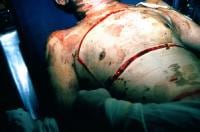
A military study revealed troops suffering from severe burns tended to receive either no prehospital fluid or too much fluid1.
The authors point out some practical realities and an attempted solution:


For a medic potentially treating multiple casualties at once in a hostile environment, the calculation of the modified Brooke or Parkland formula may be unrealistic prior to beginning fluid resuscitation in the prehospital setting.

The USAISR’s Rule of 10 is a simplified formula to guide the initial fluid resuscitation of a burn victim. The burn size is estimated to the nearest 10% TBSA. For patients weighing 40 to 80 kg, the burn size is then multiplied by 10 to give the initial fluid rate in milliliters per hour. The rate is increased by 100 mL/hour for every 10 kg above 80 kg in terms of the patient’s weight. For the majority of adult burn patients, the Rule of 10 approximates the initial fluid rate within accepted ABA guidelines.

A previous study on the rule of 10 showed it provided an estimate that fell between the modified Brooke and Parkland estimates 87.8% of the time, less than the modified Brooke <12% of the time, and hardly ever (>1%) exceeded the Parkland estimate2.


OBJECTIVE: The purpose of this article is to provide a descriptive study of the management of burns in the prehospital setting of a combat zone.

METHODS: A retrospective chart review was performed of U.S. casualties with >20% total-body-surface-area thermal burns, transported from the site of injury to Ibn Sina Combat Support Hospital (CSH) between January 1, 2006, and August 30, 2009.

RESULTS: Ibn Sina CSH received 225 burn casualties between January 2006 and August 2009. Of these, 48 met the inclusion criteria. The mean Injury Severity Score was 31.7 (range 4 to 75). Prehospital vascular access was obtained in 24 casualties (50%), and 20 of the casualties received fluid resuscitation. Out of the 48 casualties enrolled, 28 (58.3%) did not receive prehospital fluid resuscitation. Of the casualties who received fluid resuscitation, nearly all received volumes in excess of the guidelines established by the American Burn Association and those recommended by the Committee for Tactical Combat Casualty Care. With regard to pain management in the prehospital setting, 13 casualties (27.1%) received pain medication.

CONCLUSIONS: With regard to the prehospital fluid resuscitation of primary thermal injury in the combat zone, two extremes were noted. The first group did not receive any fluid resuscitation; the second group was resuscitated with fluid volumes higher than those expected if established guidelines were utilized. Pain management was not uniformly provided to major burn casualties, even in several with vascular access. These observations support improved education of prehospital personnel serving in a combat zone.

1. Prehospital burn management in a combat zone
Prehosp Emerg Care, 2012 vol. 16 (2) pp. 273-276
2. Simple derivation of the initial fluid rate for the resuscitation of severely burned adult combat casualties: in silico validation of the rule of 10
J Trauma. 2010 Jul;69 Suppl 1:S49-54

Specialised chemical burns

Certain chemical burns require a little extra thought than just irrigation and good wound care – which may even be contraindicated. An article in The Journal of Emergency Medicine addresses these, and some of the points are summarised below, with some additional information from Toxbase:
Hot tar (bitumen)

  • Immerse contaminated area in cool water until the bitumen has hardened and cooled.
  • Adherent material may be left in place to avoid causing further injury by removal attempts, and will spontaneously detach after a few days.
  • If a finger or limb is completely surrounded, split the bitumen to prevent a tourniquet effect.
  • To remove bitumen, apply a lipid or polysorbate based agent and a clean non-adherent dressing. Suitable products include melted butter, sunflower oil, liquid paraffin, and petroleum or polysorbate based antibiotic ointments. Solvents such as alcohol, acetone, kerosene, ether or gasoline are not suitable.
  • Change the dressing frequently, and reapply the product as necessary, until the bitumen is completely removed. This may take up to 72 hours.
  • Treat as a thermal burn.


Elemental sodium

  • – utilised in the manufacturing of methamphetamine.
  • will spontaneously ignite above 115°C
  • Contact with water releases sodium hydroxide and hydrogen gas. It is the heat released in the reaction with the water in air that then ignites locally produced hydrogen gas.
  • Burns involving the metallic forms of sodium, potassium, and lithium (alkali metals) produce both thermal and chemical injury to the tissue. The thermal tissue damage is due to the extreme exothermic reaction that metallic sodium undergoes when exposed to water.
  • At times, water, when mixed with either elemental sodium or potassium, undergoes an explosive reaction.
  • Avoid water irrigation; if metal is still present in the tissues, the added water could ignite it.
  • All clothing should first be removed from the victim. If retained metal exists, the affected area should be covered in mineral oil. Removal of embedded sodium should then be undertaken with forceps.
  • Mineral oil is a practical, and potentially safer, alternative to isopropyl alcohol for the storage of elemental sodium.

Chromic acid

  • – a corrosive, oxidizing acid. After skin has been exposed to chromium, burns covering as little as 10% of body surface area (BSA) have proven fatal.
  • Burns involving as little as 1% of total BSA have resulted in acute renal failure.
  • Wash thoroughly with copious amounts of water and treat as a thermal burn.
  • Application of 10% ascorbic acid solution at least three times per day may improve the rate of healing
  • Prompt excision of burned, contaminated areas is recommended to prevent absorption of the chemical.

White phosphorus

  • – will ignite spontaneously in 30°C air temperature; typically stored in water.
  • burns of > 10% can have associated mortality.
  • Three stages of systemic toxicity exist: (1) gastrointestinal symptoms (nausea, vomiting and “smoking stool”). Symptoms of headache, seizures, and coma, as well as the potential for cardiovascular collapse, may occur in the initial phase. Decreasing serum calcium concentrations; (2) symptom-free period; (3) (4 to 8 days post-exposure) neurological toxicity, bleeding diathesis, hepatic failure, renal failure, and shock.
  • Continuous coverage with water will protect both the patient and staff from ignition and fumes that result from white phosphorus’s contact with air.
  • Brushing particulate not incorporated in wounds can accomplish a significant amount of decontamination. This brushing should be followed by continuous irrigation until all particles are removed. Those debriding and decontaminating an exposed patient should have a safe method of disposing of particles: a container of cold water would suffice.
  • A way to identify phosphorous particles for removal is the use of a Wood’s lamp, which will cause the white phosphorous to fluoresce.
  • Excision may be necessary to remove the chemical if deeply entrenched in fascia.

Phenol

  • – a corrosive aromatic hydrocarbon that can be absorbed at toxic levels through all routes of absorption
  • causes extensive denaturisation of tissue proteins, producing an eschar with shallow ulcers
  • Rescue personnel should use butyl rubber gloves and aprons, and conduct decontamination in a well-ventilated area.
  • wipe exposed areas immediately with low-molecular-weight polytheylene glycol (PEG 300 or 400)
  • however Toxbase states: “The use of solvents (such as glycerol, polyethylene glycol and isopropanol) has been suggested. One (animal) study (Hunter et al, 1992) indicated that isopropanol was more effective than water, but there is no evidence in humans that solvents are more effective than washing with copious amounts of water.”
  • if the burn covers a large skin area, high pressure shower irrigation before PEG application is preferable
  • Any water applied must be applied in high pressure, as small amounts might dilute the phenol present on the skin and thus expand not only the involved area but also the amount of phenol absorbed.

Hydrofluoric acid

  • HF is highly corrosive and causes damage by two mechanisms. It produces a corrosive burn from the high concentration of hydrogen ions. It also penetrates tissues due to the lipophilic nature of fluoride, and causes liquefactive necrosis.
  • Tissue penetration leads to systemic reactions with effects on the cardiac, respiratory, nervous, and gastrointestinal systems. The fluoride ion precipitates calcium, leading to hypocalcemia, and may interfere with enzyme systems by binding magnesium and manganese, as well as important nerve conduction functions that depend on calcium.
  • Copious irrigation of HF-burned skin with water should begin immediately. Most HF burns will respond well to this.
  • Pain that persists after irrigation is a marker that the fluoride ion needs detoxification. This can be accomplished through superficial topical treatment, infiltrative treatment, or intra-arterial treatment.
  • The preferred topical agent is calcium gluconate gel.

Special considerations in hazardous materials burns.
J Emerg Med. 2010 Nov;39(5):544-53

Pre-hospital chest escharotomy

Two cases are described in Pre-hospital Emergency Care of severely burned patients who were impossible to adequately ventilate after tracheal intubation until they underwent escharotomy by a pre-hospital physician.
The review that follows reminds us of some intersting escharotomy facts:

  • circumferential extremity burns can cause limb ischaemia
  • abdominal burns can cause elevated intra-abdominal pressure and ischemic bowel
  • neck burns can cause tracheal and jugular venous compression
  • chest burns can cause respiratory compromise
  • one previous study showed that chest and abdominal escharotomies significantly decreased intra-abdominal pressure, retention of carbon dioxide, and central venous and inferior vena caval pressures while significantly increasing serum oxygen concentration and systolic blood pressure.
  • escharotomies may be performed on multiple body parts, including the extremities, the digits, the chest, the abdomen, the neck, and the penis
  • neck escharotomy is a relatively simple procedure that involves an incision of the skin eschar longitudinally in the anterior midline from the chin to the sternal notch
  • although different ways of doing chest escharotomies have been described, in the two reported cases in this article the procedure only involved longitudinal incisions, with good immediate effect.


Of note, neither of the physicians concerned had seen or done an escharotomy before. I’m adding this to my list of life-saving surgical interventions that are technically straightforward to perform, cannot always wait for another specialist to do, and happen too rarely to train for in the traditional way (ie being taught on a patient under supervision prior to the first time you do one).
Out-of-hospital chest escharotomy: a case series and procedure review
Prehosp Emerg Care. 2010 Jul-Sep;14(3):349-54

Antibiotics for severe burns yes or no?

Should prophylactic antibiotics be given to burns patients? A systematic review of 17 trials concludes they may reduce all-cause mortality when given for 4-14 days after admission; there was a reduction in pneumonia with systemic prophylaxis and a reduction in wound infections with perioperative prophylaxis. However the overall methodological quality of the trials was poor and in three trials, resistance to the antibiotic used for prophylaxis significantly increased. The authors consequently do not recommend prophylaxis for patients with severe burns other than perioperatively.
Take home message: not needed as part of critical care resuscitation
Prophylactic antibiotics for burns patients: systematic review and meta-analysis
BMJ. 2010 Feb 15;340:c241

Estimating burn survival

Even though we might not know it’s called that, many of us are familiar with the Baux score, defined as the sum of age in years and percent body burn, to predict percent mortality after severe burns. This is however a little out of date due to advances in burn care, and does not take into account inhalational injury.
The Baux score was modified using data on 39,888 burned patients using a logistic regression model that showed that age and percent burn contribute almost equally to mortality and that the presence of inhalation injury added the equivalent of 17 years (or 17% burn). These observations suggested a revised Baux Score:
Per Cent Mortality = Age + Percent Burn + [17 x (Inhalation Injury, 1= yes, = no)]
Simplified Estimates of the Probability of Death After Burn Injuries: Extending and Updating the Baux Score
J Trauma. 2010 Mar;68(3):690-7