Tag Archives: CT

Spinal imaging for the adult obtunded blunt trauma patient

‘You can’t clear the cervical spine until the patient wakes up!’ How often have you heard this said about a patient with severe traumatic brain injury who may not ‘wake up’ for weeks, if at all?
A controversial area, but many institutions now allow collar removal if a neck CT scan is normal. Does this rule out injury with 100% sensitivity? No – but it probably pushes the balance of risk towards removing the collar – an intervention with no evidence for benefit and plenty of reasons why it may be harmful to ventilated ICU patients. For example, clearing the cervical spine based on MDCT was associated with less delirium and less ventilator associated pneumonia, both of which have been associated with increased mortality in critically ill patients (this is referenced in the paper below).
The UK’s Intensive Care Society has had pragmatic guidelines along these lines since 2005, which can be found here. This month’s Intensive Care Medicine publishes an updated literature review providing some further support to this approach.


PURPOSE: Controversy exists over how to ‘clear’ (we mean enable the clinician to safely remove spinal precautions based on imaging and/or clinical examination) the spine of significant unstable injury among clinically unevaluable obtunded blunt trauma patients (OBTPs). This review provides a clinically relevant update of the available evidence since our last review and practice recommendations in 2004.

METHODS: Medline, Embase. Google Scholar, BestBETs, the trip database, BMJ clinical evidence and the Cochrane library were searched. Bibliographies of relevant studies were reviewed.

RESULTS: Plain radiography has low sensitivity for detecting unstable spinal injuries in OBTPs whereas multidetector-row computerised tomography (MDCT) approaches 100%. Magnetic resonance imaging (MRI) is inferior to MDCT for detecting bony injury but superior for detecting soft tissue injury with a sensitivity approaching 100%, although 40% of such injuries may be stable and ‘false positive’. For studies comparing MDCT with MRI for OBTPs; MRI following ‘normal’ CT may detect up to 7.5% missed injuries with an operative fixation in 0.29% and prolonged collar application in 4.3%. Increasing data is available on the complications associated with prolonged spinal immobilisation among a population where a minority have an actual injury.

CONCLUSIONS: Given the variability of screening performance it remains acceptable for clinicians to clear the spine of OBTPs using MDCT alone or MDCT followed by MRI, with implications to either approach. Ongoing research is needed and suggestions are made regarding this. It is essential clinicians and institutions audit their data to determine their likely screening performances in practice.

Clinical review: spinal imaging for the adult obtunded blunt trauma patient: update from 2004
Intensive Care Med. 2012 Mar 10. [Epub ahead of print]

Offensive medicine: CT before LP

I’m getting worn down by clinicians – often other specialists – who insist that CT imaging of the brain is mandatory prior to lumbar puncture in all patients. There is surely a subgroup of patients (especially young ones) in whom the benefit:harm balance of CT comes out in favour of NOT doing the imaging. In these cases, getting the scan is not ‘defensive medicine’ but ‘offensive medicine’ – offending the principle of primum non nocere. During ED shifts I have recently had to perform online searches in order to furnish colleagues and patients’ medically qualified relatives with printouts of the literature on this. This page is here to save me having to repeat those searches. Regarding the practice of performing a routine head CT prior to lumbar puncture to rule out risk of herniation:

  • Mass effect on CT does not predict herniation
  • Lack of mass effect on CT does not rule out raised ICP or herniation
  • Herniation has occurred in patients who did not undergoing lumbar puncture because of CT findings
  • Clinical predictors of raised ICP are more reliable than CT findings
  • CT may delay diagnosis and treatment of meningitis
  • Even in patients in whom LP may be considered contraindicated (cerebral abscess, mass effect on CT), complications from LP were rare in several studies

Best practice, it would seem, is the following

  • If you think CT will show a cause for the headache, do a CT
  • If a CT is indicated for other reasons (depressed conscious level, focal neurology), do a CT
  • If a GCS 15 patient is to undergo LP for suspected (or to rule out) meningitis, and they have a normal neurological exam (including fundi), and are not elderly or immunosuppressed, there is no need to do a CT first.
  • If you’re seriously worried about meningitis and are intent on getting a CT prior to LP, don’t let the imaging delay antimicrobial therapy.

Here are some useful references:

1. The CT doesn’t help

CT head before lumbar puncture in suspected meningitis BestBET evidence summary: In cases of suspected meningitis it is very unlikely that patients without clinical risk factors (immunocompromise/ history of CNS disease/seizures) or positive neurological findings will have a contraindication to lumbar puncture on their CT scan If CT scan is deemed to be necessary, administration of antibiotics should not be delayed. BestBETS website

Computed Tomography of the Head before Lumbar Puncture in Adults with Suspected Meningitis Much cited NEJM paper from 2001 which concludes: “In adults with suspected meningitis, clinical features can be used to identify those who are unlikely to have abnormal findings on CT of the headN Engl J Med. 2001 Dec 13;345(24):1727-33 Full Text

Cranial CT before Lumbar Puncture in Suspected Meningitis Correspondence in 2002 NEJM including study of 75 patients with pneumococcal meningitis: CT cannot rule out risk of herniation Cranial CT before Lumbar Puncture in Suspected Meningitis N Engl J Med. 2002 Apr 18;346(16):1248-51 Full Text

2. The CT may harm

Cancer risk from CT Paucis verbis card, from the wonderful Academic Life in EM

3. Guidelines say CT is not always needed

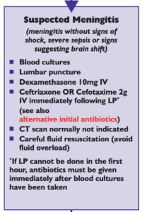
National (UK) guidelines on meningitis (community acquired meningitis in the immunocompetent host) available from meningitis.org. , including this box:

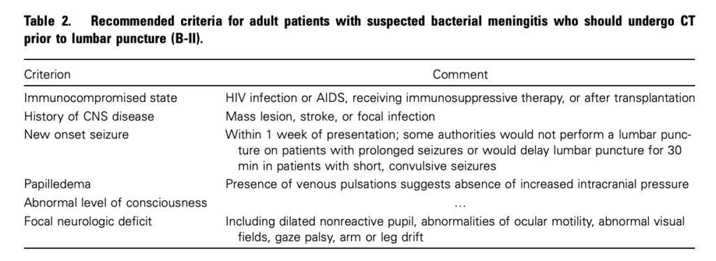
Practice Guidelines for the Management of Bacterial Meningitis These 2004 guidelines from the Infectious Diseases Society of America provide the following table listing the recommended criteria for adult patients with suspected bacterial meningitis who should undergo CT prior to lumbar puncture:

Clin Infect Dis. (2004) 39 (9): 1267-1284 Full text

4. This is potentially even more of an issue with paediatric patients

Fatal Lumbar Puncture: Fact Versus Fiction—An Approach to a Clinical Dilemma

An excellent summary of the above mentioned issues presented in a paediatric context, including the following:

On initial consideration a cranial CT would seem to be an appropriate and potentially useful diagnostic study for confirming the diagnosis of cerebral herniataion. The fallacy in this assessment has been emphasized by the finding that no clinically significant CT abnormalities are found that are not suspected on clinical assessments. Further, as previously noted, a normal CT examination may be found at about the time of a fatal herniation. Thus, the practical usefulness of a cranial CT in the majority of pediatric patients is limited to those rare patients whose increased ICP is secondary to mass lesions, not in the initial approach to acute meningitis.

Pediatrics. 2003 Sep;112(3 Pt 1):e174-6 Full Text

The last words should go to Dr Brad Spellberg, who in response to the IDSA’s guidelines wrote an excellent letter summarising much of the evidence at the time, confessed:

Why do we persist in using the CT scan for this purpose, despite the lack of supportive data? I am as guilty of this practice as anyone else, and the reason is simple: I am a chicken.

Clin Infect Dis. (2005) 40 (7): 1061 Full Text

Flat IVC on CT associated with deterioration

BACKGROUND: : We aimed to investigate the value of the diameter of the inferior vena cava (IVC) on initial computed tomography (CT) to predict hemodynamic deterioration in patients with blunt torso trauma.
METHODS: : We reviewed the initial CT scans, taken after admission to emergency room (ER), of 114 patients with blunt torso trauma who were consecutively admitted during a 24-month period. We measured the maximal anteroposterior and transverse diameters of the IVC at the level of the renal vein. Flat vena cava (FVC) was defined as a maximal transverse to anteroposterior ratio of less than 4:1. According to the hemodynamic status, the patients were categorized into three groups. Patients with hemodynamic deterioration after the CT scans were defined as group D (n = 37). The other patients who remained hemodynamically stable after the CT scans were divided into two groups: patients who were hemodynamically stable on ER arrival were defined as group S (n = 60) and those who were in shock on ER arrival and responded to the fluid resuscitation were defined as group R (n = 17).

RESULTS: : The anteroposterior diameter of the IVC in group D was significantly smaller than those in groups R and S (7.6 mm ± 4.4 mm, 15.8 mm ± 5.5 mm, and 15.3 mm ± 4.2 mm, respectively; p < 0.05). Of the 93 patients without FVC, 16 (17%) were in group D, 14 (15%) required blood transfusion, and 8 (9%) required intervention. However, of the 21 patients with FVC, all patients were in group D, 20 (95%) required blood transfusion, and 17 (80%) required intervention. The patients with FVC had higher mortality (52%) than the other patients (2%). CONCLUSION: : In cases of blunt torso trauma, patients with FVC on initial CT may exhibit hemodynamic deterioration, necessitating early blood transfusion and therapeutic intervention. Predictive Value of a Flat Inferior Vena Cava on Initial Computed Tomography for Hemodynamic Deterioration in Patients With Blunt Torso Trauma
J Trauma. 2010 Dec;69(6):1398-402

CT cervical spine in obtunded trauma patients

Prolonged collar use and spinal immobilisation in ICU patients can contribute to pressure sores, increased intracranial pressure, venous obstruction, difficulties with airway management, difficulties with central venous access, respiratory complications, and DVT, so a reliable investigation to rule out unstable cervical spine injury is required. Several studies demonstrate the high sensitivity of CT, and now a prospective study from Canada attempts to lend further support to this.
Comparing against their chosen gold standard of dynamic radiography, ie. flexion/extension views (F/E) in 402 patients who received both tests, there was one case of injury detected by F/E but not by CT, leading to quoted sensitivity of 99.75%. However this negative CT turned out to be a reporting error – the scan, which the authors include in their article, was clearly abnormal.
One weakness of this study is that they excluded patients who died on ICU. More worrying are the stats quoted. The authors stat ‘four hundred one patients (99.75%) had normal CT and F-E images facilitating clinical clearance of their C-spine and discontinuation of spinal precautions‘. So in other words, there was only one patient in their series of 402 with an injury (according to the gold standard), and this was missed. The sensitivity is therefore zero percent, not 99.75%. What seems to be a further error is the reporting in a table of 401 patients who had ‘Positive CT and Negative F-E’, which if true, would give a specificty of zero too!
This paper covers an important topic for intensivists but it seems to me to be too flawed to add meaningfully to the existing evidence that necks can be ‘cleared’ by CT in patients without signs of cervical spine injury, in whom it has been said that the risks of prolonged collar use and immobilisation may outweigh the risks of missed cervical injury.
Cervical spine clearance in obtunded blunt trauma patients: a prospective study
J Trauma. 2010 Mar;68(3):576-82

Whole-body CT during trauma resuscitation

German trauma patients are more likely to survive if they have a whole body CT rather than selective scans. Or that’s what this paper would have you believe IF you’re happy with the retrospective comparison, multivariate adjustments, and potential confounders. Still, if it helps you get your radiologists to play ball, the reference is…
Effect of whole-body CT during trauma resuscitation on survival: a retrospective, multicentre studyLancet. 2009 Apr 25;373(9673):1455-61