Category Archives: ICU

Stuff relevant to patients on ICU

Finally I understand ultrasound physics

Ever waste time trying to shake some ultrasound gel out the bottle, like a resistant blob of ketchup?
Sydney emergency medicine registrar Dr Steve Skinner demonstrates how to solve this. With physics.

This really does work, and has saved me a lot of time already. I now do a somewhat less ballistic version than the one demonstrated, so that patients don’t think I’m mad.


This video is for entertainment purposes only. We accept no responsibility for injuries sustained as a result of unaccustomed shoulder activity or inadvertently launched plastic projectiles.

What's with all the chloride? An assault on salt

I continue to be bewildered at my ED colleagues’ overwhelming preference for 0.9% saline as a resuscitation fluid regardless of clinical presentation. However, I have to acknowledge a lack of robust high level clinical evidence demonstrating its relative harm compared with more balanced solutions such as Hartmann’s / Ringer’s lactate or one of the more scarcely available Plasma-Lyte solutions.
Human and animal studies have demonstrated that saline exacerbates hyperchloraemia and metabolic acidosis and has renal effects including renal vasoconstriction and decreased glomerular filtration rate. A large observational study on surgical patients suggested that saline therapy increases the risk of patients requiring acute dialysis compared with Plasma-Lyte administration(1).
A new study in JAMA provides some further clinical evidence that saline has harmful renal effects(2). It was a before-and-after observational study, in which the change was a restriction in chloride-rich fluids so that they were made available only after prescription by the attending specialist for specific conditions (eg, hyponatraemia, traumatic brain injury, and cerebral oedema). Four of the authors published another study on the metabolic effects of this changed fluid strategy, presumably on the same or an overlapping cohort of patients, which I blogged about here.
Significant findings were that the chloride-restrictive strategy was associated with a significantly lower increase in serum creatinine level during ICU stay, a decrease in the incidence of renal injury and failure (according to the RIFLE definitions), and a decrease in renal replacement therapy. These effects persisted after adjusting for known contributors to acute kidney injury.
As this is not a randomised trial cause and effect cannot be assumed, but this is consistent with other work.
In summary, keep pushing the saline if you want to increase your patients’ risk of acute kidney injury and the need for dialysis, whilst rendering them acidotic. You may even decrease their gut perfusion(3) and give them abdominal discomfort and subjective decreased cognitive ability(4). Alternatively, give Hartmann’s / Ringer’s lactate… although bear in mind that might not be such a good choice in the context of hyponatraemia, alkalaemia, cerebral oedema, or traumatic brain injury.
1. Major complications, mortality, and resource utilization after open abdominal surgery: 0.9% saline compared to Plasma-Lyte
Ann Surg. 2012 May;255(5):821-9
[EXPAND Click for abstract]


OBJECTIVE: To assess the association of 0.9% saline use versus a calcium-free physiologically balanced crystalloid solution with major morbidity and clinical resource use after abdominal surgery.

BACKGROUND: 0.9% saline, which results in a hyperchloremic acidosis after infusion, is frequently used to replace volume losses after major surgery.

METHODS: An observational study using the Premier Perspective Comparative Database was performed to evaluate adult patients undergoing major open abdominal surgery who received either 0.9% saline (30,994 patients) or a balanced crystalloid solution (926 patients) on the day of surgery. The primary outcome was major morbidity and secondary outcomes included minor complications and acidosis-related interventions. Outcomes were evaluated using multivariable logistic regression and propensity scoring models.

RESULTS: For the entire cohort, the in-hospital mortality was 5.6% in the saline group and 2.9% in the balanced group (P < 0.001). One or more major complications occurred in 33.7% of the saline group and 23% of the balanced group (P < 0.001). In the 3:1 propensity-matched sample, treatment with balanced fluid was associated with fewer complications (odds ratio 0.79; 95% confidence interval 0.66-0.97). Postoperative infection (P = 0.006), renal failure requiring dialysis (P < 0.001), blood transfusion (P < 0.001), electrolyte disturbance (P = 0.046), acidosis investigation (P < 0.001), and intervention (P = 0.02) were all more frequent in patients receiving 0.9% saline.

CONCLUSIONS: Among hospitals in the Premier Perspective Database, the use of a calcium-free balanced crystalloid for replacement of fluid losses on the day of major surgery was associated with less postoperative morbidity than 0.9% saline.

[/EXPAND]
2. Association Between a Chloride-Liberal vs Chloride-Restrictive Intravenous Fluid Administration Strategy and Kidney Injury in Critically Ill Adults
JAMA. 2012 Oct 17;308(15):1566-72
[EXPAND Click for abstract]


CONTEXT: Administration of traditional chloride-liberal intravenous fluids may precipitate acute kidney injury (AKI).

OBJECTIVE: To assess the association of a chloride-restrictive (vs chloride-liberal) intravenous fluid strategy with AKI in critically ill patients.

DESIGN, SETTING, AND PATIENTS: Prospective, open-label, sequential period pilot study of 760 patients admitted consecutively to the intensive care unit (ICU) during the control period (February 18 to August 17, 2008) compared with 773 patients admitted consecutively during the intervention period (February 18 to August 17, 2009) at a university-affiliated hospital in Melbourne, Australia.

INTERVENTIONS: During the control period, patients received standard intravenous fluids. After a 6-month phase-out period (August 18, 2008, to February 17, 2009), any use of chloride-rich intravenous fluids (0.9% saline, 4% succinylated gelatin solution, or 4% albumin solution) was restricted to attending specialist approval only during the intervention period; patients instead received a lactated solution (Hartmann solution), a balanced solution (Plasma-Lyte 148), and chloride-poor 20% albumin.

MAIN OUTCOME MEASURES: The primary outcomes included increase from baseline to peak creatinine level in the ICU and incidence of AKI according to the risk, injury, failure, loss, end-stage (RIFLE) classification. Secondary post hoc analysis outcomes included the need for renal replacement therapy (RRT), length of stay in ICU and hospital, and survival.

RESULTS Chloride administration decreased by 144 504 mmol (from 694 to 496 mmol/patient) from the control period to the intervention period. Comparing the control period with the intervention period, the mean serum creatinine level increase while in the ICU was 22.6 μmol/L (95% CI, 17.5-27.7 μmol/L) vs 14.8 μmol/L (95% CI, 9.8-19.9 μmol/L) (P = .03), the incidence of injury and failure class of RIFLE-defined AKI was 14% (95% CI, 11%-16%; n = 105) vs 8.4% (95% CI, 6.4%-10%; n = 65) (P < .001), and the use of RRT was 10% (95% CI, 8.1%-12%; n = 78) vs 6.3% (95% CI, 4.6%-8.1%; n = 49) (P = .005). After adjustment for covariates, this association remained for incidence of injury and failure class of RIFLE-defined AKI (odds ratio, 0.52 [95% CI, 0.37-0.75]; P < .001) and use of RRT (odds ratio, 0.52 [95% CI, 0.33-0.81]; P = .004). There were no differences in hospital mortality, hospital or ICU length of stay, or need for RRT after hospital discharge.
CONCLUSION The implementation of a chloride-restrictive strategy in a tertiary ICU was associated with a significant decrease in the incidence of AKI and use of RRT.

[/EXPAND]
3. The effects of balanced versus saline-based hetastarch and crystalloid solutions on acid-base and electrolyte status and gastric mucosal perfusion in elderly surgical patients
Anesth Analg. 2001 Oct;93(4):811-6
Free full text
[EXPAND Click for abstract]


The IV administration of sodium chloride solutions may produce a metabolic acidosis and gastrointestinal dysfunction. We designed this trial to determine whether, in elderly surgical patients, crystalloid and colloid solutions with a more physiologically balanced electrolyte formulation, such as Hartmann’s solution and Hextend, can provide a superior metabolic environment and improved indices of organ perfusion when compared with saline-based fluids. Forty-seven elderly patients undergoing major surgery were randomly allocated to one of two study groups. Patients in the Balanced Fluid group received an intraoperative fluid regimen that consisted of Hartmann’s solution and 6% hetastarch in balanced electrolyte and glucose injection (Hextend). Patients in the Saline group were given 0.9% sodium chloride solution and 6% hetastarch in 0.9% sodium chloride solution (Hespan). Biochemical indices and acid-base balance were determined. Gastric tonometry was used as a reflection of splanchnic perfusion. Postoperative chloride levels demonstrated a larger increase in the Saline group than the Balanced Fluid group (9.8 vs 3.3 mmol/L, P = 0.0001). Postoperative standard base excess showed a larger decline in the Saline group than the Balanced Fluid group (-5.5 vs -0.9 mmol/L, P = 0.0001). Two-thirds of patients in the Saline group, but none in the Balanced Fluid group, developed postoperative hyperchloremic metabolic acidosis (P = 0.0001). Gastric tonometry indicated a larger increase in the CO2 gap during surgery in the Saline group compared with the Balanced Fluid group (1.7 vs 0.9 kPa, P = 0.0394). In this study, the use of balanced crystalloid and colloid solutions in elderly surgical patients prevented the development of hyperchloremic metabolic acidosis and resulted in improved gastric mucosal perfusion when compared with saline-based solutions.
IMPLICATIONS: This prospective, randomized, blinded trial showed that, in elderly surgical patients, the use of balanced IV solutions can prevent the development of hyperchloremic metabolic acidosis and provide better gastric mucosal perfusion compared with saline-based fluids.

[/EXPAND]
4. The effect of intravenous lactated Ringer’s solution versus 0.9% sodium chloride solution on serum osmolality in human volunteers.
Anesth Analg. 1999 May;88(5):999-1003
Free full text
[EXPAND Click for abstract]


Animal studies have shown that large volumes of IV lactated Ringer’s solution (LR) decrease serum osmolality, thereby increasing cerebral water. These studies have led to recommendations to limit LR to avoid cerebral edema in neurosurgical patients. Eighteen healthy human volunteers aged 20-48 yr received 50 mL/kg LR over 1 h on one occasion and 0.9% sodium chloride (NS) on another. Venous samples were taken at baseline (T1), at infusion end (T2), and 1 h after T2 (T3). Time until first urination was noted. With LR, serum osmolality decreased by 4+/-3 mOsm/kg from T1 to T2 and increased insignificantly with NS. At T3, osmolality returned almost to baseline in the LR group. Blood pH increased from T1 to T2 with LR by 0.04+/-0.04 and decreased with NS by 0.04+/-0.04. These pH changes persisted at T3. Subjective mental changes occurred only with NS. Abdominal discomfort was more common with NS. Time until first urination was longer with NS (106+/-11 min) than with LR (75+/-10 min) (P < 0.001). In healthy humans, an infusion of large volumes of LR, but not NS, transiently decreased serum osmolality, whereas acidosis associated with NS persisted and urinary output was slower with NS.
IMPLICATIONS: Large volumes of lactated Ringer’s solution administered to healthy humans produced small transient changes in serum osmolality. Large volumes of sodium chloride did not change osmolality but resulted in lower pH.

[/EXPAND]

Infectious biomarkers in the critically ill

A study examining patterns of procalcitonin in a group of critically ill patients(1) showed some interesting findings:

Shock was associated with higher procalcitonin values independent of the presence of infection

Procalcitonin (PCT) levels were less in patients who developed infections later during their ICU stay compared with those who had infections when admitted to ICU.

The accompanying editorial(2) reminds us about commonly used inflammatory biomarkers.

White blood cells are influenced by almost every inflammatory stimulus, rendering them unhelpful in the management of severely ill patients.

Daily monitoring of CRP levels can identify ICU-acquired infections early, and some prognostic information can be provided by how rapidly CRP levels respond to treatment.

PCT rises early in severe sepsis, mainly by pneumonia and bloodstream infections, and can reflect the severity of the systemic inflammatory response syndrome to infection. PCT is more specific than CRP for infection compared with non-infectious causes of systemic inflammatory response syndrome. However PCT can also be increased in noninfectious diseases such as acute pancreatitis and cardiogenic shock.

1. Longitudinal changes in procalcitonin in a heterogenous group of critically ill patients
Crit Care Med. 2012 Oct;40(10):2781-2787
[EXPAND Click for Abstract]


OBJECTIVE: The utility of procalcitonin for the diagnosis of infection in the critical care setting has been extensively investigated with conflicting results. Herein, we report procalcitonin values relative to baseline patient characteristics, presence of shock, intensive care unit time course, infectious status, and Gram stain of infecting organism.

DESIGN: Prospective, multicenter, observational study of critically ill patients admitted to intensive care unit for >24 hrs. SETTING:: Three tertiary care intensive care units.

PATIENTS: All consenting patients admitted to three mixed medical-surgical intensive care units. Patients who had elective surgery, overdoses, and who were expected to stay <24 hrs were excluded.

INTERVENTIONS: Patients were followed prospectively to ascertain the presence of prevalent (present at admission) or incident (developed during admission) infections and clinical outcomes. Procalcitonin levels were measured daily for 10 days and were analyzed as a function of the underlying patient characteristics, presence of shock, time of infection, and pathogen isolated.

MAIN RESULTS: Five hundred ninety-eight patients were enrolled. Medical and surgical infected cohorts had similar baseline procalcitonin values (3.0 [0.7-15.3] vs. 3.7 [0.6-9.8], p = .68) and peak procalcitonin (4.5 [1.0-22.9] vs. 5.0 [0.9-16.0], p = .91). Infected patients were sicker than their noninfected counterparts (Acute Physiology and Chronic Health Evaluation II 22.9 vs. 19.3, p < .001); those with infection at admission had a trend toward higher peak procalcitonin values than did those whose infection developed in the intensive care unit (4.9 vs. 1.4, p = .06). The presence of shock was significantly associated with elevations in procalcitonin in cohorts who were and were not infected (both groups p < .003 on days 1-5).

CONCLUSIONS: Procalcitonin dynamics were similar between surgical and medical cohorts. Shock had an association with higher procalcitonin values independent of the presence of infection. Trends in differences in procalcitonin values were seen in patients who had incident vs. prevalent infections.

[/EXPAND]
2. The many facets of procalcitonin in the critically ill population
Crit Care Med. 2012 Oct;40(10):2903-5

ECMO for paediatric cardiac arrest

The Taiwanese are at it again with their extracorporeal life support. This time, they report their outcomes in children who received ECMO for in-hospital cardiac arrest. Interestingly, the patients with pure cardiac causes of cardiac arrest had a survival rate similar to patients with non-cardiac causes.


PURPOSE: The study aims to describe 11 years of experience with extracorporeal cardiopulmonary resuscitation (ECPR) for in-hospital paediatric cardiac arrest in a university affiliated tertiary care hospital.

METHODS: Paediatric patients who received extracorporeal membrane oxygenation (ECMO) during active extracorporeal cardiopulmonary resuscitation (ECPR) at our centre from 1999 to 2009 were included in this retrospective study. The results from three different cohorts (1999-2001, 2002-2005 and 2006-2009) were compared. Survival rates and neurological outcomes were analysed. Favourable neurological outcome was defined as paediatric cerebral performance categories (PCPC) 1, 2 and 3.

RESULTS: We identified 54 ECPR events. The survival rate to hospital discharge was 46% (25/54), and 21 (84%) of the survivors had favourable neurological outcomes. The duration of CPR was 39±17 min in the survivors and 52±45 min in the non-survivors (p=NS). The patients with pure cardiac causes of cardiac arrest had a survival rate similar to patients with non-cardiac causes (47% (18/38) vs. 44% (7/16), p=NS). The non-survivors had higher serum lactate levels prior to ECPR (13.4±6.4 vs. 8.8±5.1 mmol/L, p<0.01) and more renal failure after ECPR (66% (19/29) vs. 20% (5/25), p<0.01). The patients resuscitated between 2006 and 2009 had shorter durations of CPR (34±13 vs. 78±76 min, p=0.032) and higher rates of survival (55% (16/29) vs. 0% (0/8), p=0.017) than those resuscitated between 1999 and 2002.

CONCLUSIONS: In our single-centre experience with ECPR for paediatric in-hospital cardiac arrest, the duration of CPR has become shorter and outcomes have improved in recent years. Higher pre-ECPR lactate levels and the presence of post-ECPR renal failure were associated with increased mortality. The presence of non-cardiac causes of cardiac arrest did not preclude successful ECPR outcomes. The duration of CPR was not significantly associated with poor outcomes in this study.

Eleven years of experience with extracorporeal cardiopulmonary resuscitation for paediatric patients with in-hospital cardiac arrest
Resuscitation. 2012 Jun;83(6):710-4

Surgical treatment for acute massive pulmonary embolism

A recent paper reminds us that surgery is an option in the management of massive pulmonary embolism(1), to be considered in the patient for whom thrombolysis has failed or is contraindicated. Good outcomes were produced when surgery was performed in a centre capable of cardiopulmonary bypass (6% 30-day postoperative mortality), but is surgery an option when these facilities are unavailable?

The “venous inflow occlusion” technique involves clamping the venae cavae prior to removing clot directly from the pulmonary artery and its branches after median sternotomy, and can be performed in any hospital with surgical facilities. Under normothermic conditions, speed is of the essence once cardiac arrest occurs, since the irreversible anoxic cerebral injury will occur after just a few minutes.
Clarke and Abrams wrote in the Lancet in 1972(2):

Our use of venous inflow-occlusion has given results which compare well with those obtained with extracorporeal circulation. 50% of our patients survived. All patients who had emboli removed without an episode of ventricular asystole survived surgery. Late deaths in 3 patients were from causes unrelated to pulmonary embolism, and from a further massive pulmonary embolus a week later. The technique has been applied with equal success in a major hospital fully equipped for cardiac surgery and in hospitals where resident and nursing staff had no experience of either thoracic or cardiac surgery. The simplicity and speed of the method has enabled the obstructed right ventricle to be relieved within thirty minutes of the onset of symptoms. The interval between induction of anaesthesia and the skin incision should be kept as short as possible, and drugs to maintain the blood pressure should be given. The period between skin incision and the restoration of the circulation has, with practice, been reduced to ten minutes.

But this clearly still requires surgical expertise and facilities. Emergency physicians can open the chest to deal with penetrating trauma. Could an ED thoracotomy facilitate clot removal from the pulmonary artery?

In 1969, a lady in her 50s arrested on the ward after an operation to remove a mass via a left lateral thoractomy. Pulmonary embolism was suspected and her thoracotomy wound was re-opened and the pulmonary artery incised, resulting in the removal of large amounts of clot. Return of spontaneous circulation resulted after a brief period of internal cardiac massage. Her case was written up decades later, in 1998(3):

The patient recovered rapidly and left the hospital on the 21st day without signs of cerebral damage. This patient is now 86 years old, mentally normal, living alone, and doing her own housekeeping. She remembers the hospital stay and the past years as worth living

Some patients may be considered too high risk for surgery and in some centres Extracorporeal Membrane Oxygenation (ECMO) is an option. It has been used both as life support pending surgery(4), or as an alternative to surgery to allow heparinisation to be used(5,6).

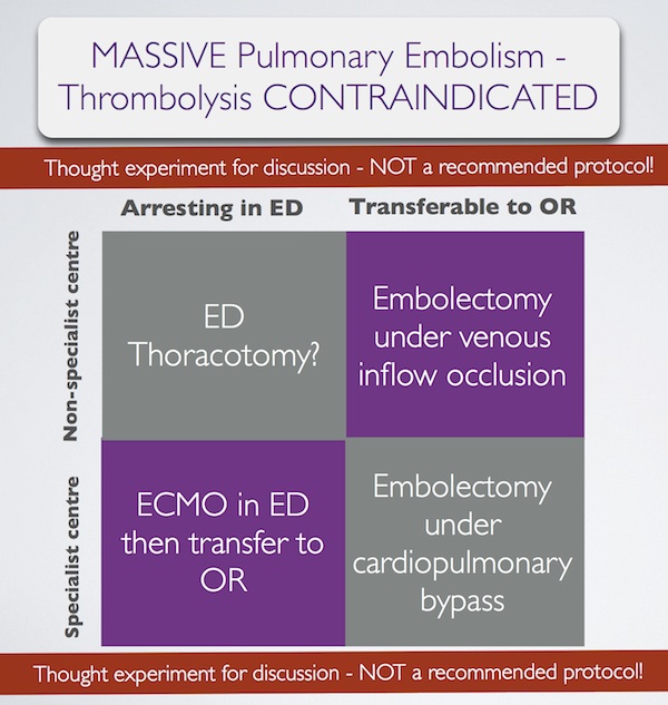
In summary, some patients with massive pulmonary embolism may benefit from surgery (contraindication to ‘lysis or failed ‘lysis). Getting them to surgery alive, or operating on them during cardiac arrest, is a challenge. Ideally they would undergo embolectomy under cardopulmonary bypass in the operating room, or could be placed on ECMO in the ED prior to going to the OR. If they present to a centre without these facilities, then the venous inflow occlusion technique could be used in the OR without bypass. Just rarely a patient may present in extremis with PE to an ED without these options. If that patient has major contraindications to thrombolysis, would an ED thoracotomy be something you would entertain?

I have done several thoracotomies for penetrating trauma but never for PE. I do not pretend to know how, and cannot find a case report of ED thoracotomy for pulmonary embolism in the literature. I’m therefore NOT recommending it. However, I would love to know people’s views on its feasibility. A possible approach could be summarised as:
Massive pulmonary embolism fascinates me, because it’s seen in the ‘talk and die’ patient. It is a single, treatable pathology that if diagnosed and treated appropriately truly makes the difference between life and death. When medicine presents us with an opportunity ‘on a plate’ like that to save a life, we need to be prepared. I have had great saves with this diagnosis and sadly have seen disastrous failures to act. When the time comes, we need to ask: ‘have we explored all options?’.

1. Surgical treatment of acute pulmonary embolism–a 12-year retrospective analysis.
Scand Cardiovasc J. 2012 Jun;46(3):172-6. Epub 2012 Mar 27.

OBJECTIVES: Surgical embolectomy for acute pulmonary embolism (PE) is considered to be a high risk procedure and therefore a last treatment option. We wanted to evaluate the procedures role in modern treatment of acute PE.
DESIGN: All data on patients treated with surgical embolectomy for acute PE were retrieved from our clinical database. The mortality was extracted from the Danish mortality register.
RESULTS: From October 1998 to July 2010, 33 patients underwent surgical embolectomy. All procedures were done through a median sternotomy and extracorporeal circulation. Twenty-six patients were diagnosed with a high risk PE and 7 with an intermediate risk PE and intracardial pathology. Six patients had been insufficiently treated with thrombolysis. Thirteen patients had contraindication for thrombolysis. Six patients were brought to the operating theatre in cardiogenic shock, 8 needed ventilator support, and 1 was in cardiac arrest. The postoperative 30-day mortality was 6% and during the 12-year follow-up the cumulative survival was 80% with 4 late deaths.
CONCLUSION: Surgical pulmonary embolectomy can be performed with low mortality although the treated patients belong to the most compromised part of the PE population. The results support surgical embolectomy as a vital part of the treatment algorithm for acute PE.

2. Pulmonary embolectomy with venous inflow-occlusion.
The Lancet 1972;1(7754):767–769

Massive pulmonary emboli have been removed surgically from 26 patients. The technique of normothermic circulatory arrest by venous inflow-occlusion was used in 25 patients. 13 patients survived. There were 10 operative deaths and 3 hospital deaths. Diagnosis was based upon clinical findings supplemented by electrocardiography and a plain radiograph of the chest. Surgery was offered to patients having a pulmonary embolus sufficiently massive to produce sustained hypotension. All patients whose hearts stopped beating before the embolectomy died. 6 successful operations were performed in hospitals without facilities for cardiac surgery. The method is recommended for its simplicity.

3. Left Anterior Thoracotomy for Pulmonary Embolectomy With 29-Year Follow-up
The Annals of Thoracic Surgery 1998, 66(4):1420-1421

Pulmonary embolectomy is usually performed in cardiopulmonary bypass. In acute situations too much time can be lost in setting up and connecting the pump oxygenator; this delay can cause cerebral damage in a patient with circulatory arrest. In such a situation left anterior thoracotomy can provide an ideal approach. An emergency thoracotomy can be performed in a few seconds. The lung automatically retracts. The phrenic nerve, pulmonary artery, and pericardium are clearly seen, and they outline the area for embolectomy. A case in which such an approach was successfully used is described.

4.ECMO treatment saved life of a young woman with acute pulmonary embolism
Lakartidningen 2004, 101(44):3420-3421

A 42-year old obese female using contraceptive medication was admitted to the emergency room because of sudden onset of dyspnoea and hypoxia. Computed tomography showed massive pulmonary emboli. Despite initial treatment with thrombolysis her condition deteriorated further and she was referred for acute surgery to our clinic. Before putting the patient to sleep extracorporeal circulation was instituted with access from the groin. After anaesthesia a median sternotomy was performed. With the heart beating, the main pulmonary artery was incised and a 9 cm long thrombus was removed. Immediate weaning from the heart-lung machine was not possible, mainly because of bleeding to the airways. The right atrium and the aorta was therefore cannulated and an extracorporeal circulation membrane oxygenator (ECMO) was used for three days. The patient required several re-entries for bleeding and a tracheotomy during the postoperative course. She was fully recovered three months after the operation.

5. Extracorporeal membrane oxygenator for pulmonary embolism.
The Annals of Thoracic Surgery 1997, 64(3):883-884 Free full text

6. Peripheral Extracorporeal Membrane Oxygenation: Comprehensive Therapy for High-Risk Massive Pulmonary Embolism
Ann Thorac Surg 2012;94:104–8

Background: Although commonly reserved as a last line of defense, experienced centers have reported excellent results with pulmonary embolectomy for massive and submassive pulmonary embolism (PE). We present a contemporary surgical series for PE that demonstrates the utility of peripheral extracorporeal membrane oxygenation (pECMO) for high-risk surgical candidates.
Methods: Between June 2005 and April 2011, 29 patients were treated for massive or submassive pulmonary embolism, with surgical embolectomy performed in 26. Four high-risk patients were placed on pECMO, established by percutaneously cannulating the right atrium through a femoral vein and perfusing by a Dacron graft anastomosed to the axillary artery. A small, extracorporeal, rotary assist device was used, interposing a compact oxygenator in the circuit, and maintaining anticoagulation with heparin.
Results: Extracorporeal membrane oxygenation was weaned in 3 of 4 patients after 5.3 days (5, 5, and 6), with normalization of right ventricular dysfunction and pulmonary artery pressure (44.0 ± 2.0 to 24.5 ± 5.5 mm Hg) by ECHO. Follow-up computed tomographies showed several peripheral, nearly resorbed emboli in 1 case and complete resolution in 2 others. The fourth patient, not improving after 10 days, underwent surgery where an embolic liposarcoma was extracted. For all 29 cases, hospital and 30-day mortality was 0% and all patients were discharged, with average postoperative length of stay of 15 days for embolectomy and 17 days for pECMO.
Conclusions: Heparin therapy with pECMO support is a rapid, effective option for patients who might benefit from pulmonary embolectomy but are at high risk for surgery.

Magnesium doesn't improve SAH outcome

A multicentre RCT showed intravenous magnesium sulphate does not improve clinical outcome after aneurysmal subarachnoid haemorrhage, therefore routine administration of magnesium cannot be recommended.
Magnesium for aneurysmal subarachnoid haemorrhage (MASH-2): a randomised placebo-controlled trial
Lancet 2012 July 7; 380(9836): 44–49 Free full text
[EXPAND Click to read abstract]


Background Magnesium sulphate is a neuroprotective agent that might improve outcome after aneurysmal subarachnoid haemorrhage by reducing the occurrence or improving the outcome of delayed cerebral ischaemia. We did a trial to test whether magnesium therapy improves outcome after aneurysmal subarachnoid haemorrhage.

Methods We did this phase 3 randomised, placebo-controlled trial in eight centres in Europe and South America. We randomly assigned (with computer-generated random numbers, with permuted blocks of four, stratified by centre) patients aged 18 years or older with an aneurysmal pattern of subarachnoid haemorrhage on brain imaging who were admitted to hospital within 4 days of haemorrhage, to receive intravenous magnesium sulphate, 64 mmol/day, or placebo. We excluded patients with renal failure or bodyweight lower than 50 kg. Patients, treating physicians, and investigators assessing outcomes and analysing data were masked to the allocation. The primary outcome was poor outcome—defined as a score of 4–5 on the modified Rankin Scale—3 months after subarachnoid haemorrhage, or death. We analysed results by intention to treat. We also updated a previous meta-analysis of trials of magnesium treatment for aneurysmal subarachnoid haemorrhage. This study is registered with controlled-trials.com (ISRCTN 68742385) and the EU Clinical Trials Register (EudraCT 2006-003523-36).

Findings 1204 patients were enrolled, one of whom had his treatment allocation lost. 606 patients were assigned to the magnesium group (two lost to follow-up), 597 to the placebo (one lost to follow-up). 158 patients (26·2%) had poor outcome in the magnesium group compared with 151 (25·3%) in the placebo group (risk ratio [RR] 1·03, 95% CI 0·85–1·25). Our updated meta-analysis of seven randomised trials involving 2047 patients shows that magnesium is not superior to placebo for reduction of poor outcome after aneurysmal subarachnoid haemorrhage (RR 0·96, 95% CI 0·86–1·08).

Interpretation Intravenous magnesium sulphate does not improve clinical outcome after aneurysmal subarachnoid haemorrhage, therefore routine administration of magnesium cannot be recommended.

[/EXPAND]

More on the meaning of lactate values

A newly published study(1) reminds us that we need to do better than just identify a raised lactate in patients with sepsis; we need to make sure it’s not increasing when they leave the ED (if we can). An incremental rise is associated with mortality.

The authors comment:


We found that the prognostic value of lactate continues to rise across a wide range of values, from 0 to 20 mmol/L…. These data suggest that grouping patients into less granular and larger groups, such as low, intermediate, and high, potentially underutilizes the prognostic value of the test. Furthermore, we did not find any value of lactate, up to a maximum value of 20 mmol/L, where mortality failed to increase with an increase in lactate concentration.

The paper does not state whether the lactate was arterial or venous, although either can be used. The Surviving Sepsis Campaign provides this comment:


In the course of the Campaign the question has been raised many times as to whether an arterial or venous lactate sample is appropriate. While there is no consensus of settled literature on this question, an elevated lactate of any variety is typically abnormal, although this may be influenced by other conditions..

This relationship between lactate trend and mortality has also been demonstrated in a study of all patients admitted to hospital (with or without sepsis), which also showed good correlation between arterial and venous lactate(2).

Lactate clearance has been shown to be an acceptable alternative to central venous oxygen saturation as a goal for therapy in ED severe sepsis patients(3), which is good because it provides one less reason for a central line.

Always remember the good emergency physician / critical care practitioner will consider other causes of a raised lactate, particularly when things don’t add up. I invented the ‘LACTATES’ acronym to help me remember them(4), and it’s come in handy several times.

Craving more info on lactate? Check out the EMCrit site with its great lactate reference sheet.

1. Prognostic Value of Incremental Lactate Elevations in Emergency Department Patients With Suspected Infection
Acad Emerg Med. 2012 Aug;19(8):983-5
[EXPAND Click for abstract]


Objectives:  Previous studies have confirmed the prognostic significance of lactate concentrations categorized into groups (low, intermediate, high) among emergency department (ED) patients with suspected infection. Although the relationship between lactate concentrations categorized into groups and mortality appears to be linear, the relationship between lactate as a continuous measurement and mortality is uncertain. This study sought to evaluate the association between blood lactate concentrations along an incremental continuum up to a maximum value of 20 mmol/L and mortality.

Methods:  This was a retrospective cohort analysis of adult ED patients with suspected infection from a large urban ED during 2007–2010. Inclusion criteria were suspected infection evidenced by administration of antibiotics in the ED and measurement of whole blood lactate in the ED. The primary outcome was in-hospital mortality. Logistic and polynomial regression were used to model the relationship between lactate concentration and mortality.

Results:  A total of 2,596 patients met inclusion criteria and were analyzed. The initial median lactate concentration was 2.1 mmol/L (interquartile range [IQR] = 1.3 to 3.3 mmol/L) and the overall mortality rate was 14.4%. In the cohort, 459 patients (17.6%) had initial lactate levels >4 mmol/L. Mortality continued to rise across the continuum of incremental elevations, from 6% for lactate <1.0 mmol/L up to 39% for lactate 19–20 mmol/L. Polynomial regression analysis showed a strong curvilinear correlation between lactate and mortality (R = 0.72, p < 0.0001).
Conclusions:  In ED patients with suspected infection, we found a curvilinear relationship between incremental elevations in lactate concentration and mortality. These data support the use of lactate as a continuous variable rather than a categorical variable for prognostic purposes.

[/EXPAND]
2. Blood lactate as a predictor for in-hospital mortality in patients admitted acutely to hospital: a systematic review
Scand J Trauma Resusc Emerg Med. 2011 Dec 28;19:74 Free Full Text
[EXPAND Click for abstract]


BACKGROUND: Using blood lactate monitoring for risk assessment in the critically ill patient remains controversial. Some of the discrepancy is due to uncertainty regarding the appropriate reference interval, and whether to perform a single lactate measurement as a screening method at admission to the hospital, or serial lactate measurements. Furthermore there is no consensus whether the sample should be drawn from arterial, peripheral venous, or capillary blood. The aim of this review was: 1) To examine whether blood lactate levels are predictive for in-hospital mortality in patients in the acute setting, i.e. patients assessed pre-hospitally, in the trauma centre, emergency department, or intensive care unit. 2) To examine the agreement between arterial, peripheral venous, and capillary blood lactate levels in patients in the acute setting.

METHODS: We performed a systematic search using PubMed, Cochrane Central Register of Controlled Trials, Cochrane Database of Systematic Reviews, and CINAHL up to April 2011. 66 articles were considered potentially relevant and evaluated in full text, of these ultimately 33 articles were selected.

RESULTS AND CONCLUSION: The literature reviewed supported blood lactate monitoring as being useful for risk assessment in patients admitted acutely to hospital, and especially the trend, achieved by serial lactate sampling, is valuable in predicting in-hospital mortality. All patients with a lactate at admission above 2.5 mM should be closely monitored for signs of deterioration, but patients with even lower lactate levels should be considered for serial lactate monitoring. The correlation between lactate levels in arterial and venous blood was found to be acceptable, and venous sampling should therefore be encouraged, as the risk and inconvenience for this procedure is minimal for the patient. The relevance of lactate guided therapy has to be supported by more studies.

[/EXPAND]
3. Lactate clearance vs central venous oxygen saturation as goals of early sepsis therapy: a randomized clinical trial
JAMA. 2010 Feb 24;303(8):739-46
[EXPAND Click for abstract]


CONTEXT: Goal-directed resuscitation for severe sepsis and septic shock has been reported to reduce mortality when applied in the emergency department.

OBJECTIVE: To test the hypothesis of noninferiority between lactate clearance and central venous oxygen saturation (ScvO2) as goals of early sepsis resuscitation.

DESIGN, SETTING, AND PATIENTS: Multicenter randomized, noninferiority trial involving patients with severe sepsis and evidence of hypoperfusion or septic shock who were admitted to the emergency department from January 2007 to January 2009 at 1 of 3 participating US urban hospitals.

INTERVENTIONS: We randomly assigned patients to 1 of 2 resuscitation protocols. The ScvO2 group was resuscitated to normalize central venous pressure, mean arterial pressure, and ScvO2 of at least 70%; and the lactate clearance group was resuscitated to normalize central venous pressure, mean arterial pressure, and lactate clearance of at least 10%. The study protocol was continued until all goals were achieved or for up to 6 hours. Clinicians who subsequently assumed the care of the patients were blinded to the treatment assignment.

MAIN OUTCOME MEASURE: The primary outcome was absolute in-hospital mortality rate; the noninferiority threshold was set at Delta equal to -10%.

RESULTS: Of the 300 patients enrolled, 150 were assigned to each group and patients were well matched by demographic, comorbidities, and physiological features. There were no differences in treatments administered during the initial 72 hours of hospitalization. Thirty-four patients (23%) in the ScvO2 group died while in the hospital (95% confidence interval [CI], 17%-30%) compared with 25 (17%; 95% CI, 11%-24%) in the lactate clearance group. This observed difference between mortality rates did not reach the predefined -10% threshold (intent-to-treat analysis: 95% CI for the 6% difference, -3% to 15%). There were no differences in treatment-related adverse events between the groups.

CONCLUSION: Among patients with septic shock who were treated to normalize central venous and mean arterial pressure, additional management to normalize lactate clearance compared with management to normalize ScvO2 did not result in significantly different in-hospital mortality.

[/EXPAND]
4. Non-septic hyperlactataemia in the emergency department
Emerg Med J. 2010 May;27(5):411-2

Flumazenil for BZD overdose: getting away with it

Standard teaching in benzodiazepine (BZD) overdose is that the reversal agent flumazenil is best avoided because of the risk of seizures, particular in those patients who are BZD dependent and those who may have co-ingested proconvulsant substances such as tricyclic antidepressants.
Data from the UK’s National Poisons Information Service indicate that these principles are often flouted in the UK, and they usually get away with it, even in patients who had had seizures prior to flumazenil therapy. This will not change my practice though.


Objective Benzodiazepine (BZD) overdose (OD) continues to cause significant morbidity and mortality in the UK. Flumazenil is an effective antidote but there is a risk of seizures, particularly in those who have co-ingested tricyclic antidepressants. A study was undertaken to examine the frequency of use, safety and efficacy of flumazenil in the management of BZD OD in the UK.

Methods A 2-year retrospective cohort study was performed of all enquiries to the UK National Poisons Information Service involving BZD OD.

Results Flumazenil was administered to 80 patients in 4504 BZD-related enquiries, 68 of whom did not have ventilatory failure or had recognised contraindications to flumazenil. Factors associated with flumazenil use were increased age, severe poisoning and ventilatory failure. Co-ingestion of tricyclic antidepressants and chronic obstructive pulmonary disease did not influence flumazenil administration. Seizure frequency in patients not treated with flumazenil was 0.3%. The frequency of prior seizure in flumazenil-treated patients was 30 times higher (8.8%). Seven patients who had seizures prior to flumazenil therapy had no recurrence of their seizures. Ventilation or consciousness improved in 70% of flumazenil-treated patients. Flumazenil administration was followed by one instance each of agitation and brief seizure.

Conclusions Flumazenil is used infrequently in the management of BZD OD in the UK. It was effective and associated with a low incidence of seizure. These results compare favourably with the results of published randomised controlled trials and cohort studies, although previous studies have not reported the use of flumazenil in such a high-risk population. This study should inform the continuing review of national guidance on flumazenil therapy.

Flumazenil use in benzodiazepine overdose in the UK: a retrospective survey of NPIS data
Emerg Med J. 2012 Jul;29(7):565-9

Extracorporeal cardiopulmonary resuscitation

You have a patient in cardiac arrest who has had excellent resuscitation from the point of collapse, and who has treatable underlying pathology (eg. PE or STEMI). However you’re unable to get return of spontaneous circulation so you call it. Someone just died for whom the technology exists to save them. Extracorporeal life support (ECLS) supports heart and lung function by externally providing circulatory flow and gas exchange until the patient’s underlying cause of arrest is treated or recovers.
ECLS requires an extracorporeal membrane oxygenation (ECMO) circuit to be placed during the cardiac arrest resuscitation. This may sound like extreme stuff, but there have been some amazing saves with this technology, and large numbers of in-hospital and out-of-hospital arrest patients have been treated in Japan, Korea, and Taiwan. ECMO has even been commenced in the field by prehospital emergency physicians.
An inspiring EMCrit podcast with Dr Joe Bellezzo described how this technology is applied at Sharp Memorial Hospital in San Diego. Bellezzo and colleagues have now published a series of their out-of-hospital arrest cases who received ECLS initiated by emergency physicians(1).
Coming back to the Japanese, a multicentre prospective cohort study of ECLS for out-of hospital cardiac arrest (the ‘SAVE-J’ study) selected patients with VF or pulseless VT in whom no ROSC was achieved with standard resuscitative measures. Their striking results mirror other ECLS studies and were published in abstract form in November 2011(2).
To me, the overwhelming take home messages from what I’ve seen and read on this are:


1. ECLS can provide dramatic saves with neurologically intact survival in cardiac arrest cases that otherwise would be dead.

2. The critical factor for successful clinical outcomes and avoidance of wasted resources and clinical futility is case selection. The underlying cause of arrest needs to be reversible (eg. myocarditis) or treatable (eg. STEMI) and good resuscitation needs to have been in place prior to ECLS.

3. In the right hospital with the right resuscitation team, it can be done.

1. Emergency physician-initiated extracorporeal cardiopulmonary resuscitation
Resuscitation. 2012 Aug;83(8):966-70
[EXPAND Click for abstract]


CONTEXT: Extracorporeal cardiopulmonary resuscitation (ECPR) refers to emergent percutaneous veno-arterial cardiopulmonary bypass to stabilize and provide temporary support of patients who suffer cardiopulmonary arrest. Initiation of ECPR by emergency physicians with meaningful long-term patient survival has not been demonstrated.

OBJECTIVE: To determine whether emergency physicians could successfully incorporate ECPR into the resuscitation of patients who present to the emergency department (ED) with cardiopulmonary collapse refractory to traditional resuscitative efforts.

DESIGN: A three-stage algorithm was developed for ED ECPR in patients meeting inclusion/exclusion criteria. We report a case series describing our experience with this algorithm over a 1-year period.

RESULTS: 42 patients presented to our ED with cardiopulmonary collapse over the 1-year study period. Of these, 18 patients met inclusion/exclusion criteria for the algorithm. 8 patients were admitted to the hospital after successful ED ECPR and 5 of those patients survived to hospital discharge neurologically intact. 10 patients were not started on bypass support because either their clinical conditions improved or resuscitative efforts were terminated.

CONCLUSION: Emergency physicians can successfully incorporate ED ECPR in the resuscitation of patients who suffer acute cardiopulmonary collapse. More studies are necessary to determine the true efficacy of this therapy.

[/EXPAND]
2. Multicenter Non-Randomized Prospective Cohort Study of Extracorporeal Cardiopulmonary Resuscitation for Out-of Hospital Cardiac Arrest: Study of Advanced Life Support for Ventricular Fibrillation with Extracorporeal Circulation in Japan (SAVE-J)
Circulation 2011; 124: A18132
[EXPAND Click for abstract]


Background: This study is aimed to examine the efficacy of extracorporeal cardiopulmonary resuscitation (ECPR) for patients in out-of hospital cardiac arrest (OHCA) with ventricular fibrillation (VF) or pulseless ventricular tachycardia (VT).

Method: The design of this study is a multicenter non-randomized prospective cohort study. Hypothesis is that the outcome of OHCA with VF or pulseless VT is similar between ECPR and conventional advanced life support (ALS). During from Oct. 2008 to Dec. 2010, forty six tertiary emergency hospitals were participated in this study. Patient inclusion criteria were 1) VF or pulseless VT on scene, 2) cardiac arrest on arrival at hospital, 3) within 45 minutes from a call to an arrival of hospital, and 4) non-ROSC by conventional ALS during 15 minutes after an arrival at hospital. Exclusion criteria were 1) age: 75 yr, 2) poor activities of daily livings, 3) non-cardiac verified cardiac arrest, and 4) hypothermia. According to the inclusion criteria, ECPR was adopted for OHCA in 26 hospitals (ECPR group) and conventional ALS was planned in 20 hospitals (non-ECPR group). Both groups (Intention-to-treat) were analyzed about the proportion of patients with favorable outcome (CPC1 or 2) assessed with the Glasgow-Pittsburgh Cerebral Performance and Overall Performance Categories at 1 month by chi square test and Fisher exact probability test.

Results: One hundred and eighty patients of ECPR group and 134 patients of non-ECPR group were enrolled. There was no difference between the background of ECPR group and non-ECPR group; Average age (56.0 VS 56.9), Witnessed (72.8% VS 75.4%), Lay-rescuer CPR (49.4% VS 45.5%), Acute coronary syndrome (65.6% VS 61.4%), Minutes from collapse to emergency department (26.8 VS 30.0). The favorable outcome rate in ECPR group (12.4%, 22 patients) was statistically higher than the rate in non-ECPR group (1.6%, two patients) (p<0.001).

Conclusion: Extracorporeal cardiopulmonary resuscitation may improve the outcome of out-of hospital cardiac arrest with VF or pulseless VT without ROSC by conventional ALS during 15 minutes after an arrival at hospital.

[/EXPAND]