Dogmalysis


I made up a word a while ago: “dogmalysis”. It refers to the dissolution of authoritative tenets held as established opinion without adequate grounds.

DOGMA: something held as an established opinion; a point of view or tenet put forth as authoritative without adequate grounds

LYSIS: a process of disintegration or dissolution (as of cells)

It’s my favourite thing in medicine. I don’t know why – perhaps because of my admiration since childhood for irreverent scientists who questioned authority, like Feynman and Sagan. Or perhaps it is because I think at times we physicians need to experience the humility of having our ignorance exposed. This is necessary to keep medicine science-based.


My undergraduate and much of my postgraduate training consisted of being taught medical certainties that I was required to regurgitate under exam conditions. The reality of clinical practice then revealed the awesome irreducible complexity of biology in our patients who ‘don’t read the textbooks’. As we learn in emergency medicine to navigate the perilous Bayesian jungle to a ‘very unlikely’ or ‘very likely’ life-threatening diagnosis, and when we have to weigh up the benefit:harm equation of an intervention that could either kill or cure, we begin to appreciate that certainty without evidence – dogma, or faith – can be lethal.


The problem is, however, that our human brains seem to thrive on it. We have evolved a whole senate of cognitive biases, which enable us to function well in everyday social situations, but which prevent us from conducting an impartial analysis of objective clinical data. An enlightening example of the degree to which our interpretation of the same information can vary is illustrated by a handful of trials on fibrinolytic therapy for stroke, producing a spectrum of reactions from aggressive promotion to skeptical opposition.


Being human, I have no doubt that I am occasionally dogmatic about topics to which I erroneously believe I have applied skepticism. I appreciate the courage of trainees who have the guts to challenge my assertions and who demand the evidence to justify them. Keep doing it. Keep asking. Keep challenging.

Keep lysing the dogma.

No-one said it better than Carl:

Dogmalytic posts


Traumatic cardiac arrest outcomes

simEver heard anyone spout dogma along the lines of: “it’s a traumatic cardiac arrest – resuscitation is futile as the outcome is hopeless: survival is close to zero per cent”?
I have. Less frequently in recent years, I’ll admit, but you still hear it spout forth from the anus of some muppet in the trauma team. Here’s some recent data to add to the existing literature that challenges the ‘zero per cent survival’ proponents. A Spanish study retrospectively analysed 167 traumatic cardiac arrests (TCAs). 6.6% achieved a complete neurological recovery (CNR), which increased to 9.4% if the first ambulance to arrive contained an advanced team including a physician. Rhythm and age were important: CNR was achieved in 36.4% of VFs, 7% of PEAs, and 2.7% of those in asystole; survival rate by age groups was 23.1% in children, 5.7% in adults, and 3.7% in the elderly.
Since traumatic arrest tends to affect a younger age group than medical arrests, the authors suggest:
Avoiding the potential decrease in life expectancy in this kind of patient justifies using medical resources to their utmost potential to achieve their survival
Since 2.7% of the asystolic patients achieved a CNR, the authors challenge the practice proposed by some authors that Advanced Life Support be withheld in TCA patients with asystole as the initial rhythm:
had that indication been followed, three of our patients who survived neurologically intact would have been declared dead on-scene.”
I’d like to know what interventions were making the difference in these patients. They describe what’s on offer as:


In our EMS, all TCA patients receive ALS on-scene, which includes intubation, intravenous access, fluid and drug therapy, point-of-care blood analysis, and procedures such as chest drain insertion, pericardiocentesis, or Focused Assessment with Sonography for Trauma ultrasonography to improve the treatment of the cause of the TCA.

It appears that crystalloids and colloids are their fluid therapy of choice; unlike many British and Australian physician-based prehospital services they made no mention of the administration of prehospital blood products.
Traumatic cardiac arrest: Should advanced life support be initiated?
J Trauma Acute Care Surg. 2013 Feb;74(2):634-8
[EXPAND Abstract]


BACKGROUND: Several studies recommend not initiating advanced life support in traumatic cardiac arrest (TCA), mainly owing to the poor prognosis in several series that have been published. This study aimed to analyze the survival of the TCA in our series and to determine which factors are more frequently associated with recovery of spontaneous circulation (ROSC) and complete neurologic recovery (CNR).

METHODS: This is a cohort study (2006-2009) of treatment benefits.

RESULTS: A total of 167 TCAs were analyzed. ROSC was obtained in 49.1%, and 6.6% achieved a CNR. Survival rate by age groups was 23.1% in children, 5.7% in adults, and 3.7% in the elderly (p < 0.05). There was no significant difference in ROSC according to which type of ambulance arrived first, but if the advanced ambulance first, 9.41% achieved a CNR, whereas only 3.7% if the basic ambulance first. We found significant differences between the response time and survival with a CNR (response time was 6.9 minutes for those who achieved a CNR and 9.2 minutes for those who died). Of the patients, 67.5% were in asystole, 25.9% in pulseless electrical activity (PEA), and 6.6% in VF. ROSC was achieved in 90.9% of VFs, 60.5% of PEAs, and 40.2% of those in asystole (p < 0.05), and CNR was achieved in 36.4% of VFs, 7% of PEAs, and 2.7% of those in asystole (p < 0.05). The mean (SD) quantity of fluid replacement was greater in ROSC (1,188.8 [786.7] mL of crystalloids and 487.7 [688.9] mL of colloids) than in those without ROSC (890.4 [622.4] mL of crystalloids and 184.2 [359.3] mL of colloids) (p < 0.05).

CONCLUSION: In our series, 6.6% of the patients survived with a CNR. Our data allow us to state beyond any doubt that advanced life support should be initiated in TCA patients regardless of the initial rhythm, especially in children and those with VF or PEA as the initial rhythm and that a rapid response time and aggressive fluid replacement are the keys to the survival of these patients.

[/EXPAND]

On chicken bombs and muppets

I want to clarify some terminology I use on a day-to-day basis, which is now so ingrained in my vocabulary that I forget that its meaning may not be obvious to all.

“You go in there and it looks like a chicken bomb has gone off…”

“..external muppet factors can delay preparation for transport”

Muppets

The first is ‘muppet’. This does not refer to the much loved and trademarked invention of Jim Henson, (and now property of Disney) – a word originally thought to be a synthesis of ‘marionette’ and ‘puppet’. If I were referring to these wonderful icons of children’s televisual theatre I would capitalise the ’m’. Nope. I refer to the British meaning, which the Oxford English Dictionary lists as: ‘an incompetent or foolish person’. However I apply it in the context of behaviour rather than character. A wealth of evidence has proven that good people can do bad things given the circumstances, and situational factors can lead us to behave in a way that we would not normally consider to be correct.
Certain situations can therefore lead our behaviour to at least appear to be incompetent or foolish. So perfectly good clinicians can appear to act like muppets during a resuscitation, given the circumstances. Various environmental and psychological factors contribute to this. Those factors generated within our own brains or bodies that influence our personal behaviour and performance have been called ‘internal muppet factors’. These include various cognitive errors such as inattention or fixation, or simple physiological stresses like fatigue or hunger. Those that relate to external forces such as environmental pressures or interaction with other team members are grouped under ‘external muppet factors’. These are most often a consequence of poor leadership and communication, and a lack of a shared mental model and agreed mission trajectory.
I had the privilege of working with Norwegian critical care doctor Per Bredmose, aka Viking One. He and I coined the terms internal and external muppet factors as a framework for debriefing resuscitation cases when attempting to understand the human factors involved. This was when we worked together in the UK in Basingstoke, where for the duration of my tenure we had a sign up on the wall in the resus room saying ‘No muppets’ (this now lives in my office in Sydney).

Chicken Bombs

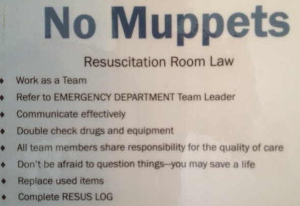
When the external muppet factor is allowed to escalate unchecked, the end result is frenetic activity and noise from the staff without coordinated meaningful intervention for the patient. Comparisons with ‘headless chickens’ are often drawn. In particularly challenging scenarios, it can appear that the panic has swelled to such magnitude that it goes nova, as though the headless chickens have actually exploded, metaphorically filling the room with a gruesome blanket of giblets and a snowstorm of feathers, clouding ones ability to assess and manage the patient effectively. This high-point of group anxiety and ineffectiveness is what I mean by the term ’chicken bomb’, and I bet most readers of this blog will have witnessed the detonation of one.
I credit the invention of this term to emergency and prehospital physician James French, a master resuscitationist and human factors wizard who also introduced the idea of clinical logistics to us.
So, next time you encounter muppets and chicken bombs, feel free to use the terminology, although preferably not during an actual resus with those who might take it personally.

The importance of first pass success

mv-vl-iconA large single-centre study in an academic tertiary care center emergency department (where residents perform most of the intubations) examined 1,828 orotracheal intubations, of which 1,333 were intubated successfully on the first attempt (72.9%).
Adverse events (AE) captured were oesophageal intubation, oxygen desaturation, witnessed aspiration, mainstem intubation, accidental extubation, cuff leak, dental trauma, laryngospasm, pneumothorax, hypotension, dysrhythmia, and cardiac arrest.
When the first pass was successful, the incidence of AEs was 14.2%. More than one attempt was associated with significantly more AEs. Patients requiring two attempts had 33% more AEs (47.2%) and as the number of attempts increased, so did the risk of AEs, with the largest increase in AEs occurring between an unsuccessful first attempt and the second intubation attempt.
This is a powerful argument in favour of optimising first pass success. In the prehospital service I work for, We like to include this in a ‘first pass, no desat, no hypotension’ package that includes team simulation training, pre-intubation briefing, checklist use, optimisation of position, ketamine induction (and avoidance of propofol), apnoeic oxygenation, bougie use, bimanual laryngoscopy, and waveform capnography.
The Importance of First Pass Success When Performing Orotracheal Intubation in the Emergency Department
Academic Emergency Medicine 2013;20(1):71–78, Free Full Text
[EXPAND Abstract]


Objectives The goal of this study was to determine the association of first pass success with the incidence of adverse events (AEs) during emergency department (ED) intubations.

Methods This was a retrospective analysis of prospectively collected continuous quality improvement data based on orotracheal intubations performed in an academic ED over a 4-year period. Following each intubation, the operator completed a data form regarding multiple aspects of the intubation, including patient and operator characteristics, method of intubation, device used, the number of attempts required, and AEs. Numerous AEs were tracked and included events such as witnessed aspiration, oxygen desaturation, esophageal intubation, hypotension, dysrhythmia, and cardiac arrest. Multivariable logistic regression was used to assess the relationship between the primary predictor variable of interest, first pass success, and the outcome variable, the presence of one or more AEs, after controlling for various other potential risk factors and confounders.

Results Over the 4-year study period, there were 1,828 orotracheal intubations. If the intubation was successful on the first attempt, the incidence of one or more AEs was 14.2% (95% confidence interval [CI] = 12.4% to 16.2%). In cases requiring two attempts, the incidence of one or more AEs was 47.2% (95% CI = 41.8% to 52.7%); in cases requiring three attempts, the incidence of one or more AEs was 63.6% (95% CI = 53.7% to 72.6%); and in cases requiring four or more attempts, the incidence of one or more AEs was 70.6% (95% CI = 56.2.3% to 82.5%). Multivariable logistic regression showed that more than one attempt at tracheal intubation was a significant predictor of one or more AEs (adjusted odds ratio [aOR] = 7.52, 95% CI = 5.86 to 9.63).

Conclusions When performing orotracheal intubation in the ED, first pass success is associated with a relatively small incidence of AEs. As the number of attempts increases, the incidence of AEs increases substantially.

[/EXPAND]

Alternative 'universal' plasma donor

The group usually considered the universal donor for fresh frozen plasma because it contains no anti-A or anti-B antibodies is Type AB. Due to its limited availability the trauma service of the Mayo Clinic in Minnesota has been issuing thawed group A plasma to its flight crews who retrieve major trauma casualties from rural centres. This is given with packed group O red cells to patients who meet their prehospital massive transfusion protocol criteria. Some patients will inevitably receive ABO-incompatible plasma (namely patients with Group B or AB blood) which could theoretically give rise to haemolytic transfusion reactions, in which donor antibodies bind host red cells, activate complement, and give rise to anaemia, disseminated intravascular coagulation, acute kidney injury, and death. However:

  • the transfusion of platelets containing ABO-incompatible plasma is common, with up to 2 units of incompatible plasma per apheresis platelet unit, whereas haemolytic reactions to platelets are rare (1 in 9,000 incompatible platelet transfusions);
  • all reports of haemolytic reactions are caused by products that contain Group O plasma and there has never been a documented case of haemolysis as a result of products containing Group A plasma

A retrospective review showed no increased rates of adverse events with Type A compared with AB or ABO-compatible plasma. Since only a small absolute number of patients received an ABO-incompatible plasma transfusion, it could be argued that the study is underpowered (a point acknowledged by the authors). However this is very important and useful information for resource-limited settings.
Emergency use of prethawed Group A plasma in trauma patients
J Trauma Acute Care Surg. 2013 Jan;74(1):69-74
[EXPAND Abstract]


BACKGROUND: Massive transfusion protocols lead to increased use of the rare universal plasma donor, Type AB, potentially limiting supply. Owing to safety data, with a goal of avoiding shortages, our blood bank exploited Group A rather than AB for all emergency release plasma transfusions. We hypothesized that ABO-incompatible plasma transfusions had mortality similar to ABO-compatible transfusions.

METHODS: Review of all trauma patients receiving emergency release plasma (Group A) from 2008 to 2011 was performed. ABO compatibility was determined post hoc. Deaths before blood typing were eliminated. p < 0.05 was considered statistically significant.

RESULTS: Of the 254 patients, 35 (14%) received ABO-incompatible and 219 (86%) received ABO-compatible transfusions. There was no difference in age (56 years vs. 59 years), sex (63% vs. 63% male), Injury Severity Score (ISS) (25 vs. 22), or time spent in the trauma bay (24 vs. 26.5 minutes). Median blood product units transfused were similar: emergency release plasma (2 vs. 2), total plasma at 24 hours (6 vs. 4), total red blood cells at 24 hours (5 vs. 4), plasma-red blood cells at 24 hours (1.3:1 vs. 1.1:1), and plasma deficits at 24 hours (2 vs. 1). Overall complications were similar (43% vs. 35%) as were rates of possible transfusion-related acute lung injury (2.9% vs. 1.8%), acute lung injury (3.7% vs. 2.5%), adult respiratory distress syndrome (2.9% vs. 1.8%), deep venous thrombosis (2.9% vs. 4.1%), pulmonary embolism (5.8% vs. 7.3%), and death (20% vs. 22%). Ventilator (6 vs. 3), intensive care unit (4 vs. 3), and hospital days (9 vs. 7) were similar. There were no hemolytic reactions. Mortality was significantly greater for the patients who received incompatible plasma if concurrent with a massive transfusion (8% vs. 40%, p = 0.044). Group AB plasma use was decreased by 96.6%.

CONCLUSION: Use of Group A for emergency release plasma resulted in ABO-incompatible transfusions; however, this had little effect on clinical outcomes. Blood banks reticent to adopt massive transfusion protocols owing to supply concerns may safely use plasma Group A, expanding the pool of emergency release plasma donors.

LEVEL OF EVIDENCE: Therapeutic study, level IV; prognostic study, level III.

[/EXPAND]

Updated Difficult Airway Guidelines

diffairwayThe American Society of Anesthesiologists has published an update to its Practice Guidelines for Management of the Difficult Airway. You can get the full PDF for free. I’m linking to it for interest, but do not expect to find anything groundbreaking for the management of critical patients.
Practice Guidelines for Management of the Difficult Airway: An Updated Report by the American Society of Anesthesiologists Task Force on Management of the Difficult Airway
Anesthesiology 2013;118:251-70

Endovascular stroke treatment

Two randomised controlled trials have been published which compare endovascular stroke treatments with intravenous tPA. Both the American Interventional Management of Stroke (IMS) III trial (1) and the Italian SYNTHESIS Expansion trial (2) had Modified Rankin Scores as their primary endpoint. No significant differences in this outcome or in mortality or intracranial haemorrhage rates were found in either trial, and IMS III was terminated early due to futility.
A third trial, from North America, called MR RESCUE, randomised patients within 8 hours after the onset of large vessel, anterior-circulation strokes to undergo mechanical embolectomy or receive standard care(3). No clinical outcome differences were demonstrated.
An accompanying editorial (4) draws the following conclusion:


“The IMS III and SYNTHESIS Expansion studies show that intravenous thrombolysis should continue to be the first-line treatment for patients with acute ischemic stroke within 4.5 hours after stroke onset, even if imaging shows an occluded major intracranial artery. Beyond 4.5 hours, the MR RESCUE trial does not provide data supporting the use of endovascular treatment in patients with an ischemic penumbra of any size.”

Many might argue that showing endovascular treatment is equivalent to thrombolysis just means endovascular treatment doesn’t work, because a significant proportion of the emergency medicine community views this as the correct interpretation of a thorough analysis of the stroke thrombolysis literature.
1. Endovascular Therapy after Intravenous t-PA versus t-PA Alone for Stroke
NEJM Feb 8, 2013 Full Text Link
2. Endovascular Treatment for Acute Ischemic Stroke
NEJM Feb 8, 2013 Full Text Link
3. A Trial of Imaging Selection and Endovascular Treatment for Ischemic Stroke
NEJM Feb 8, 2013 Full Text Link
4.Endovascular Treatment for Acute Ischemic Stroke — Still Unproven
NEJM Feb 8, 2013 Full Text Link

Ketamine & cardiovascular stability

I ‘jumped ship’ from etomidate to ketamine for rapid sequence intubation (RSI) in sick patients about seven years ago. Good thing too, since I later moved to Australia where we don’t have etomidate. I’ve been one of the aggressive influences behind my prehospital service’s switch to ketamine as the standard induction agent for prehospital RSI. It’s no secret that I think propofol has no place in RSI in the critically ill.
I love ketamine for its haemodynamic stability compared with other induction agents. In fact, I very rarely see a drop in blood pressure when I use it for RSI even in significantly shocked patients. One should however try to remain open to evidence that disconfirms ones biases, lest we allow science to be replaced by religion. I therefore was interested to read a report of two cases of cardiac arrest following the administration of ketamine for rapid sequence intubation (RSI)(1).

ketamine-arrest.003

The first case was a 25 year old with septic shock due to an intestinal perforation, with a respiratory rate of 30 ‘labored’ breaths per minute and hypoxaemia prior to intubation with 2mg/kg ketamine who became bradycardic and then had a 10-15 minute PEA arrest after ketamine administration (but prior to intubation). Pre-arrest oxygen saturation and pre-induction blood gases are not reported.
The second case was an 11 year old with septic shock and pneumonia, hypoxaemia, and a severe metabolic acidosis. She arrested with bradycardia then a brief period of asystole one minute after receiving 2.4 mg/kg ketamine with rocuronium for intubation.
Was the ketamine responsible for the arrests? Ketamine usually exhibits a stimulatory effect on the cardiovascular system, through effects which are incompletely understood but include a centrally mediated sympathetic response and probable inhibition of norepinephrine (noradrenaline) reuptake. However ketamine can have a direct depressant effect on cardiac output which is usually overridden by the sympathetic stimulation. In critically ill severely stressed patients the depressant effect may predominate. In a study on 12 critically ill surgical patients, haemodynamic indices were measured using pulmonary artery catheters within 5 minutes of ketamine administration (at a mean of 70 mg)(2). Six patients demonstrated decreases in ventricular contractility, and four had decreases in cardiac output. Mean arterial blood pressure decreased in four patients. The authors commented:


The patients..were septic, hypovolemic, or cirrhotic, and had severe stress preoperatively. It is possible that in these ill patients adrenocortical and catechol stores had been depleted prior to ketamine administration. Alternatively, in the setting of prolonged preoperative stress, there may be resistance to further sympathetic and/or adrenocotical stimulation by ketamine. In either case, preoperative stress may blunt the usual physiologic responses to ketamine, setting the stage for possible adverse effects.

The negative cardiovascular effects of ketamine may also be precipitated by larger doses or repeated doses of ketamine(3).
While this small case series of cardiac arrest following ketamine administration is interesting, we should bear in mind the other possible precipitants of arrest in these patients, which are not all discussed by the authors:
i) Both patients were hypoxaemic prior to induction and their peri-intubation oxygen saturations are not reported. Arrests following bradycardia at the time of induction in the critically ill are frequently related to hypoxaemia.
ii) The second patient had a severe metabolic acidosis and the first – an abdominal sepsis patient with a labored respiratory rate of 30 – very probably did too. A failure to match a patient’s compensatory respiratory alkalosis with hyperventilation after anaesthesia is known to precipitate arrest in acidaemic patients.
iii) Finally, if the ketamine was responsible for the arrests, one should consider that the doses given to these shocked and highly unstable patients were well in excess of what many of us would recommend, and doses in the range of 0.5-1 mg/kg might not have been associated with adverse effects.
The takehome points for me are that this report is a helpful reminder that the cardiovascular stimulation-inhibition balance of ketamine may be altered by severe critical illness, and that doses of any induction agent should be significantly reduced in the critically ill patient. In no way does this convince me that I should discard ketamine as my preferred choice for RSI in such patients.
1. Cardiac Arrest Following Ketamine Administration for Rapid Sequence Intubation
J Intensive Care Med. 2012 May 29. [Epub ahead of print]
[EXPAND Abstract]


Given their relative hemodynamic stability, ketamine and etomidate are commonly chosen anesthetic agents for sedation during the endotracheal intubation of critically ill patients. As the use of etomidate has come into question particularly in patients with sepsis, due to its effect of adrenal suppression, there has been a shift in practice with more reliance on ketamine. However, as ketamine relies on a secondary sympathomimetic effect for its cardiovascular stability, cardiovascular and hemodynamic compromise may occur in patients who are catecholamine depleted. We present 2 critically ill patients who experienced cardiac arrest following the administration of ketamine for rapid sequence intubation (RSI). The literature regarding the use of etomidate and ketamine for RSI in critically ill patients is reviewed and options for sedation during endotracheal intubation in this population are discussed.

[/EXPAND]
2. Cardiovascular effects of anesthetic induction with ketamine
Anesth Analg. 1980 May;59(5):355-8
[EXPAND Abstract]


Anesthetic induction with ketamine has been reported to maintain or improve cardiovascular performance in severely ill patients. Using invasive cardiovascular monitoring, we studied physiologic responses to a single dose of ketamine in 12 critically ill patients. Six patient demonstrated decreases in ventricular contractility, and four had decreases in cardiac output. Mean arterial blood pressure decreased in four patients. Pulmonary venous admixture increased in four of six patients, while oxygen consumption decreased in eight of 11 patients. Thus, a single dose of ketamine produced decreases in cardiac and pulmonary performance and in peripheral oxygen transport in this group of patients. It is proposed that in severely ill patients, preoperative stress may alter the usual physiologic responses to ketamine administration, and adverse effects may predominate. Ketamine, therefore, should be used with caution for induction of anesthesia in critically ill and in acutely traumatized patients until additional studies and further information on cardiovascular responses to ketamine are available.

[/EXPAND]
3. A comparison of some cardiorespiratory effects of althesin and ketamine when used for induction of anaesthesia in patients with cardiac disease
Br J Anaesth. 1976 Nov;48(11):1071-81
[EXPAND Abstract]


Cardiorespiratory effects of ketamine and Althesin were measured in two groups of premedicated patients with cardiac disease. The drugs were given in clinically equivalent doses with a second dose administered about 10 min after induction. The first dose of ketamine caused a marked increase in systemic and pulmonary arterial pressure, heart rate, and central venous and wedge pressures and cardiac index. The first dose of Althesin caused a decrease in systemic arterial pressure, central venous pressure, cardiac index and heart work, but little change in heart rate. The second dose of induction agent was administered before the cardiorespiratory effects of the initial dose had resolved. The second dose of Althesin caused changes similar to those following the first dose, but less marked. The changes following the second dose of ketamine were opposite to those following the first dose.

[/EXPAND]