Tag Archives: cardiology

Spot the WOBBLER in syncope!

Syncope is a common ED presentation. An ECG is a critical investigation in syncope to identify the cause, including rare conditions associated with risk of sudden cardiac death.
So we should be really grateful when we are invited to interpret an ECG while we’re in the middle of six other tasks.

The problem with syncope is that some of the important life-threatening causes have fairly obscure ECG features that might be hard to remember. Some of these disorders and their ECG features are not entirely familiar to the clinicians who first screen the ECG.

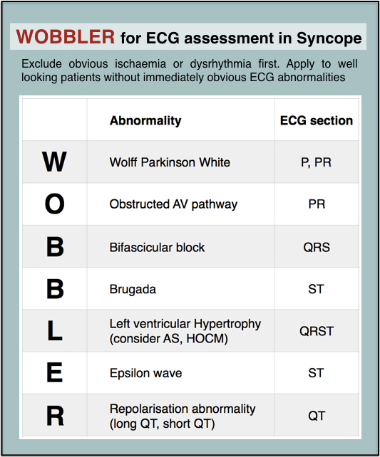
When you’re busy and cognitively stretched you can save time and reduce the risk of missing important findings by having a structured, memorable checklist. I use the acronym WOBBLER, because I don’t want these people to wobble and kiss the dirt again.

The nice thing about WOBBLER is that it uses the sequence that you follow when you look at an ECG, ie from left to right, or from P wave to T wave.

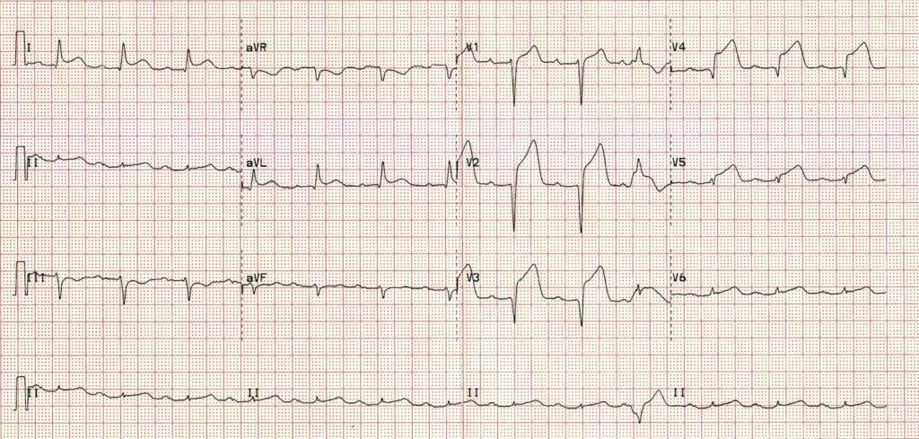
The key is that this is for ECGs without obvious ischaemia or dysrhythmia. If you see something like this (STEMI):
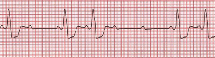
or this (VT):

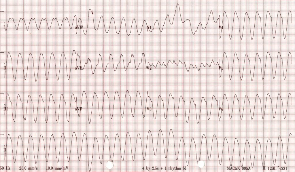
you don’t need WOBBLER, you need to be treating that patient.

So we apply WOBBLER to screen the ECG of well-looking syncope patients without obvious ischaemia or dysrhythmia.

Here goes…

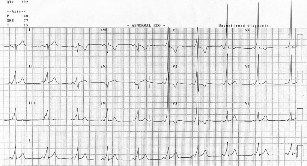
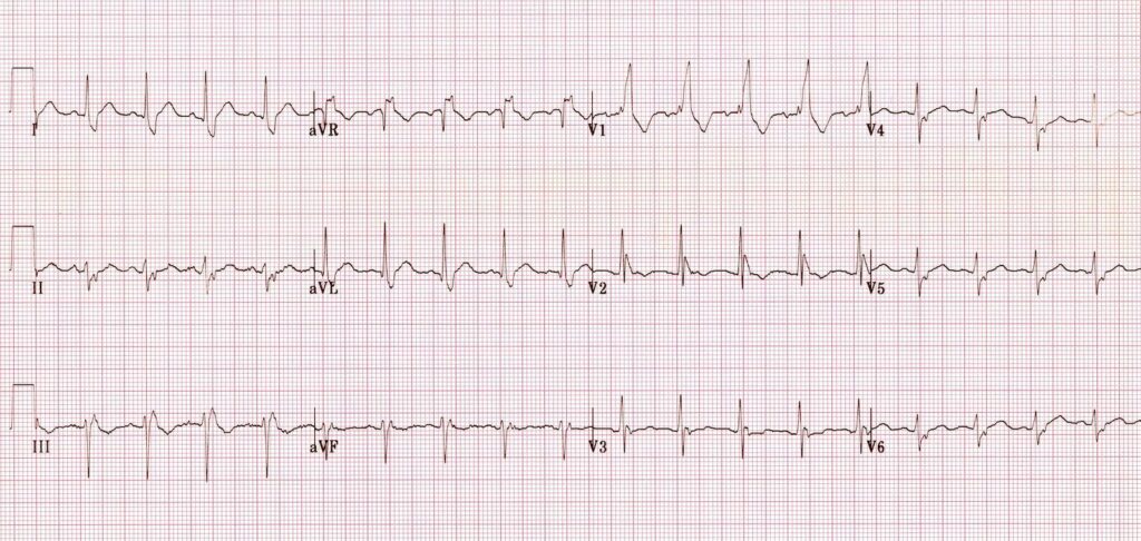
W is Wolff-Parkinson-White syndrome – look for a short PR interval or delta wave:

O is obstructed AV pathway – look for 2nd or 3rd degree block:

or axis deviation:

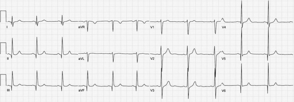
…which is the first step in looking for B bifascicular block, or the combination of axis deviation and right bundle branch block:

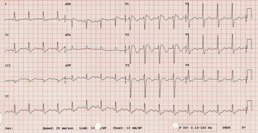
the second B is Brugada, looking for characteristic morphology of the ST segment, so called coved ST elevation:

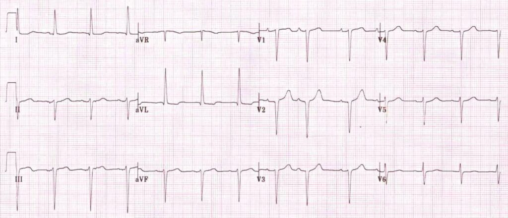
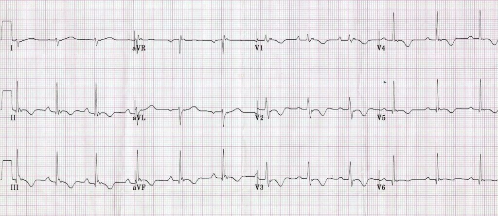
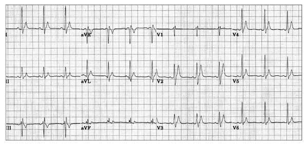
Now syncope, especially exertional syncope, can be caused by left ventricular outflow tract obstruction. Two conditions not to be missed associated with LVOTO (and exertional syncope) are hypertrophic cardiomyopathy and aortic stenosis. These both characteristically cause Lleft ventricular hypertrophy:

This example of Hypertrophic Cardiomyopathy has voltage criteria for LVH in the precordial leads as well as narrow, “dagger-like” Q waves in lateral and inferior leads

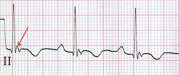
E– stands for epsilon wave, a feature of arrythmogenic right ventricular dysplasia, a rare disorder associated with sudden cardiac death. The epsilon wave looks a bit like the J wave of hypothermia and may be associated with other T wave abnormalities in V1-V3:

Epsilon wave in more detail

Finally, R stands for Repolarisation abnormality, particularly delayed Repolarisation as found in long QT syndrome:

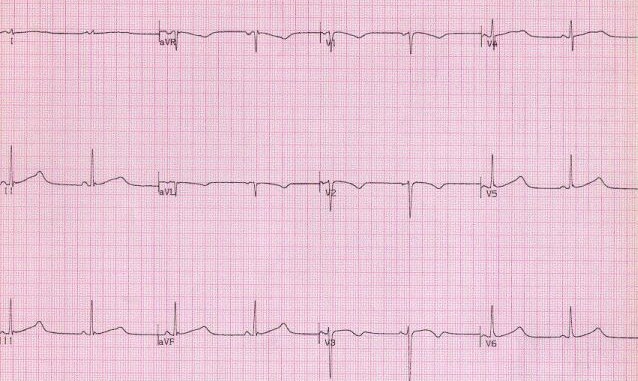
but remember there is also a short QT syndrome too:

So WOBBLER may help you find the important and rare abnormalities not to be missed in the syncope patient, going from left to right from P wave through to T wave, in the patient that does not have obvious dysrhythmia or ischaemia. I hope you find it a helpful cognitive forcing tool.

All ECGs reproduced with kind permission of Life in the Fast Lane

Non-ST-Elevation Acute Coronary Syndromes

The latest AHA/ACC guidelines on NSTEACS have been published ahead of print in Circulation.
Full text is available, and the Executive Summary is available here
Amsterdam EA, Wenger NK, Brindis RG, Casey DE, Ganiats TG, Holmes DR, et al.
2014 AHA/ACC Guideline for the Management of Patients With Non-ST-Elevation Acute Coronary Syndromes: Executive Summary: A Report of the American College of Cardiology/American Heart Association Task Force on Practice Guidelines.
Circulation. 2014 Sep 23. [Epub ahead of print]

Double balloon pump fail

IABPicon
Two recent trials question the ongoing use of intra-aortic balloon pumps: in patients with acute myocardial infarction with cardiogenic shock undergoing revascularisation(1), and patients with poor left ventricular function undergoing coronary artery bypass surgery(2).
Editorialists Krischan D Sjauw and Jan J Piek from the Netherlands make the following commentary(3) in reference to one of the studies:
Although the results of IABP-SHOCK II question the usefulness of IABP therapy in cardiogenic shock, there still might be an indication for initial stabilisation of severely compromised patients, especially in centres without facilities for early revascularisation, as an adjunct to thrombolytic therapy, or to allow transport to specialised tertiary centres.
So retrieval specialists like me may still be up in the night transferring patients with balloon pumps, but these studies suggest this should be restricted to those with cardiogenic shock pending corrective therapy (eg. revascularisation for AMI or surgery for acute mitral valvular dysfunction). Unless the ECMO team gets to them first, of course.

1. Intra-aortic balloon counterpulsation in acute myocardial infarction complicated by cardiogenic shock (IABP-SHOCK II): final 12 month results of a randomised, open-label trial
The Lancet, Volume 382, Issue 9905, Pages 1638 – 1645
[EXPAND Abstract]


BACKGROUND: In current international guidelines the recommendation for intra-aortic balloon pump (IABP) use has been downgraded in cardiogenic shock complicating acute myocardial infarction on the basis of registry data. In the largest randomised trial (IABP-SHOCK II), IABP support did not reduce 30 day mortality compared with control. However, previous trials in cardiogenic shock showed a mortality benefit only at extended follow-up. The present analysis therefore reports 6 and 12 month results.

METHODS: The IABP-SHOCK II trial was a randomised, open-label, multicentre trial. Patients with cardiogenic shock complicating acute myocardial infarction who were undergoing early revascularisation and optimum medical therapy were randomly assigned (1:1) to IABP versus control via a central web-based system. The primary efficacy endpoint was 30 day all-cause mortality, but 6 and 12 month follow-up was done in addition to quality-of-life assessment for all survivors with the Euroqol-5D questionnaire. A masked central committee adjudicated clinical outcomes. Patients and investigators were not masked to treatment allocation. Analysis was by intention to treat. This trial is registered at ClinicalTrials.gov, NCT00491036.

FINDINGS: Between June 16, 2009, and March 3, 2012, 600 patients were assigned to IABP (n=301) or control (n=299). Of 595 patients completing 12 month follow-up, 155 (52%) of 299 patients in the IABP group and 152 (51%) of 296 patients in the control group had died (relative risk [RR] 1·01, 95% CI 0·86-1·18, p=0·91). There were no significant differences in reinfarction (RR 2·60, 95% CI 0·95-7·10, p=0·05), recurrent revascularisation (0·91, 0·58-1·41, p=0·77), or stroke (1·50, 0·25-8·84, p=1·00). For survivors, quality-of-life measures including mobility, self-care, usual activities, pain or discomfort, and anxiety or depression did not differ significantly between study groups.

INTERPRETATION: In patients undergoing early revascularisation for myocardial infarction complicated by cardiogenic shock, IABP did not reduce 12 month all-cause mortality.

[/EXPAND]
2. A Randomized Controlled Trial of Preoperative Intra-Aortic Balloon Pump in Coronary Patients With Poor Left Ventricular Function Undergoing Coronary Artery Bypass Surgery
Crit Care Med. 2013 Nov;41(11):2476-83
[EXPAND Abstract]


BACKGROUND: Preoperative intra-aortic balloon pump use in high-risk patients undergoing surgical coronary revascularization is still a matter of debate. The objective of this study is to determine whether the preoperative use of an intra-aortic balloon pump improves the outcome after coronary operations in high-risk patients.

DESIGN: Single-center prospective randomized controlled trial.

SETTING: Tertiary cardiac surgery center, research hospital.

PATIENTS: One hundred ten subjects undergoing coronary operations, with a poor left ventricular ejection fraction (< 35%) and no hemodynamic instability.
INTERVENTIONS:
Patients randomized to receive preincision intra-aortic balloon pump or no intervention.

MEASUREMENTS AND MAIN RESULTS: The primary outcome measurement was postoperative major morbidity rate, defined as one of prolonged mechanical ventilation, stroke, acute kidney injury, surgical revision, mediastinitis, and operative mortality. There was no difference in major morbidity rate (40% in intra-aortic balloon pump group and 31% in control group; odds ratio, 1.49 [95% CI, 0.68-3.33]). No differences were observed for cardiac index before and after the operation; at the arrival in the ICU, patients in the intra-aortic balloon pump group had a significantly (p = 0.01) lower mean systemic arterial pressure (80.1 ± 15.1 mm Hg) versus control group patients (89.2 ± 17.9 mm Hg). Fewer patients in the intra-aortic balloon pump group (24%) than those in the control group (44%) required dopamine infusion (p = 0.043).

CONCLUSIONS: This study demonstrates that in patients undergoing nonemergent coronary operations, with a stable hemodynamic profile and a left ventricular ejection fraction less than 35%, the preincision insertion of intra-aortic balloon pump does not result in a better outcome. Given the possible complications of intra-aortic balloon pump insertion, and the additional cost of the procedure, this approach is not justified.

[/EXPAND]
3. Is the intra-aortic balloon pump leaking?
Lancet 2013;382:1616-7

Guidelines for the Management of Heart Failure

Some new guidelines to be aware of are the AHA Guidelines for the Management of Heart Failure. Full text is available free and while comprehensively covering chronic heart failure there is an interesting section on acute decompensated heart failure.
Evidence-based medicine enthusiasts might be interested in recommendations to consider dopamine, nesiritide, and ultrafiltration. These therapies also get a mention in the 2012 European Guidelines. I recommend you review the articles cited in the guidelines to make your own mind up.
Here are a couple of snippets you may find useful:


Snippet from American Guidelines: intravenous loop diuretic doses

“HF patients receiving loop diuretic therapy should receive an initial parenteral dose greater than or equal to their chronic oral daily dose; then dose should be serially adjusted.”


Snippet from European Guidelines: management algorithm for acute heart failure

Click to enlarge

AHF-ESC

2013 ACCF/AHA Guideline for the Management of Heart Failure: Executive Summary
Circulation. 2013 Oct 15;128(16):1810-52 Free Full Text
ESC Guidelines for the diagnosis and treatment of acute and chronic heart failure 2012
Eur Heart J. 2012 Jul;33(14):1787-847 Free Full Text

Isolated LAFB and outcome

A small study followed up an older cohort of patients with isolated left anterior fascicular block on their ECG (isolated left axis deviation), without clinically manifest cardiovascular disease.

LAFB-litfl
Learn about LAFB at Life In The Fast Lane

LAFB was associated with an increased risk of atrial fibrillation, heart failure and death. LAFB is caused by conduction tissue fibrosis, and is a marker of other left heart fibrosis. The patients did not go on to develop left bundle branch block, and only 2 of 39 required pacing in 10 years, suggesting these outcomes were not due to progression of conduction disease.

Long-term Outcomes of Left Anterior Fascicular Block in the Absence of Overt Cardiovascular Disease
JAMA. 2013 Apr 17;309(15):1587-8

Potential new therapy for acute heart failure

Interesting new drug to know about: Serelaxin, recombinant human relaxin-2. It’s hard to assess the clinical significance of the statistically significant findings. Let’s see if a benefit is replicated in future studies. It’s hard to imagine a normotensive patient that can’t be fixed with existing therapies though.
Serelaxin, recombinant human relaxin-2, for treatment of acute heart failure (RELAX-AHF): a randomised, placebo-controlled trial.
Lancet. 2013 Jan 5;381(9860):29-39
[EXPAND Abstract]


BACKGROUND: Serelaxin, recombinant human relaxin-2, is a vasoactive peptide hormone with many biological and haemodynamic effects. In a pilot study, serelaxin was safe and well tolerated with positive clinical outcome signals in patients with acute heart failure. The RELAX-AHF trial tested the hypothesis that serelaxin-treated patients would have greater dyspnoea relief compared with patients treated with standard care and placebo.

METHODS: RELAX-AHF was an international, double-blind, placebo-controlled trial, enrolling patients admitted to hospital for acute heart failure who were randomly assigned (1:1) via a central randomisation scheme blocked by study centre to standard care plus 48-h intravenous infusions of placebo or serelaxin (30 μg/kg per day) within 16 h from presentation. All patients had dyspnoea, congestion on chest radiograph, increased brain natriuretic peptide (BNP) or N-terminal prohormone of BNP, mild-to-moderate renal insufficiency, and systolic blood pressure greater than 125 mm Hg. Patients, personnel administering study drug, and those undertaking study-related assessments were masked to treatment assignment. The primary endpoints evaluating dyspnoea improvement were change from baseline in the visual analogue scale area under the curve (VAS AUC) to day 5 and the proportion of patients with moderate or marked dyspnoea improvement measured by Likert scale during the first 24 h, both analysed by intention to treat. This trial is registered at ClinicalTrials.gov, NCT00520806.

FINDINGS: 1161 patients were randomly assigned to serelaxin (n=581) or placebo (n=580). Serelaxin improved the VAS AUC primary dyspnoea endpoint (448 mm × h, 95% CI 120-775; p=0·007) compared with placebo, but had no significant effect on the other primary endpoint (Likert scale; placebo, 150 patients [26%]; serelaxin, 156 [27%]; p=0·70). No significant effects were recorded for the secondary endpoints of cardiovascular death or readmission to hospital for heart failure or renal failure (placebo, 75 events [60-day Kaplan-Meier estimate, 13·0%]; serelaxin, 76 events [13·2%]; hazard ratio [HR] 1·02 [0·74-1·41], p=0·89] or days alive out of the hospital up to day 60 (placebo, 47·7 [SD 12·1] days; serelaxin, 48·3 [11·6]; p=0·37). Serelaxin treatment was associated with significant reductions of other prespecified additional endpoints, including fewer deaths at day 180 (placebo, 65 deaths; serelaxin, 42; HR 0·63, 95% CI 0·42-0·93; p=0·019).

INTERPRETATION: Treatment of acute heart failure with serelaxin was associated with dyspnoea relief and improvement in other clinical outcomes, but had no effect on readmission to hospital. Serelaxin treatment was well tolerated and safe, supported by the reduced 180-day mortality

[/EXPAND]

Hypothermia as an inotrope

This small study supports the hypothesis that therapeutic hypothermia can have positive inotropic effects in patients with cardiogenic shock of ischaemic or non-ischaemic origin.
Cooling resulted in a temperature-dependent decrease in heart rate and temperature-dependent increases in stroke volume index, cardiac index, mean arterial pressure, and cardiac power output. These changes reversed when the patients were rewarmed.
The authors summarise as follows:


In summary, our studies demonstrate that moderate hypothermia is feasible and safe also for patients in cardiogenic shock.

Improved cardiac performance may contribute to the considerable decrease of mortality for survivors of cardiac arrest, and the use of hypothermia can be recommended for patients with a clear indication for cooling and poor cardiac performance.

Moreover, hypothermia might be considered as a positive inotropic intervention during cardiogenic shock.

Moderate hypothermia for severe cardiogenic shock (COOL Shock Study I & II)
Resuscitation. 2013 Mar;84(3):319-25.
[EXPAND Abstract]


AIM OF THE STUDY: Hypothermia exerts profound protection from neurological damage and death after resuscitation from circulatory arrest. Its application during concomitant cardiogenic shock has been discussed controversially, and still hypothermia is used with reserve when haemodynamic parameters are impaired. On the other hand hypothermia improves force development in isolated human myocardium. Thus, we hypothesized that hypothermia could beneficially affect cardiac function in patients during cardiogenic shock.

METHODS: 14 Patients, admitted to Intensive Care Unit for cardiogenic shock under inotropic support, were enrolled and moderate hypothermia (33°C) was induced for either one (n=5, short-term) or twenty-four (n=9, mid-term) hours.

RESULTS: 12 patients suffered from ischaemic cardiomyopathy, 2 were female, and 6 were included after cardiac arrest and resuscitation. Body temperature was controlled by an intravascular cooling device. Short-term hypothermia consistently decreased heart rate, and increased stroke volume, cardiac index and cardiac power output. Metabolic and electrocardiographic parameters remained constant during cooling. Improved cardiac function persisted during mid-term hypothermia, but was reversed during re-warming. No severe or persistent adverse effects of hypothermia were observed.

CONCLUSION: Moderate Hypothermia is safe and feasable in patients during cardiogenic shock. Moreover, hypothermia improved parameters of cardiac function, suggesting that hypothermia might be considered as a positive inotropic intervention rather than a risk for patients during cardiogenic shock.

[/EXPAND]

Decatecholaminization in septic shock

A subset of patients from the 2008 Vasopressin and Septic Shock Trial (VASST) trial had invasive haemodynamic monitoring measurements from pulmonary artery catheters. These data have now been analysed, revealing that vasopressin was associated with a lower heart rate compared with norepinephrine (noradrenaline) alone, without significant difference in cardiac index or stroke volume index. However, there was significantly greater use of inotropic drugs in the vasopressin group compared with the norepinephrine group.
Tachycardia and high quantities of catecholamine infusion are both associated with mortality in sepsis. The authors discuss:
“The idea of decatecholaminization, reducing both endogenous and exogenous adrenergic stimulation, is now believed to be an important treatment strategy, and the use of beta-blockers in septic shock is being considered. The early use of vasopressin or specific V1a receptor agonists in early septic shock may be another possible treatment.”
This interesting post-hoc analysis may help further define the patients in whom vasopressin is to be considered, by those clinicians who are using it in septic shock. For those that aren’t, I wouldn’t worry about it.
The cardiopulmonary effects of vasopressin compared with norepinephrine in septic shock
Chest. 2012 Sep;142(3):593-605
[EXPAND Click for abstract]


BACKGROUND: Vasopressin is known to be an effective vasopressor in the treatment of septic shock, but uncertainty remains about its effect on other hemodynamic parameters.

METHODS: We examined the cardiopulmonary effects of vasopressin compared with norepinephrine in 779 adult patients with septic shock recruited to the Vasopressin and Septic Shock Trial. More detailed cardiac output data were analyzed for a subset of 241 patients managed with a pulmonary artery catheter, and data were collected for the first 96 h after randomization. We compared the effects of vasopressin vs norepinephrine in all patients and according to severity of shock (< 15 or ≥ 15 μg/min of norepinephrine) and cardiac output at baseline.
RESULTS: Equal BPs were maintained in both treatment groups, with a significant reduction in norepinephrine requirements in the patients treated with vasopressin. The major hemodynamic difference between the two groups was a significant reduction in heart rate in the patients treated with vasopressin (P < .0001), and this was most pronounced in the less severe shock stratum (treatment × shock stratum interaction, P =.03). There were no other major cardiopulmonary differences between treatment groups, including no difference in cardiac index or stroke volume index between patients treated with vasopressin and those treated with norepinephrine. There was significantly greater use of inotropic drugs in the vasopressin group than in the norepinephrine group.
CONCLUSIONS: Vasopressin treatment in septic shock is associated with a significant reduction in heart rate but no change in cardiac output or other measures of perfusion.

[/EXPAND]

New STEMI guidelines

Primary percutaneous coronary intervention or fibrinolysis for STEMI? What if you don’t have PCI at your hospital?
The new 2013 ACCF/AHA Guideline for the Management of ST-Elevation Myocardial Infarction is out and you can get the summary here.
Here’s what they say about initial reperfusion therapy:

Onset of Myocardial Infarction: Recommendations
Regional Systems of STEMI Care, Reperfusion Therapy, and Time-to-Treatment Goals
Class I
1. All communities should create and maintain a regional system of STEMI care that includes assessment and continuous quality improvement of emergency medical services and hospital-based activities. Performance can be facilitated by participating in programs such as Mission: Lifeline and the Door-to-Balloon Alliance.(Level of Evidence: B)
2. Performance of a 12-lead electrocardiogram (ECG) by emergency medical services personnel at the site of first medical contact (FMC) is recommended in patients with symptoms consistent with STEMI.(Level of Evidence: B)
3. Reperfusion therapy should be administered to all eligible patients with STEMI with symptom onset within the prior 12 hours. (Level of Evidence: A)
4. Primary PCI is the recommended method of reper- fusion when it can be performed in a timely fashion by experienced operators. (Level of Evidence: A)
5. Emergency medical services transport directly to a PCI-capable hospital for primary PCI is the recommended triage strategy for patients with STEMI, with an ideal FMC-to-device time system goal of 90 minutes or less.(Level of Evidence: B)
6. Immediate transfer to a PCI-capable hospital for primary PCI is the recommended triage strategy for patients with STEMI who initially arrive at or are transported to a non–PCI-capable hospital, with an FMC-to-device time system goal of 120 minutes or less.(Level of Evidence: B)
7. In the absence of contraindications, fibrinolytic therapy should be administered to patients with STEMI at non–PCI-capable hospitals when the anticipated FMC-to-device time at a PCI-capable hospital exceeds 120 minutes because of unavoidable delays.(Level of Evidence: B)
8. When fibrinolytic therapy is indicated or chosen as the primary reperfusion strategy, it should be administered within 30 minutes of hospital arrival.(Level of Evidence: B)
Class IIa
1. Reperfusion therapy is reasonable for patients with STEMI and symptom onset within the prior 12 to 24 hours who have clinical and/or ECG evidence of ongoing ischemia. Primary PCI is the preferred strategy in this population. (Level of Evidence: B)

2013 ACCF/AHA Guideline for the Management of ST-Elevation Myocardial Infarction: Executive Summary: A Report of the American College of Cardiology Foundation/American Heart Association Task Force on Practice Guidelines.
Circulation. 2012 Dec 17. [Epub ahead of print]