Tag Archives: procedures

Cricoid can worsen VL View

It is known that cricoid pressure can hinder laryngoscopic view of the cords during direct laryngoscopy. Using a Pentax-AWS Video laryngoscope, these authors have demonstrated that cricoid pressure can also worsen glottic view during video laryngoscopy.
Videographic Analysis of Glottic View With Increasing Cricoid Pressure Force
Ann Emerg Med. 2013 Apr;61(4):407-13
[EXPAND Abstract]


BACKGROUND:Cricoid pressure may negatively affect laryngeal view and compromise airway patency, according to previous studies of direct laryngoscopy, endoscopy, and radiologic imaging. In this study, we assess the effect of cricoid pressure on laryngeal view with a video laryngoscope, the Pentax-AWS.

METHODS: This cross-sectional survey involved 50 American Society of Anesthesiologists status I and II patients who were scheduled to undergo elective surgery. The force measurement sensor for cricoid pressure and the video recording system using a Pentax-AWS video laryngoscope were newly developed by the authors. After force and video were recorded simultaneously, 11 still images were selected per 5-N (Newton; 1 N = 1 kg·m·s(-2)) increments, from 0 N to 50 N for each patient. The effect of cricoid pressure was assessed by relative percentage compared with the number of pixels on an image at 0 N.

RESULTS: Compared with zero cricoid pressure, the median percentage of glottic view visible was 89.5% (interquartile range [IQR] 64.2% to 117.1%) at 10 N, 83.2% (IQR 44.2% to 113.7%) at 20 N, 76.4% (IQR 34.1% to 109.1%) at 30 N, 51.0% (IQR 21.8% to 104.2%) at 40 N, and 47.6% (IQR 15.2% to 107.4%) at 50 N. The number of subjects who showed unworsened views was 20 (40%) at 10 N, 17 (34%) at 20 and 30 N, and 13 (26%) at 40 and 50 N.

CONCLUSION: Cricoid pressure application with increasing force resulted in a worse glottic view, as examined with the Pentax-AWS Video laryngoscope. There is much individual difference in the degree of change, even with the same force. Clinicians should be aware that cricoid pressure affects laryngeal view with the Pentax-AWS and likely other video laryngoscopes.

[/EXPAND]

Endovascular stroke treatment

Two randomised controlled trials have been published which compare endovascular stroke treatments with intravenous tPA. Both the American Interventional Management of Stroke (IMS) III trial (1) and the Italian SYNTHESIS Expansion trial (2) had Modified Rankin Scores as their primary endpoint. No significant differences in this outcome or in mortality or intracranial haemorrhage rates were found in either trial, and IMS III was terminated early due to futility.
A third trial, from North America, called MR RESCUE, randomised patients within 8 hours after the onset of large vessel, anterior-circulation strokes to undergo mechanical embolectomy or receive standard care(3). No clinical outcome differences were demonstrated.
An accompanying editorial (4) draws the following conclusion:


“The IMS III and SYNTHESIS Expansion studies show that intravenous thrombolysis should continue to be the first-line treatment for patients with acute ischemic stroke within 4.5 hours after stroke onset, even if imaging shows an occluded major intracranial artery. Beyond 4.5 hours, the MR RESCUE trial does not provide data supporting the use of endovascular treatment in patients with an ischemic penumbra of any size.”

Many might argue that showing endovascular treatment is equivalent to thrombolysis just means endovascular treatment doesn’t work, because a significant proportion of the emergency medicine community views this as the correct interpretation of a thorough analysis of the stroke thrombolysis literature.
1. Endovascular Therapy after Intravenous t-PA versus t-PA Alone for Stroke
NEJM Feb 8, 2013 Full Text Link
2. Endovascular Treatment for Acute Ischemic Stroke
NEJM Feb 8, 2013 Full Text Link
3. A Trial of Imaging Selection and Endovascular Treatment for Ischemic Stroke
NEJM Feb 8, 2013 Full Text Link
4.Endovascular Treatment for Acute Ischemic Stroke — Still Unproven
NEJM Feb 8, 2013 Full Text Link

Lifting the Fogg on ED Intubaton

Fellow retrieval specialist and Royal North Shore Hospital emergency physician Dr Toby Fogg and coauthors have published their audit of intubations in an Australian Emergency Department(1). More important than the results themselves is that the process of monitoring ones practice inevitably leads to improvements. For example, at Toby’s institution an intubation checklist has been introduced since the audit began. Other Australasian EDs are encouraged to participate using the free resources at airwayregistry.org.au.
Recently we have also seen the publication of Korean registry data on paediatric intubations performed in 13 academic EDs over 5 years(2), in which first pass success rates (overall 67.6%) were higher with emergency physicians compared with paediatricians. Interestingly, a rapid sequence intubation technique was only used in 22.4% of intubations, which was more likely to be used by emergency physicians and was associated with a greater likelihood of first pass success.
This relatively low first pass success rate is reminiscent of the American study published in September(3) which raised some eyebrows with its 52% first pass intubation success rates in a paediatric ED, and which also showed that attending-level providers were 10 times more likely to be successful on the first attempt than all trainees combined. Possible reasons for such a low first pass success rate compared with adult registry data include the rigorous video analysis method used, or perhaps more likely that paediatric emergency subspecialists are exposed to fewer critical procedures, resuscitations, and intubations than their general emergency medicine counterparts(4).
As a specialty we must continue to seek to do better, and I salute all these brave authors who are telling it like it is. Particularly with children, whose airways are relatively easy, we have to develop the training, preparation, supervision, monitoring and feedback to aim for as high a success rate as possible.

Study authors Toby Fogg and Nick Annesley demonstrate the 'Happiness Triad'

1. Prospective observational study of the practice of endotracheal intubation in the emergency department of a tertiary hospital in Sydney, Australia
Emerg Med Australas. 2012 Dec;24(6):617-24
[EXPAND Click to read abstract]


OBJECTIVE: To describe the practice of endotracheal intubation in the ED of a tertiary hospital in Australia, with particular emphasis on the indication, staff seniority, technique, number of attempts required and the rate of complications.

METHODS: A prospective observational study.

RESULTS: Two hundred and ninety-five intubations occurred in 18 months. Trauma was the indication for intubation in 30.5% (95% CI 25.3-36.0) and medical conditions in 69.5% (95% CI 64.0-74.5). Emergency physicians were team leaders in 69.5% (95% CI 64.0-74.5), whereas ED registrars or senior Resident Medical Officers made the first attempt at intubation in 88.1% (95% CI 83.9-91.3). Difficult laryngoscopy occurred in 24.0% (95% CI 19.5-29.3) of first attempts, whereas first pass success occurred in 83.4% (95% CI 78.7-87.2). A difficult intubation occurred in 3.4% (95% CI 1.9-6.1) and all patients were intubated orally in five or less attempts. A bougie was used in 30.9% (95% CI 25.8-36.5) of first attempts, whereas a stylet in 37.5% (95% CI 32.1-43.3). Complications occurred in 29.0% (95% CI 23.5-34.1) of the patients, with desaturation the commonest in 15.7% (95% CI 11.9-20.5). Cardiac arrest occurred in 2.2% (95% CI 0.9-4.4) after intubation. No surgical airways were undertaken.

CONCLUSION: Although the majority of results are comparable with overseas data, the rates of difficult laryngoscopy and desaturation are higher than previously reported. We feel that this data has highlighted the need for practice improvement within our department and we would encourage all those who undertake emergent airway management to audit their own practice of this high-risk procedure.

[/EXPAND]
2. The factors associated with successful paediatric endotracheal intubation on the first attempt in emergency departments: a 13-emergency-department registry study
Resuscitation. 2012 Nov;83(11):1363-8
[EXPAND Click to read abstract]


BACKGROUND: We investigated which factors are associated with successful paediatric endotracheal intubation (ETI) on the first attempt in emergency department (EDs) from multicentre emergency airway registry data.

METHODS: We created a multicentre registry of intubations at 13 EDs and performed surveillance over 5 years. Each intubator filled out a data form after an intubation. We defined “paediatric patients” as patients younger than 10 years of age. We assessed the specialty and level of training of intubator, the method, the equipment, and the associated adverse events. We analysed the intubation success rates on the first attempt (first-pass success, FPS) based on these variables.

RESULTS: A total of 430 ETIs were performed on 281 children seen in the ED. The overall FPS rate was 67.6%, but emergency medicine (EM) physicians showed a significantly greater success rate of 74.4%. In the logistic regression analysis, the intubator’s specialty was the only independent predictive factor for paediatric FPS. In the subgroup analysis, the EM physicians used the rapid sequence intubation/intubation (RSI) method and Macintosh laryngoscope more frequently than physicians of other specialties. ETI-related adverse events occurred in 21 (7.2%) out of the 281 cases. The most common adverse event in the FPS group was mainstem bronchus intubation, and vomiting was the most common event in the non-FPS group. The incidence of adverse events was lower in the FPS group than in the non-FPS group, but this difference was not statistically significant.

CONCLUSIONS: The intubator’s specialty was the major factor associated with FPS in emergency department paediatric ETI, The overall ETI FPS rate among paediatric patients was 67.6%, but the EM physicians had a FPS rate of 74.4%. A well structured airway skill training program, and more actively using the RSI method are important and this could explain this differences.

[/EXPAND]
3.Rapid sequence intubation for pediatric emergency patients: higher frequency of failed attempts and adverse effects found by video review.
Ann Emerg Med. 2012 Sep;60(3):251-9
[EXPAND Click to read abstract]


STUDY OBJECTIVE: Using video review, we seek to determine the frequencies of first-attempt success and adverse effects during rapid sequence intubation (RSI) in a large, tertiary care, pediatric emergency department (ED).

METHODS: We conducted a retrospective study of children undergoing RSI in the ED of a pediatric institution. Data were collected from preexisting video and written records of care provided. The primary outcome was successful tracheal intubation on the first attempt at laryngoscopy. The secondary outcome was the occurrence of any adverse effect during RSI, including episodes of physiologic deterioration. We collected time data from the RSI process by using video review. We explored the association between physician type and first-attempt success.

RESULTS: We obtained complete records for 114 of 123 (93%) children who underwent RSI in the ED during 12 months. Median age was 2.4 years, and 89 (78%) were medical resuscitations. Of the 114 subjects, 59 (52%) were tracheally intubated on the first attempt. Seventy subjects (61%) had 1 or more adverse effects during RSI; 38 (33%) experienced oxyhemoglobin desaturation and 2 required cardiopulmonary resuscitation after physiologic deterioration. Fewer adverse effects were documented in the written records than were observed on video review. The median time from induction through final endotracheal tube placement was 3 minutes. After adjusting for patient characteristics and illness severity, attending-level providers were 10 times more likely to be successful on the first attempt than all trainees combined.

CONCLUSION: Video review of RSI revealed that first-attempt failure and adverse effects were much more common than previously reported for children in an ED.

[/EXPAND]
4. A is for airway: a pediatric emergency department challenge.
Ann Emerg Med. 2012 Sep;60(3):261-3

Which pelvic compression device is best?

Two popular pelvic compression devices are the SAM Pelvic Sling II and the T-POD®.
In a direct comparison 50 health care volunteers secured both devices correctly 100% of the time.
The SAM Pelvic Sling II was quicker to apply, but participants preferred the T-POD®.
The authors conclude that very little separates the devices.
Pelvic circumferential compression devices (PCCDs): a best evidence equipment review
Eur J Trauma Emerg Surg (2012) 38:439–442
[EXPAND Click for abstract]


Purpose Traumatic disruption of the pelvis can lead to significant morbidity and mortality. ATLS® guidance advocates temporary stabilisation or ‘closure’ of the disrupted pelvis with a compression device or sheet. We undertook a best evidence equipment review to assess the ease and efficacy of the application of two leading commercially available devices, the T-POD® and the SAM Pelvic Sling™ II.

Methods Fifty health care professionals and medical students participated in pelvic circumferential compression device (PCCD) education and assessment. Participants received a 10-min lecture on the epidemiology and aetiology of pelvic fractures and the principles of circumferential compression, followed by a practical demonstration. Three volunteers acted as trauma victims. Assessment included the time taken to secure the devices and whether this was achieved correctly. All participants completed a post-assessment survey.

Results Both devices were applied correctly 100% of the time. The average time taken to secure the SAM Pelvic Sling™ II was 18 s and for the T-POD®, it was 31 s (p ≤ 0.0001). Forty-four participants (88%) agreed or strongly agreed that the SAM Pelvic Sling™ II was easy to use compared to 84% (n = 42) for the T-POD®. Thirty-nine participants (78%) reported that they preferred and, given the choice in the future, would select the T-POD® over the SAM Pelvic Sling™ II (n = 11, 22%).

Conclusions The results of this study indicate that both PCCDs are easy and acceptable to use and, once learned, can be applied easily and rapidly. Participants applied both devices correctly 100% of the time, with successful application taking, on average, less than 60 s.

[/EXPAND]

What happened to HIFU?

High intensity focused ultrasound (HIFU) was hailed as the ‘surgery of the future’ a few years ago(1). As it’s now the future, where is it?
HIFU uses ultrasound to increase the heat within tissues at a specific area, causing local necrosis and cautery without injuring surrounding tissues. It is used to treat some cancers, but has shown promise in haemorrhage control. In animal studies it reduced or stopped bleeding in liver(2), spleen(3), and vascular injuries(4).
It has been proposed to offer a promising method for hemorrhage control in both civilivan and miltary trauma(5). Automated systems have been developed and tested that identify bleeding using Doppler ultrasound techniques that then allow targeting of the HIFU beam to the bleeding tissue(6). The United States Army has identified the need for a such systems and has designed a remotely operated robotic haemostatic system to save lives of soldiers. This was presented in 2006(7).
I would love to know where we are with this technology, and why nothing seems to have appeared about it in the literature for the last few years. If you have any information, please fill us in via the comments box.
1. High intensity focused ultrasound: surgery of the future?
Br J Radiol. 2003 Sep;76(909):590-9 Full text
2. Liver hemostasis using high-intensity focused ultrasound
Ultrasound Med Biol. 1997;23(9):1413-20
3. Control of splenic bleeding by using high intensity ultrasound
J Trauma. 1999 Sep;47(3):521-5
4. Hemostasis of punctured blood vessels using high-intensity focused ultrasound
Ultrasound Med Biol. 1998 Jul;24(6):903-10
5. Hemorrhage control using high intensity focused ultrasound
Int J Hyperthermia. 2007 Mar;23(2):203-11
6. Focused ultrasound: concept for automated transcutaneous control of hemorrhage in austere settings.
Aviat Space Environ Med. 2009 Apr;80(4):391-4
7. Remotely Operated Robotic High Intensity Focused Ultrasound (HIFU) Manipulator System for Critical Systems for Trauma and Transport (CSTAT)
Presented at the IEEE Ultrasonics Symposium, October 3-6, 2006, Vancouver, Canada – Full Text Here

Perimortem Caesarean Delivery: Late is Better than Not


“To date, approximately one-third of the women who die during pregnancy remain undelivered at the time of death”

Guidelines recommend cardiac arrest in pregnant women beyond 20 weeks gestation should be treated with perimortem caesarean delivery (PMCD) commenced within 4 minutes of arrest and completed within 5. These time intervals come from two papers, neither of which is current or used robust review methodology.
To address this, an up-to-date fairly comprehensive review was undertaken of published cases of maternal cardiac arrests occurring prior to delivery. The primary outcome measures were maternal and neonatal survival to hospital discharge and the relationship between PMCD and this outcome.

The Arrests

94 cases were included in the final analysis.Most pregnancies were singleton (90.4%, n = 85) with an average gestational age at the time of the arrest of 33 ± 7 weeks (median 35, range 10–42).
The most common causes of arrest were trauma, maternal cardiac problems, severe pre-eclampsia and amniotic fluid embolism, together comprising about 70% of arrests; two thirds occurred in hospital.

The Outcomes

Overall, return of spontaneous circulation (ROSC) was achieved more often than not (60.6%) and overall survival to hospital discharge was 54.3%
Only 57 cases (75%) reported the time from arrest to delivery; the average time was 16.6 ± 12.5 min (median 10, range 1–60), with only 4 cases making it under the advocated 4-min time limit.

Timing of PMCD and Maternal Survival

In cases undergoing PMCD the average time elapsing from arrest to PMCD was significantly different between surviving (27/57) and non-surviving (30/57) mothers [10.0 ± 7.2 min (median 9, range 1–37) and 22.6 ± 13.3 min (median 20, range 4–60) respectively (p < 0.001, 95%CI 6.9–18.2)].

Timing of PMCD and Neonatal Survival

Mean times to PMCD were 14±11min (median=10, range=1–47) and 22 ± 13 min (median = 20, range = 4–60) in neonatal survivors and non-survivors respectively (p=0.016)
In cases with PMCD which reported outcome, the overall neonatal survival rate was 63.6% (42/66).


“The 4-min time frame advocated for PMCD usually remains unmet yet neonatal survival is still likely if delivery occurs within 10 or even 15 min of arrest”

Both maternal & neonatal mortality were higher with prehospital arrest location.

Summary

The study may be limited by recall bias, under-reporting and publication bias, but provides a more comprehensive evidence base on which to base resuscitation recommendations. The authors provide a useful warning against becoming fixated with the recommended four minute window, which may lead teams to fail to attempt a potentially life-saving intervention:


“Fixation on specific time frames for PMCD may not be ideal. It may be more important to focus on event recognition and good overall performance…. It may be wise to advocate a short time frame for performance of PMCD in order to achieve better outcomes; however, blanket endorsement of an unrealistic time frame may well create a defeatist attitude when that time frame cannot be met.”

Maternal cardiac arrest and perimortem caesarean delivery: Evidence or expert-based?
Resuscitation. 2012 Oct;83(10):1191-200
[EXPAND Click for abstract]


AIM: To examine the outcomes of maternal cardiac arrest and the evidence for the 4-min time frame from arrest to perimortem caesarean delivery (PMCD) recommended in current resuscitation and obstetric guidelines.

DATA SOURCES AND METHODS: Review and data extraction from all reported maternal cardiac arrests occurring prior to delivery (1980-2010). Cases were included if they provided details regarding both the event and outcomes. Outcomes of arrest were assessed using survival, Cerebral Performance Category (CPC) and maternal/neonatal harm/benefit from PMCD. Outcome measures were maternal and neonatal survival.

RESULTS: Of 1594 manuscripts screened, 156 underwent full review. Data extracted from 80 relevant papers yielded 94 included cases. Maternal outcome: 54.3% (51/94) of mothers survived to hospital discharge, 78.4% (40/51) with a CPC of 1/2. PMCD was determined to have been beneficial to the mother in 31.7% of cases and was not harmful in any case. In-hospital arrest and PMCD within 10 min of arrest were associated with better maternal outcomes (ORs 5.17 and 7.42 respectively, p<0.05 both). Neonatal outcome: mean times from arrest to delivery were 14±11 min and 22±13 min in survivors and non-survivors respectively (receiver operating area under the curve 0.729). Neonatal survival was only associated with in-hospital maternal arrest (OR 13.0, p<0.001).

CONCLUSIONS: Treatment recommendations should include a low admission threshold to a highly monitored area for pregnant women with cardiorespiratory decompensation, good overall performance of resuscitation and delivery within 10 min of arrest. Cognitive dissonance may delay both situation recognition and the response to maternal collapse.

[/EXPAND]

London Calling – part 3

Notes from Day 4 of the London Trauma Conference
The highlight for me was Mr Jonny Morrison speaking on Resuscitative Emergency Balloon Occlusion of the Aorta (REBOA). He is a British military surgeon currently out in Texas studying balloon occlusion of the aorta on pigs. Looking at trauma deaths, the next unexpected survivors will come from the uncontrollable haemorrhage group (truncal and junctional zones). This is by no means a new technique – described in the 1950’s during the Korean War – but like the early Star Wars chapters, needed to wait for technology to advance to make it feasible. It has the effect of cross clamping the aorta which provides afterload support, increases cerebral and coronary perfusion and provides proximal inflow control – without the mess of a resuscitative thoracotomy and greater access.
The placement of the balloon is determined by the location of the injury (see photo) and falls into two zones. Zone 1 is the thoracic aorta and is used for truncal haemorrhage control, avoid Zone 2 where the celiac axis etc originates and Zone 3 is infrarenal, used for junctional bleeding and pelvic haemorrhage.
His studies have determined that for Zone 3 amenable bleeds balloon occlusion up to 60min is the optimal time. Any longer and the debt of the metabolic load is paid by increased inotropic support requirements. He also compared REBOA to the current standard treatment for junctional injuries, Celox™ gauze. If coagulation is normal then both treatments perform similarly, the benefit is seen in coagulopathic patients where REBOA outperforms the gauze.
Has REBOA been used on humans? Yes a case series of 13 – the technique improved the BP allowing time to get to definitive surgery (blogged here 2.5 years ago!).
The Zone 1 studies are looking at continuous vs intermittent balloon occlusion. The jury is still out as to which is better. With the intermittent occlusion (20min on, 1min off) there are inevitably some losses when the balloon is deflated, conversely the metabolic debt generated by continuous occlusion is too great in some also leading to deaths.
What was very clear is that for this technique to have an impact it must be delivered proactively and pre-hospital. The challenges that need to be overcome are access to the femoral artery and blind accurate placement.
Prof Karim Brohi brought the conference to a close with a summary of what we have learned about coagulation in trauma this year. Here are three things;

  • FFP is good but as 43% deaths due to trauma in the UK are secondary to bleeding and occur in the first 3hr we are failing our patients by administering the treatment on average at 2.5hrs.
  • Fibrinogen levels are low in coagulopathic trauma patients; we should give cryoprecipitate early and aim for Fib ≥2.0
  • And finally whilst TEG is recommended to guide treatment and can provide results within 5 min, there are some aspects of coagulation it does not detect i.e. fibrinolysis was only detected in 8% of coagulopathic trauma patients – when measured in the plasma it was then detectable in 80%.

These are the highlights of the 2012 London Trauma Conference. I hope this whistle stop tour through these days has been informative and though provoking. I can assure you telephone hacking was not used to bring you this information and to my knowledge is correct.
This is Lou Chan, roving reporter for Resus ME! signing off.


‘London raises her head, shakes off the debris of the night from her hair, and takes stock of the damage done. The sign of a great fighter in the ring is can he get up from a fall after being knocked down… London does this every morning.’


Transtracheal airways in kids. Well, pigs' kids anyway

Ever had to do a surgical airway in a child? Thought not. They’re pretty rare. Bill Heegaard MD from Henepin County Medical Center taught me a few approaches (with the help of an anaesthetised rabbit) which really got me thinking. It’s something I’d often trained for in my internal simulator, and I even keep the equipment for it in my house (listen out for an upcoming podcast on that). Research and experience has demonstrated that open surgical airway techniques are more reliable than transtracheal needle techniques in adults, but what about kids, in whom traditional teaching cautions against open techniques?

Australian investigators who were experienced airway proceduralists evaluated transtracheal needle techniques using a rabbit model (an excellent model for the infant airway). Their success rate was only 60% and they perforated the posterior tracheal wall in 42% of attempts. Of 13 attempts to insert a dedicated paediatric tracheotomy device, the Quicktrach Child, none were successful(1) (they did not use the Quicktrach Infant model as it is not available in Australia).

Danish investigators used fresh piglet cadavers weighing around 8 kg to assess two transtracheal cannulas, in which they achieved success rates of 65.6% and 68.8%(2). There was also a very high rate of posterior tracheal wall perforation. Using an open surgical tracheostomy technique, they were successful in 97% of attempts. These were also experienced operators, with a median anaesthetic experience of 12.5 years.
Their tracheotomy technique was nice and simple, and used just a scalpel, scissors, and surgical towel clips. Here’s their technique:

Simple tracheotomy procedure described by Holm-Knudsen et al

  1. Identify larynx and proximal trachea by palpation
  2. Vertical incision through the skin and subcutaneous tissue from the upper part of larynx to the sternal notch
  3. Grasp strap muscles with two towel forceps and separate in the midline
  4. Palpate and identify the trachea (palpate rather than look for tracheal rings, as in a live patient one would expect bleeding to obscure the view)
  5. Stabilise the trachea by grasping it with a towel forceps
  6. Insert sharp tip of the scissors between two tracheal rings and lift the trachea anteriorly to avoid damage to the posterior wall
  7. Cut vertically in the midline of the trachea with the scissors – they chose to use the scissors to cut the tracheal rings to facilitate tube insertion
  8. Insert the tracheal tube

Using ultrasound and CT to evaluate comparative airway dimensions, the authors concluded that the pig model is most useful for training emergency airway management in older children aged 5–10 years.
Why were they doing a tracheotomy rather than a cricothyroidotomy? Reasons given by the authors include:

  • The infant cricothyroid membrane is very small
  • Palpation of the thyroid notch may be hindered by the overlying hyoid bone
  • The mandible may obstruct needle access to the cricothyroid membrane given the cephalad position in the neck of the infant larynx.

From an emergency medicine point of view, there are a couple of other reasons why we need to be able to access the trachea lower than the cricothyroid membrane. One is fractured larynx or other blunt or penetrating airway injury where there may be anatomical disruption at the cricothyroid level. The other situation is foreign body airway obstruction, when objects may lodge at the level of the cricoid ring which is functionally the narrowest part of the pediatric upper airway. Of course, alternative methods might be considered to remove the foreign body prior to tracheotomy, such as employing basic choking algorithms, and other techniques depending on whether you do or don’t have equipment.

Take home messages

  • Transtracheal airways in kids are so rare, we can’t avoid extrapolating animal data
  • Whichever infant or paediatric model is used, transtracheal needle techniques have a high rate of failure even by ‘experienced’ operators
  • The small size and easy compressibility of the airway probably contributes to this failure rate, including the high rate of posterior wall puncture
  • In keeping with adult audit data, open surgical techniques may have a higher success rate
  • Tracheotomy may be necessary rather than cricothyroidotomy in infants and children depending on clinical scenario and accessibility of anatomy
  • The stress and blood that is not simulated in cadaveric animal models will make open tracheotomy harder in a live patient, and so these success rates may not translate. However these factors do mean that whatever technique is used must be kept simple and should employ readily available and familiar equipment
  • Something to maintain control and anterior position of the anterior trachea wall should be used during incision and intubation of the trachea. The study reported here used towel clips; sutures around the tracheal rings may also be used (see image below)
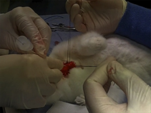
I recommend you add ‘paediatric tracheotomy’ to the list of procedures you might need to do (if it’s not already there). Identify what equipment you would use and run the simulation in your head and in your work environment.
Have fun.

1. The ‘Can’t Intubate Can’t Oxygenate’ scenario in Pediatric Anesthesia: a comparison of different devices for needle cricothyroidotomy
Paediatr Anaesth. 2012 Dec;22(12):1155-8

BACKGROUND: Little evidence exists to guide the management of the ‘Can’t Intubate, Can’t Oxygenate’ (CICO) scenario in pediatric anesthesia.

OBJECTIVES: To compare two intravenous cannulae for ease of use, success rate and complication rate in needle tracheotomy in a postmortem animal model of the infant airway, and trial a commercially available device using the same model.

METHODS: Two experienced proceduralists repeatedly attempted cannula tracheotomy in five postmortem rabbits, alternately using 18-gauge (18G) and 14-gauge (14G) BD Insyte(™) cannulae (BD, Franklin Lakes, NJ, USA). Attempts began at the first tracheal cartilage, with subsequent attempts progressively more caudad. Success was defined as intratracheal cannula placement. In each rabbit, an attempt was then made by each proceduralist to perform a cannula tracheotomy using the Quicktrach Child(™) device (VBM Medizintechnik GmbH, Sulz am Neckar, Germany).

RESULTS: The rabbit tracheas were of similar dimensions to a human infant. 60 attempts were made at cannula tracheotomy, yielding a 60% success rate. There was no significant difference in success rate, ease of use, or complication rate between cannulae of different gauge. Successful aspiration was highly predictive (positive predictive value 97%) and both sensitive (89%) and specific (96%) for tracheal cannulation. The posterior tracheal wall was perforated in 42% of tracheal punctures. None of 13 attempts using the Quicktrach Child(™) were successful.

CONCLUSION: Cannula tracheotomy in a model comparable to the infant airway is difficult and not without complication. Cannulae of 14- and 18-gauge appear to offer similar performance. Successful aspiration is the key predictor of appropriate cannula placement. The Quicktrach Child was not used successfully in this model. Further work is required to compare possible management strategies for the CICO scenario

2. Emergency airway access in children – transtracheal cannulas and tracheotomy assessed in a porcine model
Paediatr Anaesth. 2012 Dec;22(12):1159-65

OBJECTIVES: In the rare scenario when it is impossible to oxygenate or intubate a child, no evidence exists on what strategy to follow.

AIM: The aim of this study was to compare the time and success rate when using two different transtracheal needle techniques and also to measure the success rate and time when performing an emergency tracheotomy in a piglet cadaver model.

METHODS: In this randomized cross-over study, we included 32 anesthesiologists who each inserted two transtracheal cannulas (TTC) using a jet ventilation catheter and an intravenous catheter in a piglet model. Second, they performed an emergency tracheotomy. A maximum of 2 and 4 min were allowed for the procedures, respectively. The TTC procedures were recorded using a video scope.

RESULTS: Placement of a transtracheal cannula was successful in 65.6% and 68.8% of the attempts (P = 0.76), and the median duration of the attempts was 69 and 42 s (P = 0.32), using the jet ventilation catheter and the intravenous catheter, respectively. Complications were frequent in both groups, especially perforation of the posterior tracheal wall. Performing an emergency tracheotomy was successful in 97%, in a median of 88 s.

CONCLUSIONS: In a piglet model, we found no significant difference in success rates or time to insert a jet ventilation cannula or an intravenous catheter transtracheally, but the incidence of complications was high. In the same model, we found a 97% success rate for performing an emergency tracheotomy within 4 min with a low rate of complications.

Externally rotate leg for femoral vein access

Want to access the femoral vein? Externally rotate the leg at the hip and things might be a bit easier. This study was done in adult patients, with the knee straight and no abduction applied. External rotation is also helpful in kids, with abduction up to sixty degrees.


Objective: To determine if external rotation of the leg increases the size and accessibility of the femoral vein compared with a neutral position.

Methods: One hundred patients presenting to a tertiary teaching hospital were prospectively recruited. The right common femoral vein of each subject was scanned with a linear probe (5–10 MHz) inferior to the inguinal ligament, with the leg in a neutral position and then in the externally rotated position. The transverse diameter of the femoral vein, the accessible diameter of the vein (lying medial to the femoral artery) and the depth of the vein were measured.

Results: The mean diameter of the femoral vein in the externally rotated leg was greater than with the leg in the neutral position (15.4 mm vs 13.8 mm); the mean difference was 1.6 mm (95% CI 1.3–1.9). The mean accessible diameter of the femoral vein was larger with the leg externally rotated (13.8 mm vs 11.7 mm, mean difference 2.1 mm, 95% CI 1.8–2.5). The depth from the skin to the femoral vein was less with the leg in external rotation (20.9 mm vs 22.6 mm, mean difference 1.7 mm, 95% CI 1.2–2.2). The mean diameter and depth were greater in patients with overweight or obese body mass index (BMI) measurements in both leg positions. The increase in femoral vein diameter and accessibility with external rotation was observed in all BMI groups.

Conclusion: The total and accessible femoral vein diameter is increased and the surface depth of the vein is decreased by placing the leg in external rotation compared with the neutral position.

Simple external rotation of the leg increases the size and accessibility of the femoral vein
Emerg Med Australas. 2012 Aug;24(4):408-13

Needle decompression: it's still not going to work

A pet topic that keeps coming up here is management of tension pneumothorax. Plenty of studies demonstrate that traditionally taught needle thoracostomy may fail, and open, or ‘finger’ thoracostomy is recommended for the emergency management of tension pneumothorax in a patient who is being ventilated with positive pressure (including those patients in cardiac arrest).
A recent CT scan-based study of adult trauma patients makes the case that needle decompression with a standard iv cannula would be expected to fail in 42.5% of cases at the second intercostal space (ICS) compared with 16.7% at the fifth ICS at the anterior axillary line (AAL).
The authors add an important point: “As BMI increases, there is a stepwise increase in chest wall thickness, further compounding the difficulty of needle placement in all but the lowest BMI quartile for the second ICS.”
An accompanying editorial cautions that the proximity of the heart may confer a safety issue if a needle is inserted blindly into the left 5th ICS at the AAL.


Objective To compare the distance to be traversed during needle thoracostomy decompression performed at the second intercostal space (ICS) in the midclavicular line (MCL) with the fifth ICS in the anterior axillary line (AAL).

Design Patients were separated into body mass index (BMI) quartiles, with BMI calculated as weight in kilograms divided by height in meters squared. From each BMI quartile, 30 patients were randomly chosen for inclusion in the study on the basis of a priori power analysis (n = 120). Chest wall thickness on computed tomography at the second ICS in the MCL was compared with the fifth ICS in the AAL on both the right and left sides through all BMI quartiles.

Setting Level I trauma center.

Patients Injured patients aged 16 years or older evaluated from January 1, 2009, to January 1, 2010, undergoing computed tomography of the chest.

Results A total of 680 patients met the study inclusion criteria (81.5% were male and mean age was 41 years [range, 16-97 years]). Of the injuries sustained, 13.2% were penetrating, mean (SD) Injury Severity Score was 15.5 (10.3), and mean BMI was 27.9 (5.9) (range, 15.4-60.7). The mean difference in chest wall thickness between the second ICS at the MCL and the fifth ICS at the AAL was 12.9 mm (95% CI, 11.0-14.8; P < .001) on the right and 13.4 mm (95% CI, 11.4-15.3; P < .001) on the left. There was a stepwise increase in chest wall thickness across all BMI quartiles at each location of measurement. There was a significant difference in chest wall thickness between the second ICS at the MCL and the fifth ICS at the AAL in all quartiles on both the right and the left. The percentage of patients with chest wall thickness greater than the standard 5-cm decompression needle was 42.5% at the second ICS in the MCL and only 16.7% at the fifth ICS in the AAL.

Conclusions In this computed tomography–based analysis of chest wall thickness, needle thoracostomy decompression would be expected to fail in 42.5% of cases at the second ICS in the MCL compared with 16.7% at the fifth ICS in the AAL. The chest wall thickness at the fifth ICS AAL was 1.3 cm thinner on average and may be a preferred location for needle thoracostomy decompression.

Radiologic evaluation of alternative sites for needle decompression of tension pneumothorax
Arch Surg. 2012 Sep 1;147(9):813-8