
Should we shock with 2J/kg or 4J/kg in Paediatric Defibrillation? The answer seems to be ‘we still don’t know’. Don’t worry – just follow the guidelines (reproduced for you at the bottom)
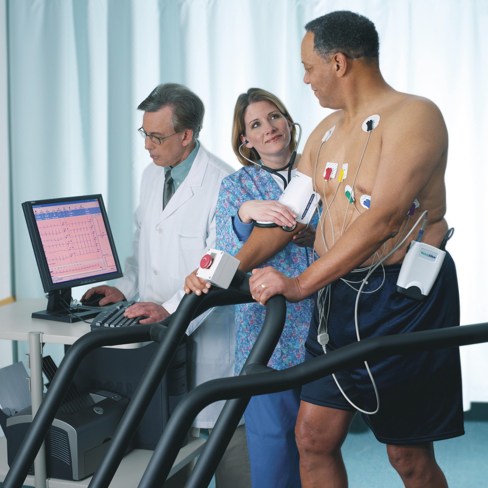
OBJECTIVE To examine the effectiveness of initial defibrillation attempts. We hypothesized that (1) an initial shock dose of 2 ± 10 J/kg would be less effective for terminating fibrillation than suggested in published historical data and (2) a 4 J/kg shock dose would be more effective.
PATIENTS AND METHODS This was a National Registry of Cardiopulmonary Resuscitation prospective, multisite, observational study of in-hospital pediatric (aged 18 years) ventricular fibrillation or pulseless ventricular tachycardia cardiac arrests from 2000–2008. Termination of ventricular fibrillation or pulseless ventricular tachycardia and event survival after initial shocks of 2 J/kg were compared with historic controls and a 4 J/kg shock dose.

RESULTS Of 266 children with 285 events, 173 of 285 (61%) survived the event and 61 of 266 (23%) survived to discharge. Termination of fibrillation after initial shock was achieved for 152 of 285 (53%) events. Termination of fibrillation with 2 ± 10 J/kg was much less frequent than that seen among historic control subjects (56% vs 91%; P < .001), but not different than 4 J/kg. Compared with 2 J/kg, an initial shock dose of 4 J/kg was associated with lower rates of return of spontaneous circulation (odds ratio: 0.41 [95% confidence interval: 0.21–0.81]) and event survival (odds ratio: 0.42 [95% confidence interval: 0.18–0.98]).
CONCLUSIONS The currently recommended 2 J/kg initial shock dose for in-hospital cardiac arrest was substantially less effective than previously published. A higher initial shock dose (4 J/kg) was not associated with superior termination of ventricular fibrillation or pulseless ventricular tachycardia or improved survival rates. The optimal pediatric defibrillation dose remains unknown.
Effect of defibrillation energy dose during in-hospital pediatric cardiac arrest
Pediatrics. 2011 Jan;127(1):e16-23
Here’s what the guidelines say:
Many AEDs have high specificity in recognizing pediatric shockable rhythms, and some are equipped to decrease (or attenuate) the delivered energy to make them suitable for infants and children <8 years of age. For infants a manual defibrillator is preferred when a shockable rhythm is identified by a trained healthcare provider (Class IIb, LOE C). The recommended first energy dose for defibrillation is 2 J/kg. If a second dose is required, it should be doubled to 4 J/kg. If a manual defibrillator is not available, an AED equipped with a pediatric attenuator is preferred for infants. An AED with a pediatric attenuator is also preferred for children <8 year of age. If neither is available, an AED without a dose attenuator may be used (Class IIb, LOE C). AEDs that deliver relatively high energy doses have been successfully used in infants with minimal myocardial damage and good neurological outcomes
Pediatric Basic Life Support: 2010 American Heart Association Guidelines for Cardiopulmonary Resuscitation and Emergency Cardiovascular Care
Full text document
Category Archives: Guidelines
Guidelines relevant to the critically ill patient
Testing of Low-Risk Chest Pain Patients
A summary of the literature on low risk chest pain, including history, physical exam, ECG, biomarkers, and investigations such as exercise tolerance testing, myocardial perfusion imaging, and other investigations, is provided in the American Heart Association’s recently published scientific statement.
The document contains a number of useful statistics on the limitations of clinical assessment in ruling out coronary artery disease, such as these:
“…the Multicenter Chest Pain Study found that 22% of patients presenting with symptoms described as sharp or stabbing pain (13% with pleuritic pain and 7% with pain reproduced on palpation) were eventually diagnosed with ACS.”
Testing of Low-Risk Patients Presenting to the Emergency Department With Chest Pain
A Scientific Statement From the American Heart Association
Circulation. 2010;122:1756-1776 – Full Text
UK children sedation guideline
 Despite the huge number of articles in the literature on paediatric sedation, one still encounters acrimonious debates about the appropriateness of non-anaesthetists doing it. How refreshing then, to see that the UK’s National Institute for Health & Clinical Excellence (“NICE”) has tackled this subject and come up with some reasonable recommendations. I’ve as yet only read the summary, but some of the good things are:
- No unachievable ‘two doctors present’ rule: ‘Two trained healthcare professionals should be available during sedation‘
- Differentiating painless imaging from painful procedures
- Monitoring standards that are appropriate for the age of child and depth of sedation (no mandatory blood pressure or ECG monitoring unless deep sedation; end-tidal capnography in deep sedation).
- Acknowledgement of the special features of ketamine: ‘Ketamine is a dissociative agent: the state of dissociative sedation cannot be readily categorised as either moderate or deep sedation; the drug is considered to have a wide margin of safety.’
- Recognition that specialists other than anaesthetists may have specialist sedation and airway skills
There are some rather conservative recommendations on fasting, although the wording of the guideline in my interpretation allows some flexibility if ketamine is used for an emergency procedure.
Sedation in children and young people
National Institute for Health & Clinical Excellence
Cervical spine guideline
The UK College of Emergency Medicine has produced guidelines on the management of cervical spine injury in the ED

Since I have a bit of a ‘thing’ about the obsession with cervical immobilisation, I’m reproducing here an excerpt from the guideline regarding this topic:
In 1998, Hauswald published retrospective data that compared the neurological outcomes of 334 patients with blunt traumatic cervical spinal injury who all had spinal immobilisation performed (New Mexico) with 120 patients with blunt traumatic cervical spinal injury that had no spinal immobilisation performed (Malaya). There was a non-significant increase in neurological disability in the immobilised group. Though this comparison is flawed, the author’s argument that any cord injury from blunt trauma occurs at the time of the impact, that subsequent movement is very unlikely to cause further damage, and that alert patient will develop a position of comfort with muscle spasm protecting the spine appears credible. It is widely accepted that it may be harmful for patients with pre-existing vertebral anatomical abnormalities eg ankylosing spondylitis to have their neck forced into an unnatural position and such patients usually have their neck supported in a position of comfort with or without a collar.
A Cochrane review updated in 2009 by Kwan et al concluded that in the absence of any randomised controlled trials the low incidence of unstable injuries of the cervical spine amongst those immobilised raised the possibility that immobilisation may be associated with a higher morbidity and mortality than non-immobilisation. In a recent literature review, Benger and Blackman concluded that alert, co-operative trauma patients do not require cervical spine immobilisation unless their conscious level deteriorates or they find short-term support of a collar helpful.
The evidence both for and against cervical spine immobilisation is weak. Although Hauswald’s study is intriguing, if we accept a 1-2% prevalence of unstable cervical spine injury following blunt trauma and hypothesise that 1 in 10 patients with unstable cervical spinal injuries would suffer a spinal cord injury as a consequence of non-immobilisation of their neck then only 1 in 500 -1,000 patients would be harmed as a result, which exceeds Hauswald’s study population. There is a need for large randomised multi-centre trials to determine the risk:benefit ratio of neck immobilisation. However, the current practice of cervical spine immobilisation has been so widely adopted and the consequence of causing or exacerbating a spinal injury so catastrophic that such trials may not be supported by ethical committees….Though evidence that the use of cervical collars prevents secondary injury is lacking, no evidence could be found to contradict this statement and it is, therefore, supported.
The guideline does not specify what exactly they mean by cervical spine immobilisation. Clinical practice ranges from one-piece hard or semi-rigid collars (eg. Stifneck) to more comfortable two-piece collars (eg. Philadelphia), tape and sandbags alone, or ‘triple immobilisation’ (collar, sandbags and tape). It is perhaps the obsessive adherence to the latter in the absence of a single piece of supportive evidence that I find bewildering.
Fortunately most Australian practice I’ve witnessed settles on a collar or manual immobilisation, with early application of a two-piece collar in those patients who require prolonged immobilisation.
The College guideline provides a helpful and pragmatic summary of the evidence to date and a digestible list of recommendations that could guide both departmental practice and postgraduat exam revision.
Guideline on the management of alert, adult patients with potential cervical spine injury in the Emergency Department
College of Emergency Medicine 2010 (PDF)
Algorithm for Body Packers
‘Mules’ or body packers are people who transport illegal drugs by packet ingestion into the gastrointestinal tract. A large study of body packers apprehended by United State Customs officials at JFK International Airport, New York describes experience with body packers and an algorithm for conservative and surgical management.

Of 56 patients requiring admission out of a total of 1250 subjects confirmed to be body packers, 25 patients (45%) required surgical intervention, whereas 31 patients (55%) were successfully managed conservatively.
Diagnosis:
- Plain abdominal x-ray was diagnostic in 49 patients (88% of all hospitalised patients).
- Non-contrast CT of the abdomen and pelvis is required if AXR is negative
- Forty-eight per cent of body packers had positive urine toxicology for illicit substances.

Management:
- Indications for intervention included:
- bowel obstruction
- packet rupture/toxicity
- delayed progression of packet transit on conservative management.
- Patients with packets found predominantly in the proximal gastrointestinal tract failed conservative management more frequently than those with packets found in the distal gastrointestinal tract.
Multiple intraoperative manoeuvres were used to remove the foreign bodies:
- gastrotomy
- enterotomy
- colotomy.
Wound infection was the most common complication and is associated with distal enterotomy and colotomy.
The authors recommend a confirmatory radiological study to demonstrate complete clearance of packets
Establishment of a definitive protocol for the diagnosis and management of body packers (drug mules).
Emerg Med J 2011;28:98-10

Percutaneous Dilatational Tracheostomy
In ICU, Percutaneous Dilatational Tracheostomy (PDT) is often performed to facilitate weaning from mechanical ventilation, reduce anatomical dead space, avoid laryngeal injury and aid in management of tracheobronchial and pulmonary secretions.
There is still controversy over optimal timing and case selection for PDT. Some organisations have helped to clarify the situation for practicing intensivists.
In 2010 the Australian and New Zealand Intensive Care Society (ANZICS) produced its Percutaneous Dilatational Tracheostomy Consensus Statement, to represent best current practice in Australia and New Zealand.
This can be downloaded from here.
Australasian resus guidelines
Australian and New Zealand resuscitation councils have now revealed their resuscitation guidelines for adults and children. The index of guidelines can be found here

The Australian Resuscitation Council Online Index of Guidelines December 2010
H1N1 Update from UK Intensive Care
H1N1 Update 16 December 2010 sent from the UK Intensive Care Society
As many of you will already be aware, the predicted second wave of swine flu seems to becoming a reality. The HPA have received information that there has been a rise in the number of confirmed H1N1 cases and has restarted regular teleconferences to discuss the current situation and to disseminate the latest advice and information. The initial teleconference was held last Friday and the first question asked was how many cases have units seen. Although the total numbers were not high, the fact that there are confirmed cases throughout the UK gave support to the decision that hospitals should prepare for an increase in the numbers.
Subsequent updates have confirmed that the case numbers are rising and although not all patients admitted to ICUs with a suspected diagnosis of H1N1 have required mechanical ventilation or had H1N1 confirmed. As of Wednesday this week the numbers of H1N1 related ICU cases had risen to 140. An additional concern is that the number of cases with probable H1N1 referred for ECMO is now 13 and this has resulted in a policy that there should be support for all the centers in the UK who can provide ECMO.
It is still too early to predict what the level escalation is going to be required, but there are real concerns that the combination of adverse weather conditions, the current financial restrictions in the NHS, and an H1N1 peak could place ICUs in a more seriously challenging situation than occurred in the previous outbreak.
For this reason, it is recommended that clinicians should once again have a low threshold for considering the possibility of H1N1 in patients who are referred to intensive care. Trusts should reconvene regular meetings to plan for any necessary expansion of critical care services. It is important that staff have up to date training in the use of personal protection equipment. One of the most important points learned from the first outbreak was that early antiviral therapy can reduce the need for mechanical ventilation and it is recommended that any patients admitted to hospital with a history and symptoms suggestive of an influenza-like illness should be given antiviral therapy.
The following points were made in the HPA–led teleconference on 10 December:
- be vigilant: have a low threshold for considering the diagnosis.
- start antivirals whenever there is a suspicion of flu (oseltamivir 75or 150 mg bd po).
- In patients with resistance or not tolerating NG medication, there is an IV preparation which is currently undergoing clinical trial. GSK produces it (zanamavir) and may provide it on patient-name compassionate grounds.
- Use ARDSnet ventilation especially for those with normal lung compliance.
- Consider HFO for those with poor compliance
- Fluid restrict patients
- Consider referral for ECMO early if conventional ventilation is failing. ECMO beds are occupied almost all occupied by ‘flu patients and elective surgery has been curtailed to accomodate them. Surge funding has been agreed for extra ECMO. In cases where conventional ventilation is failing and there are no other options, patients should be referred to Glenfield before seven days of MV.
- HPA adviced has not changed with respect to infection control measures; these can be found here:http://www.dh.gov.uk/prod_consum_dh/groups/dh_digitalassets/@dh/@en/@ps/documents/digitalasset/dh_110899.pdf
- The RCoA site still has an adult practice note from last year which will be updated
- The HPA link to seasonal flu can be found here:http://www.hpa.org.uk/Topics/InfectiousDiseases/InfectionsAZ/SeasonalInfluenza/Guidelines/
- There will be advice re pregnant women after discussion with the RCOG
- In some cases, URT specimens may be negative in severe cases and LRT samples may be needed for the diagnosis.
- Point of care testing may have inadequate sensitivity for this strain of H1N1
The current rate is 21.5/100,000.
We aim to provide updates on the ICS website and copy of this document is available to download via http://www.ics.ac.uk/ under ‘Latest News – H1N1 Latest News’.
Update by the Executive Committee of the Intensive Care Society.
Sent from the email of:
Pauline Kemp
Head of Secretariat
UK Military Clinical Guidelines
In the United Kingdom, The Academic Department of Military Emergency Medicine at the Royal Centre for Defence Medicine produces Clinical Guidelines for Operations on behalf of Surgeon General under the direction of Defence Professor of Emergency Medicine.
These guidelines, last updated in May 2010, are available on line here:

**UPDATE JUNE 2011** I have received correspondence that this document is now out of date. The link is however still active and the document makes for interesting reading.
Massive haemorrhage guideline
The Association of Anaesthetists of Great Britain and Ireland has published guidelines on the management of massive haemorrhage. Their summary:
- Hospitals must have a major haemorrhage protocol in place and this should include clinical, laboratory and logistic responses.
- Immediate control of obvious bleeding is of paramount importance (pressure, tourniquet, haemostatic dressings).
- The major haemorrhage protocol must be mobilised immediately when a massive haemorrhage situation is declared.
- A fibrinogen < 1 g.l)1 or a prothrombin time (PT) and activated partial thromboplastin time (aPTT) of > 1.5 times normal represents established haemostatic failure and is predictive of microvascular bleeding. Early infusion of fresh frozen plasma (FFP; 15 ml.kg)1) should be used to prevent this occurring if a senior clinician anticipates a massive haemorrhage.
- Established coagulopathy will require more than 15 ml.kg)1 of FFP to correct. The most effective way to achieve fibrinogen replacement rapidly is by giving fibrinogen concentrate or cryoprecipitate if fibrinogen is unavailable.
- 1:1:1 red cell:FFP:platelet regimens, as used by the military, are reserved for the most severely traumatised patients.
- A minimum target platelet count of 75 · 109.l)1 is appropriate in this clinical situation.
- Group-specific blood can be issued without performing an antibody screen because patients will have minimal circulating antibodies. O negative blood should only be used if blood is needed immediately.
- In hospitals where the need to treat massive haemorrhage is frequent, the use of locally developed shock packs may be helpful.
- Standard venous thromboprophylaxis should be commenced as soon as possible after haemostasis has been secured as patients develop a prothrombotic state following massive haemorrhage.
Blood transfusion and the anaesthetist: management of massive haemorrhage – full document