Category Archives: Kids

Acute Paediatrics

Ketamine & cardiovascular stability

I ‘jumped ship’ from etomidate to ketamine for rapid sequence intubation (RSI) in sick patients about seven years ago. Good thing too, since I later moved to Australia where we don’t have etomidate. I’ve been one of the aggressive influences behind my prehospital service’s switch to ketamine as the standard induction agent for prehospital RSI. It’s no secret that I think propofol has no place in RSI in the critically ill.
I love ketamine for its haemodynamic stability compared with other induction agents. In fact, I very rarely see a drop in blood pressure when I use it for RSI even in significantly shocked patients. One should however try to remain open to evidence that disconfirms ones biases, lest we allow science to be replaced by religion. I therefore was interested to read a report of two cases of cardiac arrest following the administration of ketamine for rapid sequence intubation (RSI)(1).

ketamine-arrest.003

The first case was a 25 year old with septic shock due to an intestinal perforation, with a respiratory rate of 30 ‘labored’ breaths per minute and hypoxaemia prior to intubation with 2mg/kg ketamine who became bradycardic and then had a 10-15 minute PEA arrest after ketamine administration (but prior to intubation). Pre-arrest oxygen saturation and pre-induction blood gases are not reported.
The second case was an 11 year old with septic shock and pneumonia, hypoxaemia, and a severe metabolic acidosis. She arrested with bradycardia then a brief period of asystole one minute after receiving 2.4 mg/kg ketamine with rocuronium for intubation.
Was the ketamine responsible for the arrests? Ketamine usually exhibits a stimulatory effect on the cardiovascular system, through effects which are incompletely understood but include a centrally mediated sympathetic response and probable inhibition of norepinephrine (noradrenaline) reuptake. However ketamine can have a direct depressant effect on cardiac output which is usually overridden by the sympathetic stimulation. In critically ill severely stressed patients the depressant effect may predominate. In a study on 12 critically ill surgical patients, haemodynamic indices were measured using pulmonary artery catheters within 5 minutes of ketamine administration (at a mean of 70 mg)(2). Six patients demonstrated decreases in ventricular contractility, and four had decreases in cardiac output. Mean arterial blood pressure decreased in four patients. The authors commented:


The patients..were septic, hypovolemic, or cirrhotic, and had severe stress preoperatively. It is possible that in these ill patients adrenocortical and catechol stores had been depleted prior to ketamine administration. Alternatively, in the setting of prolonged preoperative stress, there may be resistance to further sympathetic and/or adrenocotical stimulation by ketamine. In either case, preoperative stress may blunt the usual physiologic responses to ketamine, setting the stage for possible adverse effects.

The negative cardiovascular effects of ketamine may also be precipitated by larger doses or repeated doses of ketamine(3).
While this small case series of cardiac arrest following ketamine administration is interesting, we should bear in mind the other possible precipitants of arrest in these patients, which are not all discussed by the authors:
i) Both patients were hypoxaemic prior to induction and their peri-intubation oxygen saturations are not reported. Arrests following bradycardia at the time of induction in the critically ill are frequently related to hypoxaemia.
ii) The second patient had a severe metabolic acidosis and the first – an abdominal sepsis patient with a labored respiratory rate of 30 – very probably did too. A failure to match a patient’s compensatory respiratory alkalosis with hyperventilation after anaesthesia is known to precipitate arrest in acidaemic patients.
iii) Finally, if the ketamine was responsible for the arrests, one should consider that the doses given to these shocked and highly unstable patients were well in excess of what many of us would recommend, and doses in the range of 0.5-1 mg/kg might not have been associated with adverse effects.
The takehome points for me are that this report is a helpful reminder that the cardiovascular stimulation-inhibition balance of ketamine may be altered by severe critical illness, and that doses of any induction agent should be significantly reduced in the critically ill patient. In no way does this convince me that I should discard ketamine as my preferred choice for RSI in such patients.
1. Cardiac Arrest Following Ketamine Administration for Rapid Sequence Intubation
J Intensive Care Med. 2012 May 29. [Epub ahead of print]
[EXPAND Abstract]


Given their relative hemodynamic stability, ketamine and etomidate are commonly chosen anesthetic agents for sedation during the endotracheal intubation of critically ill patients. As the use of etomidate has come into question particularly in patients with sepsis, due to its effect of adrenal suppression, there has been a shift in practice with more reliance on ketamine. However, as ketamine relies on a secondary sympathomimetic effect for its cardiovascular stability, cardiovascular and hemodynamic compromise may occur in patients who are catecholamine depleted. We present 2 critically ill patients who experienced cardiac arrest following the administration of ketamine for rapid sequence intubation (RSI). The literature regarding the use of etomidate and ketamine for RSI in critically ill patients is reviewed and options for sedation during endotracheal intubation in this population are discussed.

[/EXPAND]
2. Cardiovascular effects of anesthetic induction with ketamine
Anesth Analg. 1980 May;59(5):355-8
[EXPAND Abstract]


Anesthetic induction with ketamine has been reported to maintain or improve cardiovascular performance in severely ill patients. Using invasive cardiovascular monitoring, we studied physiologic responses to a single dose of ketamine in 12 critically ill patients. Six patient demonstrated decreases in ventricular contractility, and four had decreases in cardiac output. Mean arterial blood pressure decreased in four patients. Pulmonary venous admixture increased in four of six patients, while oxygen consumption decreased in eight of 11 patients. Thus, a single dose of ketamine produced decreases in cardiac and pulmonary performance and in peripheral oxygen transport in this group of patients. It is proposed that in severely ill patients, preoperative stress may alter the usual physiologic responses to ketamine administration, and adverse effects may predominate. Ketamine, therefore, should be used with caution for induction of anesthesia in critically ill and in acutely traumatized patients until additional studies and further information on cardiovascular responses to ketamine are available.

[/EXPAND]
3. A comparison of some cardiorespiratory effects of althesin and ketamine when used for induction of anaesthesia in patients with cardiac disease
Br J Anaesth. 1976 Nov;48(11):1071-81
[EXPAND Abstract]


Cardiorespiratory effects of ketamine and Althesin were measured in two groups of premedicated patients with cardiac disease. The drugs were given in clinically equivalent doses with a second dose administered about 10 min after induction. The first dose of ketamine caused a marked increase in systemic and pulmonary arterial pressure, heart rate, and central venous and wedge pressures and cardiac index. The first dose of Althesin caused a decrease in systemic arterial pressure, central venous pressure, cardiac index and heart work, but little change in heart rate. The second dose of induction agent was administered before the cardiorespiratory effects of the initial dose had resolved. The second dose of Althesin caused changes similar to those following the first dose, but less marked. The changes following the second dose of ketamine were opposite to those following the first dose.

[/EXPAND]

New Sepsis Guidelines

pumpsThe latest update of the Surviving Sepsis Campaign Guidelines has been released.
There’s too much interesting stuff to easily summarise, but luckily the full text article is available at the link below.
Surviving Sepsis Campaign: International Guidelines for Management of Severe Sepsis and Septic Shock: 2012
Crit Care Med 2013 Feb;41(2):580-637 FREE FULL TEXT

Which pelvic compression device is best?

Two popular pelvic compression devices are the SAM Pelvic Sling II and the T-POD®.
In a direct comparison 50 health care volunteers secured both devices correctly 100% of the time.
The SAM Pelvic Sling II was quicker to apply, but participants preferred the T-POD®.
The authors conclude that very little separates the devices.
Pelvic circumferential compression devices (PCCDs): a best evidence equipment review
Eur J Trauma Emerg Surg (2012) 38:439–442
[EXPAND Click for abstract]


Purpose Traumatic disruption of the pelvis can lead to significant morbidity and mortality. ATLS® guidance advocates temporary stabilisation or ‘closure’ of the disrupted pelvis with a compression device or sheet. We undertook a best evidence equipment review to assess the ease and efficacy of the application of two leading commercially available devices, the T-POD® and the SAM Pelvic Sling™ II.

Methods Fifty health care professionals and medical students participated in pelvic circumferential compression device (PCCD) education and assessment. Participants received a 10-min lecture on the epidemiology and aetiology of pelvic fractures and the principles of circumferential compression, followed by a practical demonstration. Three volunteers acted as trauma victims. Assessment included the time taken to secure the devices and whether this was achieved correctly. All participants completed a post-assessment survey.

Results Both devices were applied correctly 100% of the time. The average time taken to secure the SAM Pelvic Sling™ II was 18 s and for the T-POD®, it was 31 s (p ≤ 0.0001). Forty-four participants (88%) agreed or strongly agreed that the SAM Pelvic Sling™ II was easy to use compared to 84% (n = 42) for the T-POD®. Thirty-nine participants (78%) reported that they preferred and, given the choice in the future, would select the T-POD® over the SAM Pelvic Sling™ II (n = 11, 22%).

Conclusions The results of this study indicate that both PCCDs are easy and acceptable to use and, once learned, can be applied easily and rapidly. Participants applied both devices correctly 100% of the time, with successful application taking, on average, less than 60 s.

[/EXPAND]

Reassurance: difficult laryngoscopy in children remains rare

I was taught a useful principle by a paediatric anaesthetist 10 years ago which has proven true in my experience and has contributed to keeping me calm when intubating sick kids. Unlike adults, in whom difficulty in intubation can often be unexpected, the vast majority of normal looking children are easy to intubate, and the ones who are difficult usually have obvious indicators such as dysmorphism.
This appears to be supported by recent evidence: in a large retrospective series of 11.219 anaesthesia patients, the overall incidence of difficult laryngoscopy [Cormack and Lehane (CML) grade III and IV] was only 1.35%, although was much higher in infants less than one year compared with older children. This low percentage is in the same ball park as two other paediatric studies. Besides younger age, their database suggested underweight, ASA III and IV physical status and, if obtainable, Mallampati III and IV findings as predictors for difficult laryngoscopy. The authors point out:

…the oromaxillofacial surgery department with a high proportion of cleft palate interventions and pediatric cardiac surgery contributed substantially to the total number of difficult laryngoscopies. In patients undergoing pediatric cardiac surgery, a possible explanation for the higher incidence of CML III/IV findings might be that some congenital heart defects are associated with chromosomal anomalies like microdeletion 22q11.2 syndrome. This syndrome is also associated with extracardiac anomalies like cranio-facial dysmorphism

Take home message: As a very rough rule of thumb to illustrate the difference between the ease/difficulty of laryngoscopy between adults and kids, I think it’s fair to say grade III or IV views occur in about 10% of adults but only about 1% of children.

Incidence and predictors of difficult laryngoscopy in 11.219 pediatric anesthesia procedures
Paediatr Anaesth. 2012 Aug;22(8):729-36
[EXPAND Click for abstract]


OBJECTIVE: Difficult laryngoscopy in pediatric patients undergoing anesthesia.

AIM: This retrospective analysis was conducted to investigate incidence and predictors of difficult laryngoscopy in a large cohort of pediatric patients receiving general anesthesia with endotracheal intubation.

BACKGROUND: Young age and craniofacial dysmorphy are predictors for the difficult pediatric airway and difficult laryngoscopy. For difficult laryngoscopy, other general predictors are not yet described.

METHODS: Retrospectively, from a 5-year period, data from 11.219 general anesthesia procedures in pediatric patients with endotracheal intubation using age-adapted Macintosh blades in a single center (university hospital) were analyzed statistically.

RESULTS: The overall incidence of difficult laryngoscopy [Cormack and Lehane (CML) grade III and IV] was 1.35%. In patients younger than 1 year, the incidence of CML III or IV was significantly higher than in the older patients (4.7% vs 0.7%). ASA Physical Status III and IV, a higher Mallampati Score (III and IV) and a low BMI were all associated (P < 0.05) with difficult laryngoscopy. Patients undergoing oromaxillofacial surgery and cardiac surgery showed a significantly higher rate of CML III/IV findings.

CONCLUSION: The general incidence of difficult laryngoscopy in pediatric anesthesia is lower than in adults. Our results show that the risk of difficult laryngoscopy is much higher in patients below 1 year of age, in underweight patients and in ASA III and IV patients. The underlying disease might also contribute to the risk. If the Mallampati score could be obtained, prediction of difficult laryngoscopy seems to be reliable. Our data support the existing recommendations for a specialized anesthesiological team to provide safe anesthesia for infants and neonates.

[/EXPAND]

Transtracheal airways in kids. Well, pigs' kids anyway

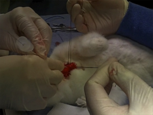
Ever had to do a surgical airway in a child? Thought not. They’re pretty rare. Bill Heegaard MD from Henepin County Medical Center taught me a few approaches (with the help of an anaesthetised rabbit) which really got me thinking. It’s something I’d often trained for in my internal simulator, and I even keep the equipment for it in my house (listen out for an upcoming podcast on that). Research and experience has demonstrated that open surgical airway techniques are more reliable than transtracheal needle techniques in adults, but what about kids, in whom traditional teaching cautions against open techniques?

Australian investigators who were experienced airway proceduralists evaluated transtracheal needle techniques using a rabbit model (an excellent model for the infant airway). Their success rate was only 60% and they perforated the posterior tracheal wall in 42% of attempts. Of 13 attempts to insert a dedicated paediatric tracheotomy device, the Quicktrach Child, none were successful(1) (they did not use the Quicktrach Infant model as it is not available in Australia).

Danish investigators used fresh piglet cadavers weighing around 8 kg to assess two transtracheal cannulas, in which they achieved success rates of 65.6% and 68.8%(2). There was also a very high rate of posterior tracheal wall perforation. Using an open surgical tracheostomy technique, they were successful in 97% of attempts. These were also experienced operators, with a median anaesthetic experience of 12.5 years.
Their tracheotomy technique was nice and simple, and used just a scalpel, scissors, and surgical towel clips. Here’s their technique:

Simple tracheotomy procedure described by Holm-Knudsen et al

  1. Identify larynx and proximal trachea by palpation
  2. Vertical incision through the skin and subcutaneous tissue from the upper part of larynx to the sternal notch
  3. Grasp strap muscles with two towel forceps and separate in the midline
  4. Palpate and identify the trachea (palpate rather than look for tracheal rings, as in a live patient one would expect bleeding to obscure the view)
  5. Stabilise the trachea by grasping it with a towel forceps
  6. Insert sharp tip of the scissors between two tracheal rings and lift the trachea anteriorly to avoid damage to the posterior wall
  7. Cut vertically in the midline of the trachea with the scissors – they chose to use the scissors to cut the tracheal rings to facilitate tube insertion
  8. Insert the tracheal tube

Using ultrasound and CT to evaluate comparative airway dimensions, the authors concluded that the pig model is most useful for training emergency airway management in older children aged 5–10 years.
Why were they doing a tracheotomy rather than a cricothyroidotomy? Reasons given by the authors include:

  • The infant cricothyroid membrane is very small
  • Palpation of the thyroid notch may be hindered by the overlying hyoid bone
  • The mandible may obstruct needle access to the cricothyroid membrane given the cephalad position in the neck of the infant larynx.

From an emergency medicine point of view, there are a couple of other reasons why we need to be able to access the trachea lower than the cricothyroid membrane. One is fractured larynx or other blunt or penetrating airway injury where there may be anatomical disruption at the cricothyroid level. The other situation is foreign body airway obstruction, when objects may lodge at the level of the cricoid ring which is functionally the narrowest part of the pediatric upper airway. Of course, alternative methods might be considered to remove the foreign body prior to tracheotomy, such as employing basic choking algorithms, and other techniques depending on whether you do or don’t have equipment.

Take home messages

  • Transtracheal airways in kids are so rare, we can’t avoid extrapolating animal data
  • Whichever infant or paediatric model is used, transtracheal needle techniques have a high rate of failure even by ‘experienced’ operators
  • The small size and easy compressibility of the airway probably contributes to this failure rate, including the high rate of posterior wall puncture
  • In keeping with adult audit data, open surgical techniques may have a higher success rate
  • Tracheotomy may be necessary rather than cricothyroidotomy in infants and children depending on clinical scenario and accessibility of anatomy
  • The stress and blood that is not simulated in cadaveric animal models will make open tracheotomy harder in a live patient, and so these success rates may not translate. However these factors do mean that whatever technique is used must be kept simple and should employ readily available and familiar equipment
  • Something to maintain control and anterior position of the anterior trachea wall should be used during incision and intubation of the trachea. The study reported here used towel clips; sutures around the tracheal rings may also be used (see image below)

I recommend you add ‘paediatric tracheotomy’ to the list of procedures you might need to do (if it’s not already there). Identify what equipment you would use and run the simulation in your head and in your work environment.
Have fun.

1. The ‘Can’t Intubate Can’t Oxygenate’ scenario in Pediatric Anesthesia: a comparison of different devices for needle cricothyroidotomy
Paediatr Anaesth. 2012 Dec;22(12):1155-8

BACKGROUND: Little evidence exists to guide the management of the ‘Can’t Intubate, Can’t Oxygenate’ (CICO) scenario in pediatric anesthesia.

OBJECTIVES: To compare two intravenous cannulae for ease of use, success rate and complication rate in needle tracheotomy in a postmortem animal model of the infant airway, and trial a commercially available device using the same model.

METHODS: Two experienced proceduralists repeatedly attempted cannula tracheotomy in five postmortem rabbits, alternately using 18-gauge (18G) and 14-gauge (14G) BD Insyte(™) cannulae (BD, Franklin Lakes, NJ, USA). Attempts began at the first tracheal cartilage, with subsequent attempts progressively more caudad. Success was defined as intratracheal cannula placement. In each rabbit, an attempt was then made by each proceduralist to perform a cannula tracheotomy using the Quicktrach Child(™) device (VBM Medizintechnik GmbH, Sulz am Neckar, Germany).

RESULTS: The rabbit tracheas were of similar dimensions to a human infant. 60 attempts were made at cannula tracheotomy, yielding a 60% success rate. There was no significant difference in success rate, ease of use, or complication rate between cannulae of different gauge. Successful aspiration was highly predictive (positive predictive value 97%) and both sensitive (89%) and specific (96%) for tracheal cannulation. The posterior tracheal wall was perforated in 42% of tracheal punctures. None of 13 attempts using the Quicktrach Child(™) were successful.

CONCLUSION: Cannula tracheotomy in a model comparable to the infant airway is difficult and not without complication. Cannulae of 14- and 18-gauge appear to offer similar performance. Successful aspiration is the key predictor of appropriate cannula placement. The Quicktrach Child was not used successfully in this model. Further work is required to compare possible management strategies for the CICO scenario

2. Emergency airway access in children – transtracheal cannulas and tracheotomy assessed in a porcine model
Paediatr Anaesth. 2012 Dec;22(12):1159-65

OBJECTIVES: In the rare scenario when it is impossible to oxygenate or intubate a child, no evidence exists on what strategy to follow.

AIM: The aim of this study was to compare the time and success rate when using two different transtracheal needle techniques and also to measure the success rate and time when performing an emergency tracheotomy in a piglet cadaver model.

METHODS: In this randomized cross-over study, we included 32 anesthesiologists who each inserted two transtracheal cannulas (TTC) using a jet ventilation catheter and an intravenous catheter in a piglet model. Second, they performed an emergency tracheotomy. A maximum of 2 and 4 min were allowed for the procedures, respectively. The TTC procedures were recorded using a video scope.

RESULTS: Placement of a transtracheal cannula was successful in 65.6% and 68.8% of the attempts (P = 0.76), and the median duration of the attempts was 69 and 42 s (P = 0.32), using the jet ventilation catheter and the intravenous catheter, respectively. Complications were frequent in both groups, especially perforation of the posterior tracheal wall. Performing an emergency tracheotomy was successful in 97%, in a median of 88 s.

CONCLUSIONS: In a piglet model, we found no significant difference in success rates or time to insert a jet ventilation cannula or an intravenous catheter transtracheally, but the incidence of complications was high. In the same model, we found a 97% success rate for performing an emergency tracheotomy within 4 min with a low rate of complications.

Externally rotate leg for femoral vein access

Want to access the femoral vein? Externally rotate the leg at the hip and things might be a bit easier. This study was done in adult patients, with the knee straight and no abduction applied. External rotation is also helpful in kids, with abduction up to sixty degrees.


Objective: To determine if external rotation of the leg increases the size and accessibility of the femoral vein compared with a neutral position.

Methods: One hundred patients presenting to a tertiary teaching hospital were prospectively recruited. The right common femoral vein of each subject was scanned with a linear probe (5–10 MHz) inferior to the inguinal ligament, with the leg in a neutral position and then in the externally rotated position. The transverse diameter of the femoral vein, the accessible diameter of the vein (lying medial to the femoral artery) and the depth of the vein were measured.

Results: The mean diameter of the femoral vein in the externally rotated leg was greater than with the leg in the neutral position (15.4 mm vs 13.8 mm); the mean difference was 1.6 mm (95% CI 1.3–1.9). The mean accessible diameter of the femoral vein was larger with the leg externally rotated (13.8 mm vs 11.7 mm, mean difference 2.1 mm, 95% CI 1.8–2.5). The depth from the skin to the femoral vein was less with the leg in external rotation (20.9 mm vs 22.6 mm, mean difference 1.7 mm, 95% CI 1.2–2.2). The mean diameter and depth were greater in patients with overweight or obese body mass index (BMI) measurements in both leg positions. The increase in femoral vein diameter and accessibility with external rotation was observed in all BMI groups.

Conclusion: The total and accessible femoral vein diameter is increased and the surface depth of the vein is decreased by placing the leg in external rotation compared with the neutral position.

Simple external rotation of the leg increases the size and accessibility of the femoral vein
Emerg Med Australas. 2012 Aug;24(4):408-13

Etomidate & sepsis

A meta-analysis attempts to quantify etomidate’s effect on mortality and adrenal suppression. Of course, we all know a meta-analysis can only be as reliable as the original data it’s analysing. I think editorialists Lauzier and Turgeon have a point with their statement:

“Given the widespread use of etomidate in the emergency room, we believe that a RCT designed to evaluate the safety of etomidate as a hypnotic agent for endotracheal intubation of patients with sepsis is not only ethical but also urgently warranted”

For a critique of the paper and subsequent discussion, check out the Academic Life in EM blog post by Brian Hayes


OBJECTIVE: To evaluate the effects of single-dose etomidate on the adrenal axis and mortality in patients with severe sepsis and septic shock.

DESIGN: A systematic review of randomized controlled trials and observational studies with meta-analysis.

SETTING: Literature search of EMBASE, Medline, Cochrane Database, and Evidence-Based Medical Reviews.

SUBJECTS: Sepsis patients who received etomidate for rapid sequence intubation.

INTERVENTIONS: None.

MEASUREMENTS AND MAIN RESULTS: We conducted a systematic review of randomized controlled trials and observational studies with meta-analysis assessing the effects of etomidate on adrenal insufficiency and all-cause mortality published between January 1950 and February 2012. We only examined studies including septic patients. All-cause mortality served as our primary end point, whereas the prevalence of adrenal insufficiency was our secondary end point. Adrenal insufficiency was determined using a cosyntropin stimulation test in all studies. We used a random effects model for analysis; heterogeneity was assessed with the I statistic. Publication bias was evaluated with Begg’s test. Five studies were identified that assessed mortality in those who received etomidate. A total of 865 subjects were included. Subjects who received etomidate were more likely to die (pooled relative risk 1.20; 95% confidence interval 1.02-1.42; Q statistic, 4.20; I2 statistic, 4.9%). Seven studies addressed the development of adrenal suppression associated with the administration of etomidate; 1,303 subjects were included. Etomidate administration increased the likelihood of developing adrenal insufficiency (pooled relative risk 1.33; 95% confidence interval 1.22-1.46; Q statistic, 10.7; I2 statistic, 43.9%).

CONCLUSIONS: Administration of etomidate for rapid sequence intubation is associated with higher rates of adrenal insufficiency and mortality in patients with sepsis.

Etomidate is associated with mortality and adrenal insufficiency in sepsis: A meta-analysis Crit Care Med. 2012 Nov;40(11):2945-53

Don't bronchodilators work in infants?

Inpatient paediatric teams can be scornful when bronchodilators are given by ED staff to wheezing infants, correctly referring to the lack of evidence of clinical benefit(1). There is however a persisting meme out there I’ve heard on a number of occasions that ‘young infants don’t have the receptors so inhaled beta agonists will never work.’ I’d love to know where this comes from.
Apparently, beta 2-receptors are present from the 16th gestational week(2). Pulmonary function testing of ventilated, very-low-birth-weight babies has shown that some consistently responded to beta-agonists whereas others did not(3). A newly published study reports that a quarter of mechanically ventilated infants with bronchiolitis were responders to inhaled albuterol, defined as a reduction in respiratory system resistance more than 30% below baseline(4).
In summary: beta-agonist bronchodilators have not been shown to improve clinical outcomes in wheezing infants. However some infants with some wheezing disorders will show a response in terms of pulmonary function. The receptors are there, and in life-threatening presentations bronchodilators should certainly be considered.
1. Short acting beta agonists for recurrent wheeze in children under 2 years of age
Cochrane Database Syst Rev. 2002;(3):CD002873
[EXPAND Click to read abstract]


BACKGROUND: Wheeze is a common symptom in infancy and is a common cause for both primary care consultations and hospital admission. Beta2-adrenoceptor agonists (b2-agonists) are the most frequently used as bronchodilator but their efficacy is questionable.
OBJECTIVES: To determine the effectiveness of b2-agonist for the treatment of infants with recurrent and persistent wheeze.
SEARCH STRATEGY: Relevant trials were identified using the Cochrane Airways Group database (CENTRAL), Medline and Pubmed. The database search used the following terms: Wheeze or asthma and Infant or Child and Short acting beta-agonist or Salbutamol (variants), Albuterol, Terbutaline (variants), Orciprenaline, Fenoterol

SELECTION CRITERIA: Randomised controlled trials comparing the effect of b2-agonist against placebo in children under 2 years of age who had had two or more previous episodes of wheeze, not related to another form of chronic lung disease.
DATA COLLECTION AND ANALYSIS: Eight studies met the criteria for inclusion in this meta-analysis. The studies investigated patients in three settings: at home (3 studies), in hospital (2 studies) and in the pulmonary function laboratory (3 studies). The main outcome measure was change in respiratory rate except for community based studies where symptom scores were used.

MAIN RESULTS: The studies were markedly heterogeneous and between study comparisons were limited. Improvement in respiratory rate, symptom score and oxygen saturation were noted in one study in the emergency department following two salbutamol nebulisers but this had no impact on hospital admission. There was a reduction in bronchial reactivity following salbutamol. There was no significant benefit from taking regular inhaled salbutamol on symptom scores recorded at home.

REVIEWER’S CONCLUSIONS: There is no clear benefit of using b2-agonists in the management of recurrent wheeze in the first two years of life although there is conflicting evidence. At present, further studies should only be performed if the patient group can be clearly defined and there is a suitable outcome parameter capable of measuring a response.

[/EXPAND]
2. The beta-2-agonists in asthma in infants and young children
Arch Pediatr. 2002 Aug;9 Suppl 3:384s-389s
[EXPAND Click to read abstract]


Beta 2-agonists, by inducing a fast and long relaxation of the bronchial smooth muscle, are considered as the more potent bronchodilators. beta 2-receptors are present from the 16th gestational week, explaining a possible bronchial response in the youngest children. beta 2-agonists do not induce any bronchodilator response in healthy children. Short-acting beta 2-agonists (salbutamol or albuterol, terbutaline) are indicated for asthma attacks, as needed in chronic asthma, and for prevention of symptoms during effort. They are safe and secure. The more efficient route of administration in preschool children is pressurized metered-dose inhaler used with a spacer device. Therefore, whatever the route of inhalation chosen (inhalation, injection, or continuous nebulization in acute asthma attack), more specified indications and doses are needed in young children. Long-acting beta 2-agonists (formoterol, salmeterol) are not authorized in France in children under 4 to 5 years of age depending on the drug used. Because of new oral formulations and recent considerations about their use in asthma attack, instead of short-acting beta 2-agonists, their indication in preschool asthmatic children might be reconsidered.

[/EXPAND]
3. Use of a beta-agonist in ventilated, very-low-birth-weight babies: a longitudinal evaluation
Dev Pharmacol Ther. 1990;15(2):61-7
[EXPAND Click to read abstract]


To determine if there is a specific postnatal (PNA) or postconceptional age (PCA) at which ventilated preterm infants respond to beta-agonists, we evaluated 15 infants with a mean gestational age of 26.5 +/- 1.5 weeks and mean birth weight of 0.89 +/- 0.23 kg who required mechanical ventilation at 10 days of age. Weekly pulmonary function testing (PFT) was performed before and 1 h after administration of albuterol. Taking the group as a whole, as well as individual babies, regression analysis showed no relationship between positive response and either PNA or PCA. Evaluation of individual infants, however, showed that some consistently responded to beta-agonists whereas others did not. We recommend individual PFT to identify those infants who will benefit from use of beta-agonists.

[/EXPAND]
4. Pulmonary mechanics following albuterol therapy in mechanically ventilated infants with bronchiolitis
J Asthma. 2012 Sep;49(7):688-96
[EXPAND Click to read abstract]


BACKGROUND AND AIMS: Bronchiolitis is a common cause of critical illness in infants. Inhaled β(2)-agonist bronchodilators are frequently used as part of treatment, despite unproven effectiveness. The purpose of this study was to describe the physiologic response to these medications in infants intubated and mechanically ventilated for bronchiolitis.

MATERIALS AND METHODS: We conducted a prospective trial of albuterol treatment in infants intubated and mechanically ventilated for bronchiolitis. Before and for 30 minutes following inhaled albuterol treatment, sequential assessments of pulmonary mechanics were determined using the interrupter technique on repeated consecutive breaths.

RESULTS: Fifty-four infants were enrolled. The median age was 44 days (25-75%; interquartile range (IQR) 29-74 days), mean hospital length of stay (LOS) was 18.3 ± 13.3 days, mean ICU LOS was 11.3 ± 6.4 days, and mean duration of mechanical ventilation was 8.5 ± 3.5 days. Fifty percent (n = 27) of the infants were male, 81% (n = 44) had public insurance, 80% (n = 41) were Caucasian, and 39% (n = 21) were Hispanic. Fourteen of the 54 (26%) had reduction in respiratory system resistance (Rrs) that was more than 30% below baseline, and were defined as responders to albuterol. Response to albuterol was not associated with demographic factors or hospitalization outcomes such as LOS or duration of mechanical ventilation. However, increased Rrs, prematurity, and non-Hispanic ethnicity were associated with increased LOS.

CONCLUSIONS: In this population of mechanically ventilated infants with bronchiolitis, relatively few had a reduction in pulmonary resistance in response to inhaled albuterol therapy. This response was not associated with improvements in outcomes.

[/EXPAND]

ECMO for paediatric cardiac arrest

The Taiwanese are at it again with their extracorporeal life support. This time, they report their outcomes in children who received ECMO for in-hospital cardiac arrest. Interestingly, the patients with pure cardiac causes of cardiac arrest had a survival rate similar to patients with non-cardiac causes.


PURPOSE: The study aims to describe 11 years of experience with extracorporeal cardiopulmonary resuscitation (ECPR) for in-hospital paediatric cardiac arrest in a university affiliated tertiary care hospital.

METHODS: Paediatric patients who received extracorporeal membrane oxygenation (ECMO) during active extracorporeal cardiopulmonary resuscitation (ECPR) at our centre from 1999 to 2009 were included in this retrospective study. The results from three different cohorts (1999-2001, 2002-2005 and 2006-2009) were compared. Survival rates and neurological outcomes were analysed. Favourable neurological outcome was defined as paediatric cerebral performance categories (PCPC) 1, 2 and 3.

RESULTS: We identified 54 ECPR events. The survival rate to hospital discharge was 46% (25/54), and 21 (84%) of the survivors had favourable neurological outcomes. The duration of CPR was 39±17 min in the survivors and 52±45 min in the non-survivors (p=NS). The patients with pure cardiac causes of cardiac arrest had a survival rate similar to patients with non-cardiac causes (47% (18/38) vs. 44% (7/16), p=NS). The non-survivors had higher serum lactate levels prior to ECPR (13.4±6.4 vs. 8.8±5.1 mmol/L, p<0.01) and more renal failure after ECPR (66% (19/29) vs. 20% (5/25), p<0.01). The patients resuscitated between 2006 and 2009 had shorter durations of CPR (34±13 vs. 78±76 min, p=0.032) and higher rates of survival (55% (16/29) vs. 0% (0/8), p=0.017) than those resuscitated between 1999 and 2002.

CONCLUSIONS: In our single-centre experience with ECPR for paediatric in-hospital cardiac arrest, the duration of CPR has become shorter and outcomes have improved in recent years. Higher pre-ECPR lactate levels and the presence of post-ECPR renal failure were associated with increased mortality. The presence of non-cardiac causes of cardiac arrest did not preclude successful ECPR outcomes. The duration of CPR was not significantly associated with poor outcomes in this study.

Eleven years of experience with extracorporeal cardiopulmonary resuscitation for paediatric patients with in-hospital cardiac arrest
Resuscitation. 2012 Jun;83(6):710-4

Superglue for CVCs

In resuscitation situations, the securing of vascular catheters is an important but sometimes cumbersome process, particular when sutures are required for central lines or arterial lines.

Medical grade ‘superglue’ (cyanoacrylate) can be used and this has been described in the anaesthetic literature before(1). Now, further in vitro work shows the glue does not weaken the intravenous catheter and is not associated with bacterial colonisation(2).

I think this is perfect for resuscitation lines. Just last night I used this technique to secure a femoral arterial line during a cardiac arrest resuscitation. It was great not to have to faff around with sharp suture needles during CPR and the line felt very secure after just a few seconds.

1. Tissue adhesive as an alternative to sutures for securing central venous catheters
Anaesthesia. 2007 Sep;62(9):969-70

2. Cyanoacrylate tissue adhesives – effective securement technique for intravascular catheters: in vitro testing of safety and feasibility
Anaesth Intensive Care. 2012 May;40(3):460-6