Category Archives: Kids

Acute Paediatrics

Caution with intraosseous adenosine

Two cases of failed cardioversion of SVT after tibial intraosseous administration of adenosine in infants are described in this month’s Pediatric Emergency Care. Both cases were subsequently cardioverted by intravenous adenosine. The maximum intraosseous dose given was 0.25 mg/kg. The successful IV doses were not higher than the IO doses.
It has been noted before that infants may require relatively higher doses of adenosine than children and that 0.2 mg/kg might even be considered a starting dose in infancy. I wonder if a bigger IO dose would have been effective, or whether a proximal humeral insertion site would make a difference. IO adenosine has been successfully used in infants and piglets.
This interesting case series provides a helpful caution in the management of paediatric SVT.

ABSTRACT: Supraventricular tachycardia (SVT) is a common tachyarrhythmia in the pediatric population that can necessitate immediate treatment. Adenosine has been well studied as a mainstay treatment, but the methods of adenosine administration have not been very well delineated. The intraosseous technique has presented itself as a possible method of administration. We describe 2 cases in which adenosine was administered through bone marrow infusion to convert SVT without success. The cases we describe show that intraosseous is not a reliable method of administering adenosine to stop SVT. Both patients presented with SVT refractory to vagal maneuvers and difficult intravenous placement. Intraosseous access was achieved, but administration of adenosine at increasing doses was unable to successfully convert the arrhythmia.

Intraosseous Infusion Is Unreliable for Adenosine Delivery in the Treatment of Supraventricular Tachycardia
Pediatr Emerg Care. 2012 Jan;28(1):47-8

A big brain saves a little one

Something I’ve been teaching for years – but never actually done – has been described in a case report from Oman.
A 2 year old child suffered a respiratory arrest due to an inhaled foreign body, which led to a bradyasystolic cardiac arrest. She was intubated by the resuscitation team who could not achieve any ventilation through the tube. The tube was removed and reinserted by an ‘expert’ (there is no mention of capnometry, for what it’s worth) and the same problem persisted.
The life-saving manouevre was to insert the tracheal tube further down into the right main bronchus and then withdraw to the trachea. This forced the obstructing object distally so that one-lung ventilation was then possible, resulting in return of spontaneous circulation and oxygen saturations in the mid-80’s. The object – a broken piece of plastic – was removed bronchoscopically and happily the child made an uneventful recovery.
Is this technique in your list of life-saving tricks? Hopefully, it is now.
A child is alive because a doctor was able to ‘think outside the guidelines’ in an incredibly high pressure situation. Rigid adherence to ACLS procedures here would have been futile. The guidelines save lives, but a few more can be saved when care can be individualised to the clinical situation by a thinking clinician.
Well done Dr Mishra and colleagues.

Sudden near-fatal tracheal aspiration of an undiagnosed nasal foreign body in a small child

Emerg Med Australas. 2011 Dec;23(6):776-8
[And here’s something else to consider if you have no airway equipment with you and your basic choking algorithm isn’t working]

Two new anaphylaxis guidelines

Many local and national guidelines for the management of anaphylaxis exist, but did you know there was a World Allergy Organization, and it has a very detailed guideline on this important life threatening condition?

Some interesting snippets from the guideline are included here

Anaphylaxis and cardiac disease

  • Anaphylaxis can precipitate acute myocardial infarction in susceptible individuals: in patients with ischemic heart disease, the number and density of cardiac mast cells is increased, including in the atherosclerotic plaques. Mediators released during anaphylaxis contribute to vasoconstriction and coronary artery spasm.
  • Epinephrine is not contraindicated in the treatment of anaphylaxis in patients with known or suspected cardiovascular disease, or in middle-aged or elderly patients without any history of coronary artery disease who are at increased risk of ACS only because of their age. Through its beta-1 adrenergic effects, epinephrine actually increases coronary artery blood flow because of an increase in myocardial contractility and in the duration of diastole relative to systole.
  • Glucagon has noncatecholamine-dependent inotropic and chronotropic cardiac effects, and is sometimes needed in patients taking a beta-adrenergic blocker who have hypotension and bradycardia and who do not respond optimally to epinephrine.
  • Anticholinergic agents are sometimes needed in beta-blocked patients, for example, atropine in those with persistent bradycardia or ipratropium in those with epinephrine-resistant bronchospasm.

How quickly can untreated anaphylaxis kill you?

Studies show median times to cardiorespiratory arrest after exposure to the offending stimulus were 5 minutes after administration of contrast media or drugs, 15 minutes after an insect sting, and 30 minutes after food ingestion.

What about confirming the diagnosis with serum tryptase measurements?

  • Blood samples for measurement of tryptase levels are optimally obtained 15 minutes to 3 hours after symptom onset.
  • Blood samples for measurement of histamine levels are optimally obtained 15–60 minutes after symptom onset. These tests are not specific for anaphylaxis.
  • Increased serum tryptase levels are often found in patients with anaphylaxis from insect stings or injected medications, and in those who are hypotensive
  • However, levels are often within normal limits in patients with anaphylaxis triggered by food and in those who are normotensive
  • Serial measurement of tryptase levels during an anaphylactic episode, and measurement of a baseline level after recovery are reported to be more useful than measurement at only one point in time.
  • Normal levels of either tryptase or histamine do not rule out the clinical diagnosis of anaphylaxis


How does epinephrine help?

  • Epinephrine is life-saving because of its alpha-1 adrenergic vasoconstrictor effects in most body organ systems (skeletal muscle is an important exception) and its ability to prevent and relieve airway obstruction caused by mucosal edema, and to prevent and relieve hypotension and shock.
  • Other relevant properties in anaphylaxis include its beta-1 adrenergic agonist inotropic and chronotropic properties leading to an increase in the force and rate of cardiac contractions, and its beta-2 adrenergic agonist properties such as decreased mediator release, bronchodilation and relief of urticaria
  • Epinephrine in a dose of 0.01 mg/kg of a 1:1,000 (1 mg/mL) solution injected promptly by the intramuscular route is effective and safe in the initial treatment of anaphylaxis. In other anaphylaxis scenarios, this low first-aid dose is unlikely to be effective. For example, if shock is imminent or has already developed, epinephrine needs to be given by slow intravenous infusion, ideally with the dose titrated according to noninvasive continuous cardiac monitoring.

What is the empty ventricle syndrome?

  • Patients with anaphylaxis should not suddenly sit, stand, or be placed in the upright position.
  • Instead, they should be placed on the back with their lower extremities elevated or, if they are experiencing respiratory distress or vomiting, they should be placed in a position of comfort with their lower extremities elevated.
  • This accomplishes 2 therapeutic goals: 1) preservation of fluid in the circulation (the central vascular compartment), an important step in managing distributive shock; and 2) prevention of the empty vena cava/empty ventricle syndrome, which can occur within seconds when patients with anaphylaxis suddenly assume or are placed in an upright position.
  • Patients with this syndrome are at high risk for sudden death. They are unlikely to respond to epinephrine regardless of route of administration, because it does not reach the heart and therefore cannot be circulated throughout the body

Should we give antihistamines, beta 2 agonists, and steroids?

The evidence base for use of these second line medications in the initial management of anaphylaxis, is extrapolated mainly from their use in treatment of other diseases such as urticaria (antihistamines) or acute asthma (beta-2 adrenergic agonists and glucocorticoids). Concerns have been raised that administering one or more second-line medications potentially delays prompt injection of epinephrine, the first-line treatment


Is ‘biphasic anaphylaxis’ a real phenomenon we should be concerned about?

  • Biphasic anaphylaxis occurs when symptoms recur within 1–72 hours (usually within 8–10 hours) after the initial symptoms have resolved, despite no further exposure to the trigger.
  • It occurs in up to 23% of adults and up to 11% of children.
  • After apparent resolution of symptoms, duration of monitoring in a medically supervised setting should be individualized. For example, patients with moderate respiratory or cardiovascular compromise should be monitored for at least 4 hours, and if indicated, for 8–10 hours or longer.
  • Protracted uniphasic anaphylaxis is uncommon, but can last for days.

World Allergy Organization Guidelines for the Assessment and Management of Anaphylaxis
World Allergy Organization Journal 2011;4(2):13-37 Full Text
[EXPAND click for abstract]

The illustrated World Allergy Organization (WAO) Anaphylaxis Guidelines were created in response to absence of global guidelines for anaphylaxis. Uniquely, before they were developed, lack of worldwide availability of essentials for the diagnosis and treatment of anaphylaxis was documented. They incorporate contributions from more than 100 allergy/immunology specialists on 6 continents. Recommendations are based on the best evidence available, supported by references published to the end of December 2010. The Guidelines review patient risk factors for severe or fatal anaphylaxis, co-factors that amplify anaphylaxis, and anaphylaxis in vulnerable patients, including pregnant women, infants, the elderly, and those with cardiovascular disease. They focus on the supreme importance of making a prompt clinical diagnosis and on the basic initial treatment that is urgently needed and should be possible even in a low resource environment. This involves having a written emergency protocol and rehearsing it regularly; then, as soon as anaphylaxis is diagnosed, promptly and simultaneously calling for help, injecting epinephrine (adrenaline) intramuscularly, and placing the patient on the back or in a position of comfort with the lower extremities elevated. When indicated, additional critically important steps include administering supplemental oxygen and maintaining the airway, establishing intravenous access and giving fluid resuscitation, and initiating cardiopulmonary resuscitation with continuous chest compressions. Vital signs and cardiorespiratory status should be monitored frequently and regularly (preferably, continuously). The Guidelines briefly review management of anaphylaxis refractory to basic initial treatment. They also emphasize preparation of the patient for self-treatment of anaphylaxis recurrences in the community, confirmation of anaphylaxis triggers, and prevention of recurrences through trigger avoidance and immunomodulation. Novel strategies for dissemination and implementation are summarized. A global agenda for anaphylaxis research is proposed.

[/EXPAND]
This month has also seen the publication guidelines from the UK’s National Institute for Health & Clinical Excellence, entitled ‘Anaphylaxis: assessment to confirm an anaphylactic episode and the decision to refer after emergency treatment for a suspected anaphylactic episode’
Their guideline summary is as follows:

After a suspected anaphylactic reaction in adults or young people aged 16 years or older, take timed blood samples for mast cell tryptase testing as follows:

  • a sample as soon as possible after emergency treatment has started
  • a second sample ideally within 1–2 hours (but no later than 4 hours) from the onset of symptoms.

After a suspected anaphylactic reaction in children younger than 16 years, consider taking blood samples for mast cell tryptase testing as follows if the cause is thought to be venom-related, drug-related or idiopathic:

  • a sample as soon as possible after emergency treatment has started
  • a second sample ideally within 1–2 hours (but no later than 4 hours) from the onset of symptoms.

Patients who have had emergency treatment for suspected anaphylaxis should be observed for 6–12 hours from the onset of symptoms, depending on their response to emergency treatment
After emergency treatment for suspected anaphylaxis, offer people a referral to a specialist allergy service (age-appropriate where possible) consisting of healthcare professionals with the skills and competencies necessary to accurately investigate, diagnose, monitor and provide ongoing management of, and patient education about, suspected anaphylaxis.
After emergency treatment for suspected anaphylaxis, offer people (or, as appropriate, their parent and/or carer) an appropriate adrenaline injector as an interim measure before the specialist allergy service appointment.
Before discharge a healthcare professional with the appropriate skills and competencies should offer people (or, as appropriate, their parent and/or carer) the following:

  • information about anaphylaxis, including the signs and symptoms of an anaphylactic reaction
  • information about the risk of a biphasic reaction
  • information on what to do if an anaphylactic reaction occurs (use the adrenaline injector and call emergency services)

Anaphylaxis: assessment to confirm an anaphylactic episode and the decision to refer after emergency treatment for a suspected anaphylactic episode
CG134 Anaphylaxis: NICE guideline

Treating sepsis – have we got it the wrong way round?

In our understanding of the pathophysiology of sepsis, we often attribute organ damage and death to the excessive host response to infection, including the popular phrase ‘cytokine storm’. This has been nicely described as ‘friendly fire’ by Prof Derek Angus, who points out that this central tenet of sepsis understanding may in some cases be flawed1; it has led to research on drugs that suppress parts of these inflammatory pathways, although none have yet proven effective. An elegant study on patients dying from sepsis showed clear evidence of immunosuppression compared with controls2.

Editorialist Peter Ward3 proposes an area for future research: whether such derangements can be reversed by treatment with agents such as interleukins 7 or 15, which might combat the T-cell depletion state in sepsis.
The authors point out that all the patients included in the study died on ICU, some after a considerable duration of illness, and they emphasise that early deaths from sepsis in previously healthy patients with infections of highly virulent organisms are associated with an extremely exuberant immunoinflammatory response.
1.The Search for Effective Therapy for Sepsis: Back to the Drawing Board?
JAMA December 21, 2011, Vol 306, No. 23, pp 2614-5
2.Immunosuppression in Patients Who Die of Sepsis and Multiple Organ Failure
JAMA December 21, 2011, Vol 306, No. 23, pp 2594-2605
[EXPAND Abstract]


Context Severe sepsis is typically characterized by initial cytokine-mediated hyperinflammation. Whether this hyperinflammatory phase is followed by immunosuppression is controversial. Animal studies suggest that multiple immune defects occur in sepsis, but data from humans remain conflicting.
Objectives To determine the association of sepsis with changes in host innate and adaptive immunity and to examine potential mechanisms for putative immunosuppression.
Design, Setting, and Participants Rapid postmortem spleen and lung tissue harvest was performed at the bedsides of 40 patients who died in intensive care units (ICUs) of academic medical centers with active severe sepsis to characterize their immune status at the time of death (2009-2011). Control spleens (n = 29) were obtained from patients who were declared brain-dead or had emergent splenectomy due to trauma; control lungs (n = 20) were obtained from transplant donors or from lung cancer resections.
Main Outcome Measures Cytokine secretion assays and immunophenotyping of cell surface receptor-ligand expression profiles were performed to identify potential mechanisms of immune dysfunction. Immunohistochemical staining was performed to evaluate the loss of immune effector cells.
Results The mean ages of patients with sepsis and controls were 71.7 (SD, 15.9) and 52.7 (SD, 15.0) years, respectively. The median number of ICU days for patients with sepsis was 8 (range, 1-195 days), while control patients were in ICUs for 4 or fewer days. The median duration of sepsis was 4 days (range, 1-40 days). Compared with controls, anti-CD3/anti-CD28–stimulated splenocytes from sepsis patients had significant reductions in cytokine secretion at 5 hours: tumor necrosis factor, 5361 (95% CI, 3327-7485) pg/mL vs 418 (95% CI, 98-738) pg/mL; interferon γ, 1374 (95% CI, 550-2197) pg/mL vs 37.5 (95% CI, −5 to 80) pg/mL; interleukin 6, 3691 (95% CI, 2313-5070) vs 365 (95% CI, 87-642) pg/mL; and interleukin 10, 633 (95% CI, −269 to 1534) vs 58 (95% CI, −39 to 156) pg/mL; (P < .001 for all). There were similar reductions in 5-hour lipopolysaccharide-stimulated cytokine secretion. Cytokine secretion in sepsis patients was generally less than 10% that in controls, independent of age, duration of sepsis, corticosteroid use, and nutritional status. Although differences existed between spleen and lung, flow cytometric analysis showed increased expression of selected inhibitory receptors and ligands and expansion of suppressor cell populations in both organs. Unique differences in cellular inhibitory molecule expression existed in immune cells isolated from lungs of sepsis patients vs cancer patients and vs transplant donors. Immunohistochemical staining showed extensive depletion of splenic CD4, CD8, and HLA-DR cells and expression of ligands for inhibitory receptors on lung epithelial cells. Conclusions Patients who die in the ICU following sepsis compared with patients who die of nonsepsis etiologies have biochemical, flow cytometric, and immunohistochemical findings consistent with immunosuppression. Targeted immune-enhancing therapy may be a valid approach in selected patients with sepsis.

[/EXPAND]
3.Immunosuppression in Sepsis
JAMA December 21, 2011, Vol 306, No. 23, pp 2618-9

2011 Asthma Guidelines

The British Thoracic Society / SIGN Guidelines on asthma have been updated for 2011. There don’t seem to be any modificiations to the sections on acute severe asthma which were updated in 2009 and blogged here, although the treatment algorithms seem to be presented in a slightly different format and therefore are reproduced here:

Management of acute severe asthma in adults in hospital

Management of acute asthma in children in hospital

Oxygen therapy for asthma can elevate CO2

Patients with acute exacerbations of asthma randomised to receive high concentration oxygen therapy showed a greater rise in CO2 than those who received titrated oxygen to keep SpO2 > 93%.
This study has a few weaknesses but raises an interesting challenge to the dogma of high flow oxygen (and oxygen driven nebulisers) for all acute asthma exacerbations.
The suggested main mechanism for the elevation in CO2 is worsening ventilation/perfusion mismatching as a result of the release of hypoxic pulmonary vasoconstriction and a consequent increase in physiological dead space. The authors remind us that this has been demonstrated in other studies on asthma and acute COPD exacerbations. The authors infer that high concentration oxygen therapy may therefore potentially increase the PaCO2 across a range of respiratory conditions with abnormal gas exchange due to ventilation/perfusion mismatching
Some of the weaknesses include lack of blinding, recruiting fewer patients than planned, and changing their primary outcome variable after commencing the study (which the authors are honest about) from absolute CO2 to increase in CO2 (since it was apparent on preliminary analysis of the first few patients that presenting CO2 was the primary determinant of subsequent CO2). Furthermore, the CO2 was measured from a transcutaneous device as opposed to the true ‘gold standard’ of arterial blood gas analysis, although good reasons are given for this.
Despite some of these drawbacks this study provides us with a further reminder that oxygen is a drug with some unwanted effects and therefore its dose needs to be individualised for the patient.


Background The effect on Paco(2) of high concentration oxygen therapy when administered to patients with severe exacerbations of asthma is uncertain.

Methods 106 patients with severe exacerbations of asthma presenting to the Emergency Department were randomised to high concentration oxygen (8 l/min via medium concentration mask) or titrated oxygen (to achieve oxygen saturations between 93% and 95%) for 60 min. Patients with chronic obstructive pulmonary disease or disorders associated with hypercapnic respiratory failure were excluded. The transcutaneous partial pressure of carbon dioxide (Ptco(2)) was measured at 0, 20, 40 and 60 min. The primary outcome variable was the proportion of patients with a rise in Ptco(2) ≥4 mm Hg at 60 min.

Results The proportion of patients with a rise in Ptco(2) ≥4 mm Hg at 60 min was significantly higher in the high concentration oxygen group, 22/50 (44%) vs 10/53 (19%), RR 2.3 (95% CI 1.2 to 4.4, p<0.006). The high concentration group had a higher proportion of patients with a rise in Ptco(2) ≥8 mm Hg, 11/50 (22%) vs 3/53 (6%), RR 3.9 (95% CI 1.2 to 13.1, p=0.016). All 10 patients with a final Ptco(2) ≥45 mm Hg received high concentration oxygen therapy, and in five there was an increase in Ptco(2) ≥10 mm Hg.
Conclusion High concentration oxygen therapy causes a clinically significant increase in Ptco(2) in patients presenting with severe exacerbations of asthma. A titrated oxygen regime is recommended in the treatment of severe asthma, in which oxygen is administered only to patients with hypoxaemia, in a dose that relieves hypoxaemia without causing hyperoxaemia.

Randomised controlled trial of high concentration versus titrated oxygen therapy in severe exacerbations of asthma
Thorax. 2011 Nov;66(11):937-41

Offensive medicine: CT before LP

I’m getting worn down by clinicians – often other specialists – who insist that CT imaging of the brain is mandatory prior to lumbar puncture in all patients. There is surely a subgroup of patients (especially young ones) in whom the benefit:harm balance of CT comes out in favour of NOT doing the imaging. In these cases, getting the scan is not ‘defensive medicine’ but ‘offensive medicine’ – offending the principle of primum non nocere. During ED shifts I have recently had to perform online searches in order to furnish colleagues and patients’ medically qualified relatives with printouts of the literature on this. This page is here to save me having to repeat those searches. Regarding the practice of performing a routine head CT prior to lumbar puncture to rule out risk of herniation:

  • Mass effect on CT does not predict herniation
  • Lack of mass effect on CT does not rule out raised ICP or herniation
  • Herniation has occurred in patients who did not undergoing lumbar puncture because of CT findings
  • Clinical predictors of raised ICP are more reliable than CT findings
  • CT may delay diagnosis and treatment of meningitis
  • Even in patients in whom LP may be considered contraindicated (cerebral abscess, mass effect on CT), complications from LP were rare in several studies

Best practice, it would seem, is the following

  • If you think CT will show a cause for the headache, do a CT
  • If a CT is indicated for other reasons (depressed conscious level, focal neurology), do a CT
  • If a GCS 15 patient is to undergo LP for suspected (or to rule out) meningitis, and they have a normal neurological exam (including fundi), and are not elderly or immunosuppressed, there is no need to do a CT first.
  • If you’re seriously worried about meningitis and are intent on getting a CT prior to LP, don’t let the imaging delay antimicrobial therapy.

Here are some useful references:

1. The CT doesn’t help

CT head before lumbar puncture in suspected meningitis BestBET evidence summary: In cases of suspected meningitis it is very unlikely that patients without clinical risk factors (immunocompromise/ history of CNS disease/seizures) or positive neurological findings will have a contraindication to lumbar puncture on their CT scan If CT scan is deemed to be necessary, administration of antibiotics should not be delayed. BestBETS website

Computed Tomography of the Head before Lumbar Puncture in Adults with Suspected Meningitis Much cited NEJM paper from 2001 which concludes: “In adults with suspected meningitis, clinical features can be used to identify those who are unlikely to have abnormal findings on CT of the headN Engl J Med. 2001 Dec 13;345(24):1727-33 Full Text

Cranial CT before Lumbar Puncture in Suspected Meningitis Correspondence in 2002 NEJM including study of 75 patients with pneumococcal meningitis: CT cannot rule out risk of herniation Cranial CT before Lumbar Puncture in Suspected Meningitis N Engl J Med. 2002 Apr 18;346(16):1248-51 Full Text

2. The CT may harm

Cancer risk from CT Paucis verbis card, from the wonderful Academic Life in EM

3. Guidelines say CT is not always needed

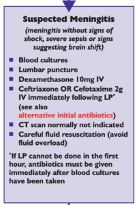
National (UK) guidelines on meningitis (community acquired meningitis in the immunocompetent host) available from meningitis.org. , including this box:

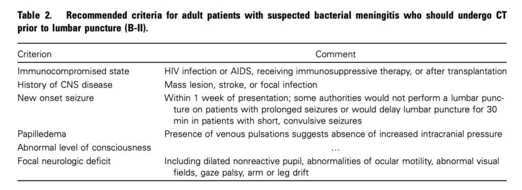
Practice Guidelines for the Management of Bacterial Meningitis These 2004 guidelines from the Infectious Diseases Society of America provide the following table listing the recommended criteria for adult patients with suspected bacterial meningitis who should undergo CT prior to lumbar puncture:

Clin Infect Dis. (2004) 39 (9): 1267-1284 Full text

4. This is potentially even more of an issue with paediatric patients

Fatal Lumbar Puncture: Fact Versus Fiction—An Approach to a Clinical Dilemma

An excellent summary of the above mentioned issues presented in a paediatric context, including the following:

On initial consideration a cranial CT would seem to be an appropriate and potentially useful diagnostic study for confirming the diagnosis of cerebral herniataion. The fallacy in this assessment has been emphasized by the finding that no clinically significant CT abnormalities are found that are not suspected on clinical assessments. Further, as previously noted, a normal CT examination may be found at about the time of a fatal herniation. Thus, the practical usefulness of a cranial CT in the majority of pediatric patients is limited to those rare patients whose increased ICP is secondary to mass lesions, not in the initial approach to acute meningitis.

Pediatrics. 2003 Sep;112(3 Pt 1):e174-6 Full Text

The last words should go to Dr Brad Spellberg, who in response to the IDSA’s guidelines wrote an excellent letter summarising much of the evidence at the time, confessed:

Why do we persist in using the CT scan for this purpose, despite the lack of supportive data? I am as guilty of this practice as anyone else, and the reason is simple: I am a chicken.

Clin Infect Dis. (2005) 40 (7): 1061 Full Text

Salicylate poisoning and pseudohyperchloraemia


Severe salicylate poisoning can cause metabolic acidosis from an accumulation of salicylic acid, lactic acid, and ketone bodies. A high anion gap acidosis is therefore the typical metabolic abnormality seen. A case series illustrates salicylate poisoning presenting with a normal gap (hyperchloraemic) acidosis – one patient had a chloride of 111 mmol/l and the other 123 mmol/l. This can occur when some analysers falsely read an elevated chloride in the presence of high concentrations of salicylate.


Severe salicylate poisoning is classically associated with an anion gap metabolic acidosis. However, high serum salicylate levels can cause false increase of laboratory chloride results on some analyzers. We present 2 cases of life-threatening salicylate poisoning with an apparently normal anion gap caused by an important laboratory interference. These cases highlight that the diagnosis of severe salicylism must be considered in all patients presenting with metabolic acidosis, even in the absence of an increased anion gap.

Falsely Normal Anion Gap in Severe Salicylate Poisoning Caused by Laboratory Interference
Ann Emerg Med. 2011 Sep;58(3):280-1

Easy rapid infusion set up

Kapoor and Singh's system from the Open Access article - click for explanation

Here’s a nice and simple set up for rapid iv infusions using simple cheap equipment

Full details at the Scandinavian Journal Site
Novel rapid infusion device for patients in emergency situations
Scand J Trauma Resusc Emerg Med. 2011 Jun 10;19:35 (Free Full Text)

Is there nothing ketamine can't do?

As well as the benefits of cardiovascular stability, maintenance of cerebral perfusion pressure, possibly lowering ICP and providing other neuroprotective benefits, ketamine may have other advantages. These are reviewed in a British Journal of Anaesthesia article from which I’ve selected those benefits of interest to practitioners of emergency medicine and critical care.


Additional Beneficial Effects of Ketamine

  • the dysphoric, or ’emergence’ reactions associated with ketamine may be reduced by pre-administration or co-administration of sedatives, such as benzodiazepines, propofol, dexmedetomidine, or droperidol.
  • ketamine potentiates opioid analgesia in multiple settings, reducing opioid total dose and in some groups of patients reducing postoperative desaturation
  • ketamine has possible anti-inflammatory effects demonstrated in some types of surgical patients
  • ketamine may prevent awareness, recall, or both during general anaesthesia

Ketamine: new uses for an old drug?
Br J Anaesth. 2011 Aug;107(2):123-6