Category Archives: Kids

Acute Paediatrics

Two hands on the jaw for mask ventilation

Elective surgery patients were anaesthetised with propofol with or without fentanyl and had an oropharyngeal airway placed. They were ventilated with pressure control ventilation via facemask held with a single handed traditional ‘EC clamp’ grip and with a two-handed jaw thrust, and compared. The order in which these two techniques were trialled was randomised. All breaths were delivered with a peak pressure of 15 cm H2O, an inspiratory-to-expiratory ratio of 1:1, at a frequency of 15 breaths per minute. Ventilation was more effective with the two handed technique.
Using a self-inflating bag for resuscitation, this would translate to a two-person technique. Of note in methodology however was use of a ‘standard pillow’ and some emphasis on head extension. Perhaps ventilation would have been more effective with either technique if they had applied the golden rule of ear-to-sternal-notch positioning: a must for effective mask ventilation and successful laryngoscopy.

BACKGROUND: Mask ventilation is considered a “basic” skill for airway management. A one-handed “EC-clamp” technique is most often used after induction of anesthesia with a two-handed jaw-thrust technique reserved for difficult cases. Our aim was to directly compare both techniques with the primary outcome of air exchange in the lungs.
METHODS: Forty-two elective surgical patients were mask-ventilated after induction of anesthesia by using a one-handed “EC-clamp” technique and a two-handed jaw-thrust technique during pressure-control ventilation in randomized, crossover fashion. When unresponsive to a jaw thrust, expired tidal volumes were recorded from the expiratory limb of the anesthesia machine each for five consecutive breaths. Inadequate mask ventilation and dead-space ventilation were defined as an average tidal volume less than 4 ml/kg predicted body weight or less than 150 ml/breath, respectively. Differences in minute ventilation and tidal volume between techniques were assessed with the use of a mixed-effects model.
RESULTS: Patients were (mean ± SD) 56 ± 18 yr old with a body mass index of 30 ± 7.1 kg/m. Minute ventilation was 6.32 ± 3.24 l/min with one hand and 7.95 ± 2.70 l/min with two hands. The tidal volume was 6.80 ± 3.10 ml/kg predicted body weight with one hand and 8.60 ± 2.31 ml/kg predicted body weight with two hands. Improvement with two hands was independent of the order used. Inadequate or dead-space ventilation occurred more frequently during use of the one-handed compared with the two-handed technique (14 vs. 5%; P = 0.013).
CONCLUSION: A two-handed jaw-thrust mask technique improves upper airway patency as measured by greater tidal volumes during pressure-controlled ventilation than a one-handed “EC-clamp” technique in the unconscious apneic person.

A Two-handed Jaw-thrust Technique Is Superior to the One-handed “EC-clamp” Technique for Mask Ventilation in the Apneic Unconscious Person
Anesthesiology. 2010 Oct;113(4):873-9

Intranasal ketamine analgesia

I published a case report in the EMJ highlighting the use of intranasal ketamine in a pre-hospital paediatric burns case.
The lad had nasty scalds but did not need iv fluids and had no other indications for an iv line. The vigorous first aid had rendered him cold and veinless and an intraosseous would have been overkill. Ketamine was perfect for the job and Ambulance Service New South Wales paramedics carry a mucosal atomisation device (MAD) for the administration of i.n. fentanyl. I used the MAD to adminster 0.5 mg/kg ketamine, but there is a dead space in the device (0.1 ml) that probably resulted in actual delivery of 0.25mg/kg. This gave great analgesia and compliance enabling us to painlessly apply polyethylene film to the burns.
I received the following email from TIm Wolfe, the inventor of the MAD nasal (reproduced with permission):

Cliff,
Nice contribution to the literature. There is a lot of interest in IN ketamine in these lower doses to treat pain but not cause sedation. You eluded to the military interest and the hospice interest. I think your insights for EMS are also cutting edge – hopefully this will lead others to design a larger trial.
Thanks
Tim Wolfe, MD

More information on the use of intranasal medication is available at www.intranasal.net. I have no conflicts of interest to declare.
Case report: prehospital use of intranasal ketamine for paediatric burn injury
Emerg Med J. 2011 Feb 3. [Epub ahead of print]

Weight formula validation

Further validation of the UK-derived Luscombe weight formula has been made in the Australian setting. The nice simple formula for estimating the weight of a child based on age is:

Weight (kg) = 3 x age(years) + 7

It was compared with other formulae including the Best Guess formula, which is a bit more difficult to apply as the formula varies according to age range. This is reported in a previous post.
The authors provide the following cautionary advice:
“Whereas age-based formulae are, in the main, easy to calculate, the evidence suggests that ethnicity and body habitus pose serious challenges to their accuracy. In comparative studies, age-based formulae were found to be less accurate than the Broselow tape and parental estimate, with parental estimate being the most accurate weight estimation method. In light of this evidence, age-based formulae should only be used when these more accurate methods are not available.”

OBJECTIVE: Several paediatric weight estimation methods have been described for use when direct weight measurement is not possible. A new age-based weight estimation method has recently been proposed. The Luscombe formula, applicable to children aged 1-10 years, is calculated as (3 × age in years) + 7. Our objective was to externally validate this formula using an existing database.
METHOD: Secondary analysis of a prospective observational cohort study. Data collected included height, age, ethnicity and measured weight. The outcome of interest was agreement between estimated weight using the Luscombe formula and measured weight. Secondary outcome was comparison with performance of Argall, APLS and Best Guess formulae. Accuracy of weight estimation methods was compared using mean difference (bias), 95% limits of agreement, root mean square error and proportion with agreement within 10%.
RESULTS: Four hundred and ten children were studied. Median age was 4 years; 54.4% were boys. Mean body mass index was 17 kg/m(2) and mean measured weight was 21.2 kg. The Luscombe formula had a mean difference of 0.66 kg (95% limits of agreement -9.9 to +11.3 kg; root mean square error of 5.44 kg). 45.4% of estimates were within 10% of measured weight. The Best Guess and Luscombe formulae performed better than Argall or APLS formulae.
CONCLUSION: The Luscombe formula is among the more accurate age-based weight estimation formulae. When more accurate methods (e.g. parental estimation or the Broselow tape) are not available, it is an acceptable option for estimating children’s weight.

Validation of the Luscombe weight formula for estimating children’s weight
Emerg Med Australas 2011 Feb;23(1):59-62

ILCOR neonatal cooling guideline

On the basis of the published data to date the Neonatal Task Force of the International Liaison Committee on Resuscitation (ILCOR) made the following recommendation on February 2010 with regard to therapeutic hypothermia:

  • Newly born infants born at term or near-term with evolving moderate to severe hypoxic-ischemic encephalopathy should be offered therapeutic hypothermia.
  • Whole-body cooling and selective head cooling are both appropriate strategies.
  • Cooling should be initiated and conducted in neonatal intensive care facilities using protocols consistent with those used in the randomized clinical trials i.e. commence within 6 h, continue for 72 h and rewarm over at least 4 h.
  • Carefully monitor for known adverse effects of cooling – thrombocytopenia and hypotension.
  • All treated infants should be followed longitudinally.

Therapeutic hypothermia following intrapartum hypoxia-ischemia. An advisory statement from the Neonatal Task Force of the International Liaison Committee on Resuscitation
Resuscitation 2010;81(11):1459-1461

Laryngospasm and ketamine

What are the factors associated with laryngospasm in ketamine sedation? A large study was unable to identify specific predictors:
Objective: The objective of this study was to assess predictors of emergency department (ED) ketamine-associated laryngospasm using case-control techniques.
Methods: We performed a matched case-control analysis of a sample of 8282 ED ketamine sedations (including 22 occurrences of laryngospasm) assembled from 32 prior published series. We sequentially studied the association of each of 7 clinical variables with laryngospasm by assigning 4 controls to each case while matching for the remaining 6 variables. We then used univariate statistics and conditional logistic regression to analyze the matched sets.

Results: We found no statistical association of age, dose, oropharyngeal procedure, underlying physical illness, route, or coadministered anticholinergics with laryngospasm. Coadministered benzodiazepines showed a borderline association in the multivariate but not univariate analysis that was considered anomalous.
Conclusions: This case-control analysis of the largest available sample of ED ketamine-associated laryngospasm did not demonstrate evidence of association with age, dose, or other clinical factors. Such laryngospasm seems to be idiosyncratic, and accordingly, clinicians administering ketamine must be prepared for its rapid identification and management. Given no evidence that they decrease the risk of laryngospasm, coadministered anticholinergics seem unnecessary.
Laryngospasm During Emergency Department Ketamine Sedation: A Case-Control Study
Pediatr Emerg Care. 2010 Nov;26(11):798-802

What's a normal newborn O2 sat?

Maybe they’re not just little adults after all: the normal reference ranges for oxygen saturation in the first few minutes of life have been defined for healthy newborns:
OBJECTIVE The goal was to define reference ranges for pulse oxygen saturation (SpO2) values in the first 10 minutes after birth for infants who received no medical intervention in the delivery room.
METHODS Infants were eligible if a member of the research team was available to record SpO2 immediately after birth. Infants were excluded if they received supplemental oxygen or any type of assisted ventilation. SpO2 was measured with a sensor applied to the right hand or wrist as soon as possible after birth; data were collected every 2 seconds.

RESULTS We studied 468 infants and recorded 61650 SpO2 data points. The infants had a mean ± SD gestational age of 38 ± 4 weeks and birth weight of 2970 ± 918 g. For all 468 infants, the 3rd, 10th, 50th, 90th, and 97th percentile values at 1 minute were 29%, 39%, 66%, 87%, and 92%, respectively, those at 2 minutes were 34%, 46%, 73%, 91%, and 95%, and those at 5 minutes were 59%, 73%, 89%, 97%, and 98%. It took a median of 7.9 minutes (interquartile range: 5.0–10 minutes) to reach a SpO2 value of >90%. SpO2 values for preterm infants increased more slowly than those for term infants. We present percentile charts for all infants, term infants of 37 weeks, preterm infants of 32 to 36 weeks, and extremely preterm infants of <32 weeks.
CONCLUSION These data represent reference ranges for SpO2 in the first 10 minutes after birth for preterm and term infants.
Defining the Reference Range for Oxygen Saturation for Infants After Birth
Pediatrics. 2010 Jun;125(6):e1340-7

US determined best LP position

Here ultrasound was used to ascertain the best position for doing a lumbar puncture in kids, where the interspinous space was maximised:
BACKGROUND Lumbar punctures are commonly performed in the pediatric emergency department. There is no standard, recommended, optimal position for children who are undergoing the procedure.
OBJECTIVE To determine a position for lumbar punctures where the interspinous space is maximized, as measured by bedside ultrasound.
METHODS A prospective convenience sample of children under age 12 was performed. Using a portable ultrasound device, the L3-L4 or L4-L5 interspinous space was measured with the subject in 5 different positions. The primary outcome was the interspinous distance between 2 adjacent vertebrae. The interspinous space was measured with the subject sitting with and without hip flexion. In the lateral recumbent position, the interspinous space wasmeasured with the hips in a neutral position as well as in flexion, both with and without neck flexion. Data were analyzed by comparing pairwise differences.

RESULTS There were 28 subjects enrolled (13 girls and 15 boys) at a median age of 5 years. The sitting-flexed position provided a significantly increased interspinous space (P < .05). Flexion of the hips increased the interspinous space in both the sitting and lateral recumbent positions (P < .05). Flexion of the neck, did not significantly change the interspinous space (P = .998).
CONCLUSIONS The interspinous space of the lumbar spine was maximally increased with children in the sitting position with flexed hips; therefore we recommend this position for lumbar punctures. In the lateral recumbent position, neck flexion does not increase the interspinous space and may increase morbidity; therefore, it is recommended to hold patients at the level of the shoulders as to avoid neckflexion.
Positioning for lumbar puncture in children evaluated by bedside ultrasound
Pediatrics. 2010 May;125(5):e1149-53
A more recent study also used ultrasound in infants to investigate the anatomic necessity and advantage derived from a tight flexed lateral recumbent position, since hypoxia has been observed in that position:
Objectives:  Hypoxia has been observed when infants undergo lumbar puncture in a tight flexed lateral recumbent position. This study used sonographic measurements of lumbar interspinous spaces to investigate the anatomic necessity and advantage derived from this tight flexed positioning in infants.
Methods:  This was a brief, prospective, observational study of a convenience sample of patients. Twenty-one healthy infants under 1 month of age were scanned in two positions: prone in a spine-neutral position and lateral recumbent with their knees bent into their chest and their neck flexed. In each position, a 5- to 10-MHz linear array transducer was used to scan midline along the lumbar spinous processes in the sagittal plane. The distances between the spinous processes were measured near the ligamentum flavum using the ultrasound machine’s calipers. Pulse oximetry was monitored on all infants during flexed positioning.
Results:  In the spine-neutral position, all studied interspinous spaces were much wider than a 22-gauge spinal needle (diameter 0.072 cm). The mean (±SD) interspinous spaces for L3-4, L4-5, and L5-S1 in a spine-neutral position were 0.42 (±0.07), 0.37 (±0.06), and 0.36 (±0.11) cm, respectively. Flexing the infants increased the mean lumbar interspinous spaces at L3-4, L4-5, and L5-S1 by 31, 51, and 44%, respectively.
Conclusions:  This study verified that tight, lateral flexed positioning substantially enhances the space between the lumbar spinous processes and that a spine-neutral position also allows for a large enough anatomic interspinous space to perform lumbar puncture. However, further clinical research is required to establish the feasibility of lumbar puncture in a spine-neutral position.
Evaluating infant positioning for lumbar puncture using sonographic measurements
Acad Emerg Med. 2011 Feb;18(2):215-8

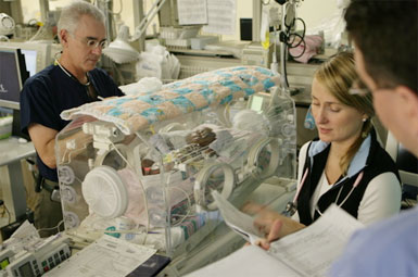
Therapeutic hypothermia for newborns

More evidence that cooling the hypoxic neonatal brain improves outcomes….
OBJECTIVE Mild hypothermia after perinatal hypoxic-ischemic encephalopathy (HIE) reduces neurologic sequelae without significant adverse effects, but studies are needed to determine the most-efficacious methods.
METHODS In the neo.nEURO.network trial, term neonates with clinical and electrophysiological evidence of HIE were assigned randomly to either a control group, with a rectal temperature of 37°C (range: 36.5–37.5°C), or a hypothermia group, cooled and maintained at a rectal temperature of 33.5°C (range: 33–34°C) with a cooling blanket for 72 hours, followed by slow rewarming. All infants received morphine (0.1 mg/kg) every 4 hours or an equivalent dose of fentanyl. Neurodevelopmental outcomes were assessed at the age of 18 to 21 months. The primary outcome was death or severe disability.
RESULTS A total of 129 newborn infants were enrolled, and 111 infants were evaluated at 18 to 21 months (53 in the hypothermia group and 58 in the normothermia group). The rates of death or severe disability were 51% in the hypothermia group and 83% in the normothermia group (P = .001; odds ratio: 0.21 [95% confidence interval [CI]: 0.09–0.54]; number needed to treat: 4 [95% CI: 3–9]). Hypothermia also had a statistically significant protective effect in the group with severe HIE (n = 77; P = .005; odds ratio: 0.17 [95% CI: 0.05–0.57]). Rates of adverse events during the intervention were similar in the 2 groups except for fewer clinical seizures in the hypothermia group.
CONCLUSION Systemic hypothermia in the neo.nEURO.network trial showed a strong neuroprotective effect and was effective in the severe HIE group.
Systemic Hypothermia After Neonatal Encephalopathy: Outcomes of neo.nEURO.network RCT
Pediatrics. 2010 Oct;126(4):e771-8
Update Dec 2014:
An RCT to determine if longer duration cooling (120 hours), deeper cooling (32.0°C), or both are superior to cooling at 33.5°C for 72 hours in neonates who are full-term with moderate or severe hypoxic ischemic encephalopathy.
Longer cooling, deeper cooling, or both compared with hypothermia at 33.5°C for 72 hours did not reduce NICU death. Small study.
Effect of depth and duration of cooling on deaths in the NICU among neonates with hypoxic ischemic encephalopathy: a randomized clinical trial
JAMA. 2014 Dec 24;312(24):2629-39

2J or 4J/kg in Paediatric Defibrillation?

Should we shock with 2J/kg or 4J/kg in Paediatric Defibrillation? The answer seems to be ‘we still don’t know’. Don’t worry – just follow the guidelines (reproduced for you at the bottom)
OBJECTIVE To examine the effectiveness of initial defibrillation attempts. We hypothesized that (1) an initial shock dose of 2 ± 10 J/kg would be less effective for terminating fibrillation than suggested in published historical data and (2) a 4 J/kg shock dose would be more effective.
PATIENTS AND METHODS This was a National Registry of Cardiopulmonary Resuscitation prospective, multisite, observational study of in-hospital pediatric (aged 18 years) ventricular fibrillation or pulseless ventricular tachycardia cardiac arrests from 2000–2008. Termination of ventricular fibrillation or pulseless ventricular tachycardia and event survival after initial shocks of 2 J/kg were compared with historic controls and a 4 J/kg shock dose.

RESULTS Of 266 children with 285 events, 173 of 285 (61%) survived the event and 61 of 266 (23%) survived to discharge. Termination of fibrillation after initial shock was achieved for 152 of 285 (53%) events. Termination of fibrillation with 2 ± 10 J/kg was much less frequent than that seen among historic control subjects (56% vs 91%; P < .001), but not different than 4 J/kg. Compared with 2 J/kg, an initial shock dose of 4 J/kg was associated with lower rates of return of spontaneous circulation (odds ratio: 0.41 [95% confidence interval: 0.21–0.81]) and event survival (odds ratio: 0.42 [95% confidence interval: 0.18–0.98]).
CONCLUSIONS The currently recommended 2 J/kg initial shock dose for in-hospital cardiac arrest was substantially less effective than previously published. A higher initial shock dose (4 J/kg) was not associated with superior termination of ventricular fibrillation or pulseless ventricular tachycardia or improved survival rates. The optimal pediatric defibrillation dose remains unknown.
Effect of defibrillation energy dose during in-hospital pediatric cardiac arrest
Pediatrics. 2011 Jan;127(1):e16-23
Here’s what the guidelines say:
Many AEDs have high specificity in recognizing pediatric shockable rhythms, and some are equipped to decrease (or attenuate) the delivered energy to make them suitable for infants and children <8 years of age. For infants a manual defibrillator is preferred when a shockable rhythm is identified by a trained healthcare provider (Class IIb, LOE C). The recommended first energy dose for defibrillation is 2 J/kg. If a second dose is required, it should be doubled to 4 J/kg. If a manual defibrillator is not available, an AED equipped with a pediatric attenuator is preferred for infants. An AED with a pediatric attenuator is also preferred for children <8 year of age. If neither is available, an AED without a dose attenuator may be used (Class IIb, LOE C). AEDs that deliver relatively high energy doses have been successfully used in infants with minimal myocardial damage and good neurological outcomes
Pediatric Basic Life Support: 2010 American Heart Association Guidelines for Cardiopulmonary Resuscitation and Emergency Cardiovascular Care
Full text document

Out of hospital monitoring in kids

I don’t have full text access to the Journal Pediatrics, so I’m not sure what I make of this small randomised trial comparing two types of blood pressure monitoring during paediatric transport:
BACKGROUND The “golden-hour” concept has led to emphasis on speed of patient delivery during pediatric interfacility transport. Timely intervention, in addition to enhanced monitoring during transport, is the key to improved outcomes in critically ill patients. Taking the ICU to the patient may be more beneficial than rapid delivery to a tertiary care center.

METHODS The Improved Monitoring During Pediatric Interfacility Transport trial was the first randomized controlled trial in the out-of-hospital pediatric transport environment. It was designed to determine the impact of improved blood pressure monitoring during pediatric interfacility transport and the effect on clinical outcomes in patients with systemic inflammatory response syndrome and moderate-to-severe head trauma. Patients in the control group had their blood pressure monitored intermittently with an oscillometric device; those in the intervention group had their blood pressure monitored every 12 to 15 cardiac contractions with a near-continuous, noninvasive device.
RESULTS Between May 2006 and June 2007, 1995, consecutive transport patients were screened, and 94 were enrolled (48 control, 46 intervention). Patients in the intervention group received more intravenous fluid (19.8 ± 22.2 vs 9.9 ± 9.9 mL/kg; P = .01), had a shorter hospital stay (6.8 ± 7.8 vs 10.9 ± 13.4 days; P = .04), and had less organ dysfunction (18 of 206 vs 32 of 202 PICU days; P = .03).
CONCLUSIONS Improved monitoring during pediatric transport has the potential to improve outcomes of critically ill children. Clinical trials, including randomized controlled trials, can be accomplished during pediatric transport. Future studies should evaluate optimal equipment, protocols, procedures, and interventions during pediatric transport, aimed at improving the clinical and functional outcomes of critically ill patients.
Enhanced Monitoring Improves Pediatric Transport Outcomes: A Randomized Controlled Trial
Pediatrics. 2011 Jan;127(1):42-8