Tag Archives: diagnosis

Offensive medicine: CT before LP

I’m getting worn down by clinicians – often other specialists – who insist that CT imaging of the brain is mandatory prior to lumbar puncture in all patients. There is surely a subgroup of patients (especially young ones) in whom the benefit:harm balance of CT comes out in favour of NOT doing the imaging. In these cases, getting the scan is not ‘defensive medicine’ but ‘offensive medicine’ – offending the principle of primum non nocere. During ED shifts I have recently had to perform online searches in order to furnish colleagues and patients’ medically qualified relatives with printouts of the literature on this. This page is here to save me having to repeat those searches. Regarding the practice of performing a routine head CT prior to lumbar puncture to rule out risk of herniation:

  • Mass effect on CT does not predict herniation
  • Lack of mass effect on CT does not rule out raised ICP or herniation
  • Herniation has occurred in patients who did not undergoing lumbar puncture because of CT findings
  • Clinical predictors of raised ICP are more reliable than CT findings
  • CT may delay diagnosis and treatment of meningitis
  • Even in patients in whom LP may be considered contraindicated (cerebral abscess, mass effect on CT), complications from LP were rare in several studies

Best practice, it would seem, is the following

  • If you think CT will show a cause for the headache, do a CT
  • If a CT is indicated for other reasons (depressed conscious level, focal neurology), do a CT
  • If a GCS 15 patient is to undergo LP for suspected (or to rule out) meningitis, and they have a normal neurological exam (including fundi), and are not elderly or immunosuppressed, there is no need to do a CT first.
  • If you’re seriously worried about meningitis and are intent on getting a CT prior to LP, don’t let the imaging delay antimicrobial therapy.

Here are some useful references:

1. The CT doesn’t help

CT head before lumbar puncture in suspected meningitis BestBET evidence summary: In cases of suspected meningitis it is very unlikely that patients without clinical risk factors (immunocompromise/ history of CNS disease/seizures) or positive neurological findings will have a contraindication to lumbar puncture on their CT scan If CT scan is deemed to be necessary, administration of antibiotics should not be delayed. BestBETS website

Computed Tomography of the Head before Lumbar Puncture in Adults with Suspected Meningitis Much cited NEJM paper from 2001 which concludes: “In adults with suspected meningitis, clinical features can be used to identify those who are unlikely to have abnormal findings on CT of the headN Engl J Med. 2001 Dec 13;345(24):1727-33 Full Text

Cranial CT before Lumbar Puncture in Suspected Meningitis Correspondence in 2002 NEJM including study of 75 patients with pneumococcal meningitis: CT cannot rule out risk of herniation Cranial CT before Lumbar Puncture in Suspected Meningitis N Engl J Med. 2002 Apr 18;346(16):1248-51 Full Text

2. The CT may harm

Cancer risk from CT Paucis verbis card, from the wonderful Academic Life in EM

3. Guidelines say CT is not always needed

National (UK) guidelines on meningitis (community acquired meningitis in the immunocompetent host) available from meningitis.org. , including this box:

Practice Guidelines for the Management of Bacterial Meningitis These 2004 guidelines from the Infectious Diseases Society of America provide the following table listing the recommended criteria for adult patients with suspected bacterial meningitis who should undergo CT prior to lumbar puncture:

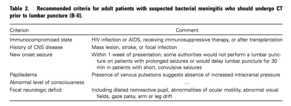
Clin Infect Dis. (2004) 39 (9): 1267-1284 Full text

4. This is potentially even more of an issue with paediatric patients

Fatal Lumbar Puncture: Fact Versus Fiction—An Approach to a Clinical Dilemma

An excellent summary of the above mentioned issues presented in a paediatric context, including the following:

On initial consideration a cranial CT would seem to be an appropriate and potentially useful diagnostic study for confirming the diagnosis of cerebral herniataion. The fallacy in this assessment has been emphasized by the finding that no clinically significant CT abnormalities are found that are not suspected on clinical assessments. Further, as previously noted, a normal CT examination may be found at about the time of a fatal herniation. Thus, the practical usefulness of a cranial CT in the majority of pediatric patients is limited to those rare patients whose increased ICP is secondary to mass lesions, not in the initial approach to acute meningitis.

Pediatrics. 2003 Sep;112(3 Pt 1):e174-6 Full Text

The last words should go to Dr Brad Spellberg, who in response to the IDSA’s guidelines wrote an excellent letter summarising much of the evidence at the time, confessed:

Why do we persist in using the CT scan for this purpose, despite the lack of supportive data? I am as guilty of this practice as anyone else, and the reason is simple: I am a chicken.

Clin Infect Dis. (2005) 40 (7): 1061 Full Text

Salicylate poisoning and pseudohyperchloraemia


Severe salicylate poisoning can cause metabolic acidosis from an accumulation of salicylic acid, lactic acid, and ketone bodies. A high anion gap acidosis is therefore the typical metabolic abnormality seen. A case series illustrates salicylate poisoning presenting with a normal gap (hyperchloraemic) acidosis – one patient had a chloride of 111 mmol/l and the other 123 mmol/l. This can occur when some analysers falsely read an elevated chloride in the presence of high concentrations of salicylate.


Severe salicylate poisoning is classically associated with an anion gap metabolic acidosis. However, high serum salicylate levels can cause false increase of laboratory chloride results on some analyzers. We present 2 cases of life-threatening salicylate poisoning with an apparently normal anion gap caused by an important laboratory interference. These cases highlight that the diagnosis of severe salicylism must be considered in all patients presenting with metabolic acidosis, even in the absence of an increased anion gap.

Falsely Normal Anion Gap in Severe Salicylate Poisoning Caused by Laboratory Interference
Ann Emerg Med. 2011 Sep;58(3):280-1

Capillary refill time

A review of capillary refill time (CRT) reveals some interesting details about this test:

  • CRT is affected by age – the upper limit of normal for neonates is 3 seconds.
  • It increases with age – one study recommended the upper limit of normal for adult women should be increased to 2.9 seconds and for the elderly to 4.5 seconds.
  • It is affected by multiple external factors (especially ambient temperature).
  • Although it is claimed to have some predictive value in the assessment of dehydration and serious infection in children, studies vary in where and for how long pressure should be applied, and there is poor interobserver reliability.

The latest (5th Edition) of the Advanced Paediatric Life Support Manual states:
Poor capillary refill and differential pulse volumes are neither sensitive nor specific indicators of shock in infants and children, but are useful clinical signs when used in conjunction with the other signs described
In my view, it is best used as a monitor of trends (in accordance with skin temperature and other markers of perfusion), rather than by placing emphasis on the exact number of seconds of a single reading. See below for a video of my perfectly happy and healthy son demonstrating a CRT of over six seconds in a cool room during an English Summer’s day.
The authors of the review caution:
Operating rooms are cold, patients are often draped, which limits access, and because most anesthetics are potent vasodilators, the use of CRT to guide practice is not justified. The possibility of a false-positive or false-negative assessment is simply too great.


Capillary refill time (CRT) is widely used by health care workers as part of the rapid, structured cardiopulmonary assessment of critically ill patients. Measurement involves the visual inspection of blood returning to distal capillaries after they have been emptied by pressure. It is hypothesized that CRT is a simple measure of alterations in peripheral perfusion. Evidence for the use of CRT in anesthesia is lacking and further research is required, but understanding may be gained from evidence in other fields. In this report, we examine this evidence and factors affecting CRT measurement. Novel approaches to the assessment of CRT are under investigation. In the future, CRT measurement may be achieved using new technologies such as digital videography or modified oxygen saturation probes; these new methods would remove the limitations associated with clinical CRT measurement and may even be able to provide an automated CRT measurement.

Capillary Refill Time: Is It Still a Useful Clinical Sign?
Anesth Analg. 2011 Jul;113(1):120-3
The Capillary Refill Video

Surgeons and trauma teams

There can be issues associated with calling surgeons to trauma team activations in the ED, including interruption to the surgeon’s other duties, and the absence of anything useful for the surgeon to do, when most blunt trauma patients are managed by emergency physicians, intensivists, and orthopaedic surgeons, with a growing input from interventional radiologists. At one American major trauma centre for example, emergency operation by a trauma surgeon for blunt trauma averages once every 7 weeks for adults and less than once every 3 years for children1.
While there are many surgeons who are passionate about trauma care and excellent in the non-operative aspects of trauma management, there are probably more who would welcome measures to reduce the need to attend ED for all trauma team activations. Of course no triage tool is perfect: they will always have to trade sensitivity against specificity. One such tool from the Loma Linda University Medical Center uses the simple criteria of penetrating trauma, systolic blood pressure, and heart rate. These pertain to pre-hospital measurement and therefore the surgeon can be activated prior to patient arrival.
This triage tool performed better than the American College of Surgeons’ “major resuscitation” trauma triage criteria2:


STUDY OBJECTIVE: Trauma centers use “secondary triage” to determine the necessity of trauma surgeon involvement. A clinical decision rule, which includes penetrating injury, an initial systolic blood pressure less than 100 mm Hg, or an initial pulse rate greater than 100 beats/min, was developed to predict which trauma patients require emergency operative intervention or emergency procedural intervention (cricothyroidotomy or thoracotomy) in the emergency department. Our goal was to validate this rule in an adult trauma population and to compare it with the American College of Surgeons’ major resuscitation criteria.

METHODS: We used Level I trauma center registry data from September 1, 1995, through November 30, 2008. Outcomes were confirmed with blinded abstractors. Sensitivity, specificity, and 95% confidence intervals (CIs) were calculated.

RESULTS: Our patient sample included 20,872 individuals. The median Injury Severity Score was 9 (interquartile range 4 to 16), 15.3% of patients had penetrating injuries, 13.5% had a systolic blood pressure less than 100 mm Hg, and 32.5% had a pulse rate greater than 100 beats/min. Emergency operative intervention or procedural intervention was required in 1,099 patients (5.3%; 95% CI 5.0% to 5.6%). The sensitivities and specificities of the rule and the major resuscitation criteria for predicting emergency operative intervention or emergency procedural intervention were 95.6% (95% CI 94.3% to 96.8%) and 56.1% (95% CI 55.4% to 56.8%) and 85.5% (95% CI 83.3% to 87.5%) and 80.9% (95% CI 80.3% to 81.4%), respectively.

CONCLUSION: This new rule was more sensitive for predicting the need for emergency operative intervention or emergency procedural intervention directly compared with the American College of Surgeons’ major resuscitation criteria, which may improve the effectiveness and efficiency of trauma triage.

Although not mentioned in the abstract, the study also included assessment of refinements of the Loma Linda Rule based on different cutoffs of heart rate and blood pressure. Once such refinement that included penetrating injury to the torso and less conservative physiological criteria (systolic blood pressure <90 mm Hg and pulse rate >110 beats/min) resulted in a slightly lower sensitivity, with a dramatic improvement in specificity compared with the original Loma Linda Rule.
A good point is made by Steve Green in his accompanying editoral3:


A possibility is that emergency physicians supervising out-of-hospital radio calls can predict the need for surgeon presence just as accurately (or perhaps more accurately) as any of these rules. After all, judgment is the time-tested mechanism by which emergency physicians summon all other consultants for all other conditions.

Unfortunately for many UK and Australasian centres, the challenge that remains is not deciding when to call the surgeon, but getting one when you do call, preferable one who is not committed to an elective operating list and one who has some training and experience in trauma surgery.
1. Clinical decision rules for secondary trauma triage: predictors of emergency operative management.
Ann Emerg Med. 2006 Feb;47(2):135
2. Validation and refinement of a rule to predict emergency intervention in adult trauma patients
Ann Emerg Med. 2011 Aug;58(2):164-71
3. Trauma is occasionally a surgical disease: how can we best predict when?
Ann Emerg Med. 2011 Aug;58(2):172-177

Ultrasound to detect difficult laryngoscopy

A pilot study suggests sonographic measurements of neck soft tissue thickness may predict difficult laryngoscopy. Laryngoscopy was difficult in patients with increased thickness of the anterior neck soft tissue at the level of the hyoid bone and thyrohyoid membrane. The authors suggest that anterior neck soft tissue thickness cutoff value of 2.8 cm at the thyrohyoid membrane level can potentially be used to detect difficult laryngoscopy, but that this would require further validation since in this pilot study there were only six subjects in the difficult laryngoscopy group.


Objectives:  Prediction of difficult laryngoscopy in emergency care settings is challenging. The preintubation clinical screening tests may not be applied in a large number of emergency intubations due to the patient’s clinical condition. The objectives of this study were 1) to determine the utility of sonographic measurements of thickness of the tongue, anterior neck soft tissue at the level of the hyoid bone, and thyrohyoid membrane in distinguishing difficult and easy laryngoscopies and 2) to examine the association between sonographic measurements (thickness of tongue and anterior neck soft tissue) and difficult airway clinical screening tests (modified Mallampati score, thyromental distance, and interincisor gap).

Methods:  This was a prospective observational study at an academic medical center. Adult patients undergoing endotracheal intubation for an elective surgical procedure were included. The investigators involved in data collection were blinded to each other’s assessments. Demographic variables were collected preoperatively. The clinical screening tests to predict a difficult airway were performed. The ultrasound (US) measurements of tongue and anterior neck soft tissue were obtained. The laryngoscopic view was graded using Cormack and Lehane classification by anesthesia providers on the day of surgery. To allow for comparisons between difficult airway and easy airway groups, a two-sided Student’s t-test and Fisher’s exact test were employed as appropriate. Spearman’s rank correlation coefficients were used to examine the association between screening tests and sonographic measurements.

Results:  The mean (±standard deviation [SD]) age of 51 eligible patients (32 female, 19 male) was 53.1 (±13.2) years. Six of the 51 patients (12%, 95% confidence interval [CI] = 3% to 20%) were classified as having difficult laryngoscopy by anesthesia providers. The distribution of laryngoscopy grades for all subjects was 63, 25, 4, and 8% for grades 1, 2, 3, and 4, respectively. In this study, 83% of subjects with difficult airways were males. No other significant differences were noted in the demographic variables and difficult airway clinical screening tests between the two groups. The sonographic measurements of anterior neck soft tissue were greater in the difficult laryngoscopy group compared to the easy laryngoscopy group at the level of the hyoid bone (1.69, 95% CI = 1.19 to 2.19 vs. 1.37, 95% CI = 1.27 to 1.46) and thyrohyoid membrane (3.47, 95% CI = 2.88 to 4.07 vs. 2.37, 95% CI = 2.29 to 2.44). No significant correlation was found between sonographic measurements and clinical screening tests.

Conclusions:  This pilot study demonstrated that sonographic measurements of anterior neck soft tissue thickness at the level of hyoid bone and thyrohyoid membrane can be used to distinguish difficult and easy laryngoscopies. Clinical screening tests did not correlate with US measurements, and US was able to detect difficult laryngoscopy, indicating the limitations of the conventional screening tests for predicting difficult laryngoscopy.

Pilot Study to Determine the Utility of Point-of-care Ultrasound in the Assessment of Difficult Laryngoscopy
Acad Emerg Med. 2011 Jul;18(7):754-8

Early CT may rule out subarachnoid haemorrhage


A multicentre Canadian study challenges the practice of routine lumbar puncture after negative CT in patients with suspected subarachnoid haemorrhage. CT scanning within six hours was highly sensitive, although a few cases of initially misinterpreted CTs “illustrate the importance of having a qualified radiologist with a high level of skill interpreting the head scans in a timely manner“.

Nearly 2% of patients were lost to all follow-up; the authors point out that even if a quarter of these patients could have experienced a subarachnoid haemorrhage, the corresponding negative likelihood ratio for a computed tomography performed within six hours rises to only 0.024 (0.007 to 0.07). They assert:

Such a likelihood ratio could be incorporated into the informed discussion surrounding the risks and benefits of lumbar puncture after negative results on computed tomography for this diagnosis

They point out that when CT imaging is obtained more than six hours after headache onset, clinicians should continue to be cautious because of the decreasing sensitivity for subarachnoid haemorrhage beyond this time.


Objective To measure the sensitivity of modern third generation computed tomography in emergency patients being evaluated for possible subarachnoid haemorrhage, especially when carried out within six hours of headache onset.

Design Prospective cohort study. Setting 11 tertiary care emergency departments across Canada, 2000-9.

Participants Neurologically intact adults with a new acute headache peaking in intensity within one hour of onset in whom a computed tomography was ordered by the treating physician to rule out subarachnoid haemorrhage.

Main outcome measures Subarachnoid haemorrhage was defined by any of subarachnoid blood on computed tomography, xanthochromia in cerebrospinal fluid, or any red blood cells in final tube of cerebrospinal fluid collected with positive results on cerebral angiography.

Results Of the 3132 patients enrolled (mean age 45.1, 2571 (82.1%) with worst headache ever), 240 had subarachnoid haemorrhage (7.7%). The sensitivity of computed tomography overall for subarachnoid
haemorrhage was 92.9% (95% confidence interval 89.0% to 95.5%), the specificity was 100% (99.9% to 100%), the negative predictive value was 99.4% (99.1% to 99.6%), and the positive predictive value was 100% (98.3% to 100%). For the 953 patients scanned within six hours of headache onset, all 121 patients with subarachnoid haemorrhage were identified by computed tomography, yielding a sensitivity of 100% (97.0% to 100.0%), specificity of 100% (99.5% to 100%), negative predictive value of 100% (99.5% to 100%), and positive predictive value of 100% (96.9% to 100%).

Conclusion Modern third generation computed tomography is extremely sensitive in identifying subarachnoid haemorrhage when it is carried out within six hours of headache onset and interpreted by a qualified radiologist

Sensitivity of computed tomography performed within six hours of onset of headache for diagnosis of subarachnoid haemorrhage: prospective cohort study
BMJ. 2011 Jul 18;343:d4277

What do I do with a high sensitivity troponin?

Newer high-sensitivity troponin tests can be positive in patients who would have negative tests with the ‘traditional’ assay, which can result in confusion about what to do with the patient, particularly those patients without an obvious cardiac presentation. A recent study1 shows that the majority of patients that fall into this group had non-cardiac discharge diagnoses.


Background: High sensitivity troponin T (hsTnT) detects lower levels of troponin T with greater precision than the 4th generation (cTnT) assay. However, the clinical implications of this are uncertain.

Objectives: Primary: Describe the proportion of patients who test ‘positive’ with hsTnT but negative with cTnT. Secondary: Determine proportion in each group with an adverse event (representation, AMI or died) within 90 days of the index test.

Method: 161 patients samples were tested with cTnT and hsTNT assays. McNemar’s test was used to compare paired samples. Electronic medical records were reviewed, with discharge diagnosis and 90 day outcomes determined blind to hsTnT results. Patients were then classified as ‘TnT negative’ (hsTnT was <0.014 mcg/mL), 'new positive' (hsTnT was ≥0.014 mcg/mL and cTnT <0.03 mcg/mL) and 'TnT positive' (cTNT was ≥0.03 mcg/mL)
Results: Positive results more than doubled with the hsTnT assay (50% vs 22%, P < 0.001). 81 patients were ‘TnT negative’, 44 were ‘new positive’ and 36 ‘cTnT positive’. The discharge diagnosis for ‘new positives’ was AMI in 4 (9%), other cardiac in 13 (30%) and non-cardiac in 27 (61%). At 90 days adverse events occurred in 30%, 54% and 50% of the groups respectively. There were no late cases of AMI or cardiovascular death in ‘new positive’ patients.

Conclusion: Many patients with diagnoses other than AMI will have hsTNT above the reference level. Indiscriminate testing with hsTnT might lead to more patients requiring serial troponin testing and/or invasive further tests, which will have process and resource implications for EDs and health services.

An accompanying editorial2 highlights that:

Elevations are seen in pathological conditions, including structural heart disease, renal impairment and pulmonary embolism, but might also be seen in extreme exertion, such as marathon runners. It is now clear that when using a highly sensitive assay, circulating levels of troponin will be detected in many normal people.

The editorial makes the interesting observation that the duration of rise may help elucidate the cause; ischaemic elevation of troponin falls rapidly, since the rise might be due to the release of small amounts of troponin that exist free within the cytoplasm, in contrast to the more persistent elevation seen with myocardial necrosis. The editorialist provides the following caution:


Overall, our practice for ordering troponin will need to be urgently reviewed. Single troponin values will continue to be of little to no use in defining disease states in the ED. Identifying a chronic versus an acute elevation will only be revealed by serial troponin testing. The time interval between testing is currently contentious.

High sensitivity troponins are referred to in the newly published 2011 Addendum to the National Heart Foundation of Australia/Cardiac Society of Australia and New Zealand Guidelines for the Management of Acute Coronary Syndromes (full text link below)3:

RECOMMENDED PROTOCOL FOR TROPONIN TESTING USING HIGH SENSITIVITY ASSAYS IN “RULING-OUT” ACS

  • All patients with a suspected ACS should undergo troponin testing on arrival at ED to ‘rule in’ ACS within the differential diagnosis
  • For a patient with a positive troponin result or a change in troponin levels over time, neither ACS nor other significant pathology (e.g. pulmonary embolus, aortic dissection, and sepsis) can be excluded. These patients are at higher risk of subsequent events. A positive result should be considered within the entire clinical context (history, examination, ECG findings and other investigations). Further investigations directed at all plausible clinical diagnoses should be considered and, if ACS is thought to be the likely cause, these patients may require cardiology assessment.
  • All patients with a negative result should undergo repeat testing 3–4 hours later.
  • The testing interval to ‘rule out’ MI may be reduced to 3 hours, provided that one sample is taken at least 6 hours after symptom onset:
  • Patients with a negative result at 3 hours after presentation and at least 6 hours after the onset of pain should be considered for early assessment by non-invasive anatomic or functional testing, as determined by local availability.
  • For patients presenting more than 6 hours after pain onset, a single high sensitivity troponin assay is sufficient to rule out myocardial infarction in the absence of ongoing chest pain.

High sensitivity troponin assays have an increased sensitivity for the detection of “myonecrosis”, but a reduced specificity for the diagnosis of “MI”. A positive result (≥99th centile for reference population OR where there is a change of ≥50% above an initial baseline level) should be interpreted in the context of the entire clinical presentation and does not necessarily represent an indication for coronary angiography. The management MI secondary to other conditions (e.g. anaemia, thyrotoxicosis, and sepsis) should be primarily directed at those conditions.
The finding of troponin concentrations that remain stable over time suggests that the presence of troponin is due to chronic disease. Acute exacerbations of chronic disease that result in elevated troponin levels can mimic an MI release pattern.

1. Clinical diagnosis and outcomes for Troponin T ‘positive’ patients assessed by a high sensitivity compared with a 4th generation assay
Emerg Med Australas. 2011 Aug;23(4):490-501
2. Troponin: A risk-defining biomarker for emergency department physicians
Emerg Med Australas. 2011 Aug;23(4):391-4
3. 2011 Addendum to the National Heart Foundation of Australia/Cardiac Society of Australia and New Zealand Guidelines for the Management of Acute Coronary Syndromes
Heart, Lung and Circulation 2011 Aug;28(8):487-502 Free Full Text

Open book fractures and ultrasound

For me, this is one of those ‘why didn’t I think of that?!’ studies… extending the FAST scan to measure pubic symphyseal widening to detect open-book pelvic fractures. A pubic symphysis width of 25 mm was considered positive; the authors state that this width is considered diagnostic for anterior-posterior compression fracture of the pelvis in the non-pregnant patient.
Since only four of the 23 patients studied had radiological widening, the authors’ conclusions make sense: Further study with a larger cohort is needed to confirm this technique’s validity for diagnosing PS widening in APC pelvic fractures.
A reasonable question might be: ‘so what?’, especially if pelvic binders are routinely applied to polytrauma patients and radiographs are rapidly obtained. However as a retrieval medicine doctor working in remote and austere environments I wonder whether this could be useful to us. Perhaps if combined with this intervention?

BACKGROUND: The focused abdominal sonography in trauma (FAST) examination is a routine component of the initial work-up of trauma patients. However, it does not identify patients with retroperitoneal hemorrhage associated with significant pelvic trauma. A wide pubic symphysis (PS) is indicative of an open book pelvic fracture and a high risk of retroperitoneal bleeding.

STUDY OBJECTIVES: We hypothesized that an ultrasound image of the PS as part of the FAST examination (FAST-PS) would be an accurate method to determine if pubic symphysis diastasis was present.

METHODS: This is a comparative study of a diagnostic test on a convenience sample of 23 trauma patients at a Level 1 Trauma Center. The PS was measured sonographically in the Emergency Department (ED) and post-mortem (PM) at the State Medical Examiner. The ultrasound (US) measurements were then compared with PS width on anterior-posterior pelvis radiograph.

RESULTS: Twenty-three trauma patients were evaluated with both plain radiographs and US (11 PM, 12 ED). Four patients had radiographic PS widening (3 PM, 1 ED) and 19 patients had radiographically normal PS width; all were correctly identified with US. US measurements were compared with plain X-ray study by Bland-Altman plot. With one exception, US measurements were within 2 standard deviations of the radiographic measurements and, therefore, have excellent agreement. The only exception was a patient with pubic symphysis wider than the US probe.

CONCLUSION: Bedside ultrasound examination may be able to identify pubic symphysis widening in trauma patients. This potentially could lead to faster application of a pelvic binder and tamponade of bleeding.

Ultrasonographic determination of pubic symphyseal widening in trauma: the FAST-PS study
J Emerg Med. 2011 May;40(5):528-33

E-FAST for pneumothorax

Some further evidence of the superiority of ultrasound over chest x-ray for the detection of pneumothorax (although it’s not perfect):

INTRODUCTION: Early identification of pneumothorax is crucial to reduce the mortality in critically injured patients. The objective of our study is to investigate the utility of surgeon performed extended focused assessment with sonography for trauma (EFAST) in the diagnosis of pneumothorax.
METHODS: We prospectively analysed 204 trauma patients in our level I trauma center over a period of 12 (06/2007-05/2008) months in whom EFAST was performed. The patients’ demographics, type of injury, clinical examination findings (decreased air entry), CXR, EFAST and CT scan findings were entered into the data base. Sensitivity, specificity, positive (PPV) and negative predictive values (NPV) were calculated.
RESULTS: Of 204 patients (mean age–43.01+/-19.5 years, sex–male 152, female 52) 21 (10.3%) patients had pneumothorax. Of 21 patients who had pneumothorax 12 were due to blunt trauma and 9 were due to penetrating trauma. The diagnosis of pneumothorax in 204 patients demonstrated the following: clinical examination was positive in 17 patients (true positive in 13/21, 62%; 4 were false positive and 8 were false negative), CXR was positive in 16 (true positive in 15/19, 79%; 1 false positive, 4 missed and 2 CXR not performed before chest tube) patients and EFAST was positive in 21 patients (20 were true positive [95.2%], 1 false positive and 1 false negative). In diagnosing pneumothorax EFAST has significantly higher sensitivity compared to the CXR (P=0.02).
CONCLUSIONS: Surgeon performed trauma room extended FAST is simple and has higher sensitivity compared to the chest X-ray and clinical examination in detecting pneumothorax.

Extended focused assessment with sonography for trauma (EFAST) in the diagnosis of pneumothorax: experience at a community based level I trauma center
Injury. 2011 May;42(5):511-4

CRP helpful in risk stratifying febrile kids

In febrile children, peripheral white blood cell counts were not helpful in separating children with self limiting infections from those with serious bacterial infections, but serum C reactive protein was1: febrile children with serum C reactive protein concentrations of 20 mg/L or less have a 5% risk of serious infection, whereas those with serum concentrations greater than 80 mg/L have a risk of 72%; children with intermediate values have a risk of about 15%. According to the accompanying BMJ editorial2:
This grouping, although imperfect, provides some guidance to help clinicians deciding which children may avoid extensive evaluation and treatment.

OBJECTIVE: To collate all available evidence on the diagnostic value of laboratory tests for the diagnosis of serious infections in febrile children in ambulatory settings.

DESIGN: Systematic review.

DATA SOURCES: Electronic databases, reference tracking, and consultation with experts.

STUDY SELECTION: Studies were selected on six criteria: design (studies of diagnostic accuracy or deriving prediction rules), participants (otherwise healthy children and adolescents aged 1 month to 18 years), setting (first contact ambulatory care), outcome (serious infection), features assessed (in first contact care), and data reported (sufficient to construct a 2×2 table).

DATA EXTRACTION: Quality assessment was based on the quality assessment tool of diagnostic accuracy studies (QUADAS) criteria. Meta-analyses were done using the bivariate random effects method and hierarchical summary receiver operating characteristic curves for studies with multiple thresholds.

DATA SYNTHESIS: None of the 14 studies identified were of high methodological quality and all were carried out in an emergency department or paediatric assessment unit. The prevalence of serious infections ranged from 4.5% to 29.3%. Tests were carried out for C reactive protein (five studies), procalcitonin (three), erythrocyte sedimentation rate (one), interleukins (two), white blood cell count (seven), absolute neutrophil count (two), band count (three), and left shift (one). The tests providing most diagnostic value were C reactive protein and procalcitonin. Bivariate random effects meta-analysis (five studies, 1379 children) for C reactive protein yielded a pooled positive likelihood ratio of 3.15 (95% confidence interval 2.67 to 3.71) and a pooled negative likelihood ratio of 0.33 (0.22 to 0.49). To rule in serious infection, cut-off levels of 2 ng/mL for procalcitonin (two studies, positive likelihood ratio 13.7, 7.4 to 25.3 and 3.6, 1.4 to 8.9) and 80 mg/L for C reactive protein (one study, positive likelihood ratio 8.4, 5.1 to 14.1) are recommended; lower cut-off values of 0.5 ng/mL for procalcitonin or 20 mg/L for C reactive protein are necessary to rule out serious infection. White blood cell indicators are less valuable than inflammatory markers for ruling in serious infection (positive likelihood ratio 0.87-2.43), and have no value for ruling out serious infection (negative likelihood ratio 0.61-1.14). The best performing clinical decision rule (recently validated in an independent dataset) combines testing for C reactive protein, procalcitonin, and urinalysis and has a positive likelihood ratio of 4.92 (3.26 to 7.43) and a negative likelihood ratio of 0.07 (0.02 to 0.27).

CONCLUSION: Measuring inflammatory markers in an emergency department setting can be diagnostically useful, but clinicians should apply different cut-off values depending on whether they are trying to rule in or rule out serious infection. Measuring white blood cell count is less useful for ruling in serious infection and not useful for ruling out serious infection. More rigorous studies are needed, including studies in primary care, to assess the value of laboratory tests alongside clinical diagnostic measurements, including vital signs.

1. Diagnostic value of laboratory tests in identifying serious infections in febrile children: systematic review
BMJ. 2011 Jun 8;342:d3082
2. How useful are laboratory tests in diagnosing serious infections in febrile children?
BMJ. 2011 Jun 8;342:d2782