
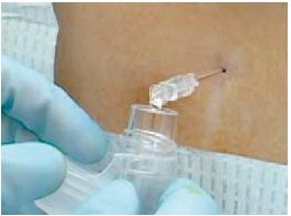
More data on the RhinoChill device from an in-hospital study of post-cardiac arrest patients in Germany. The RhinoChill device (BeneChill Inc., San Diego, USA) allows evaporative cooling by spraying an inert liquid coolant (a perfluorochemical) into the nasal cavity. The liquid evaporates instantaneously, thereby removing heat. It can make your nose discoloured, and in one patient with cardiogenic shock, tissue damage of nose and cheeks due to freezing occurred. Several of the authors are linked with the company that manufactures the device.

AIM: Mild therapeutic hypothermia improves survival and neurologic recovery in primary comatose survivors of cardiac arrest. Cooling effectivity, safety and feasibility of nasopharyngeal cooling with the RhinoChill device (BeneChill Inc., San Diego, USA) were determined for induction of therapeutic hypothermia.
METHODS: Eleven emergency departments and intensive care units participated in this multi-centre, single-arm descriptive study. Eighty-four patients after successful resuscitation from cardiac arrest were cooled with nasopharyngeal delivery of an evaporative coolant for 1h. Subsequently, temperature was controlled with systemic cooling at 33 degrees C. Cooling rates, adverse events and neurologic outcome at hospital discharge using cerebral performance categories (CPC; CPC 1=normal to CPC 5=dead) were documented. Temperatures are presented as median and the range from the first to the third quartile.
RESULTS: Nasopharyngeal cooling for 1h reduced tympanic temperature by median 2.3 (1.6; 3.0) degrees C, core temperature by 1.1 (0.7; 1.5) degrees C. Nasal discoloration occurred during the procedure in 10 (12%) patients, resolved in 9, and was persistent in 1 (1%). Epistaxis was observed in 2 (2%) patients. Periorbital gas emphysema occurred in 1 (1%) patient and resolved spontaneously. Thirty-four of 84 patients (40%) patients survived, 26/34 with favorable neurological outcome (CPC of 1-2) at discharge.
CONCLUSIONS: Nasopharyngeal evaporative cooling used for 1h in primary cardiac arrest survivors is feasible and safe at flow rates of 40-50L/min in a hospital setting.
Safety and feasibility of nasopharyngeal evaporative cooling in the emergency department setting in survivors of cardiac arrest
Resuscitation. 2010 Aug;81(8):943-9
Tag Archives: procedures
EZ-IO in pre-hospital care
French pre-hospital physicians liked the EZ-IO intraosseous drill, using it for drugs (including rapid sequence intubation drugs) and fluids in the pre-hospital setting. There was a very high insertion success rate.
OBJECTIVE: Intraosseous access is a rapid and safe alternative when peripheral vascular access is difficult. Our aim was to assess the safety and efficacy of a semi-automatic intraosseous infusion device (EZ-IO) when using a management algorithm for difficult vascular access in an out-of-hospital setting.
METHODS: This was a one-year prospective, observational study by mobile intensive care units. After staff training in the use of the EZ-IO device and provision of a management algorithm for difficult vascular access, all vehicles were equipped with the device. We determined device success rate and ease of use, resuscitation fluid volume and drugs administered by the intraosseous route, and complications at insertion site.

RESULTS: A total of 4666 patients required vascular access. The EZ-IO device was used in 30 cardiac arrest patients (25 adults; 5 children) and 9 adults with spontaneous cardiac activity. The success rate for first insertion was 84%. Overall success rate (max. 2 attempts) was 97%. The device was used for fluid resuscitation in 16 patients (mean volume: 680ml), adrenaline administration in 24 patients, and rapid sequence induction in 2 patients. There was only one local complication (transient local inflammation).
CONCLUSIONS: On implementation of an algorithm for the management of difficult vascular access, the EZ-IO device proved safe and highly effective in both adult and paediatric patients in an out-of-hospital emergency setting. It is a suitable device for consideration as a first-line option for difficult vascular access in this setting.
Efficacy and safety of the EZ-IOTM intraosseous device: Out-of-hospital implementation of a management algorithm for difficult vascular access
Resuscitation. 2011 Jan;82(1):126-9
Balloon catheters for haemorrhage control
Something I keep up my sleeve (not literally) for managing some life-threatening vascular wounds prior to surgery is the use of a balloon catheter like a foley to tamponade haemorrhage. This paper looks at series of such attempts although they state: “Except for the base of the skull (naso/oropharynx), all catheters were de- ployed in the operating room.“, so not exactly emergency medicine / pre-hospital practice, but a useful reminder that this is an option when going immediately to the operating room isn’t:

BACKGROUND: : Balloon catheter tamponade is a valuable technique for arresting exsanguinating hemorrhage. Indications include (1) inaccessible major vascular injuries, (2) large cardiac injuries, and (3) deep solid organ parenchymal bleeding. Published literature is limited to small case series. The primary goal was to review a recent experience with balloon catheter use for emergency tamponade in a civilian trauma population.
METHODS: : All patients requiring emergency use of a balloon catheter to tamponade exsanguinating hemorrhage (1998-2009) were included. Patient demographics, injury characteristics, technique, and outcomes were analyzed.
 RESULTS: : Of the 44 severely injured patients (82% presented with hemodynamic instability; mean base deficit = -20.4) who required balloon catheter tamponade, 23 of the balloons (52%) remained indwelling for more than 6 hours. Overall mortality depended on the site of injury/catheter placement and indwelling time (81% if <6 hours; 52% if ≥6 hours; p < 0.05). Physiologic exhaustion was responsible for 76% of deaths in patients with short-term balloons. Mortality among patients with prolonged balloon catheter placement was 11%, 50%, and 88% for liver, abdominal vascular, and facial/pharyngeal injuries, respectively. Mean indwelling times for iliac, liver, and carotid injuries were 31 hours, 53 hours, and 78 hours, respectively. Overall survival rates were 67% (liver), 67% (extremity vascular), 50% (abdominal vascular), 38% (cardiac), and 8% (face). Techniques included Foley, Fogarty, Blakemore, and/or Penrose drains with concurrent red rubber Robinson catheters. Initial tamponade of bleeding structures was successful in 93% of patients.
CONCLUSIONS: : Balloon catheter tamponade can be used in multiple anatomic regions and for variable patterns of injury to arrest ongoing hemorrhage. Placement for central hepatic gunshot wounds is particularly useful. This technique remains a valuable tool in a surgeon’s armamentarium.
A Decade’s Experience With Balloon Catheter Tamponade for the Emergency Control of Hemorrhage
J Trauma. 2011 Feb;70(2):330-3
Difficult tube – Easytube
French pre-hospital physicians included the Easytube, which is similar to the Combitube, in their difficult airway algorithm. They describe the insertion method as:
“ ..inserted blindly, the patient’s head must be in neutral position. Manually opening the patient’s mouth and pressing the tongue gently toward the mandible, the tube is inserted parallel to the frontal axis of the patient until the proximal black ring mark is positioned at the level of the incisors. If the EzT is inserted blindly, the tip is likely to be positioned in the esophagus with a probability of more than 95% [3]. Ventilation of the patient should be performed using a colored lumen, and the transparent lumen can then be used to insert a gastric tube or to drain gastric contents.”
The authors suggest that the main advantages of the Ezt are: shorter insertion time for Ezt than for ETI, better protection against aspiration than a laryngeal mask and the possibility of blind insertion of the Ezt in patients trapped in a sitting position.
BACKGROUND: Securing the airway in emergency is among the key requirements of appropriate prehospital therapy. The Easytube (Ezt) is a relatively new device, which combines the advantages of both an infraglottic and supraglottic airway.
AIMS: Our goal was to evaluate the effectiveness and the safety of use of Ezt by emergency physicians in case of difficult airway management in a prehospital setting with minimal training.

METHODS: We performed a prospective multi-centre observational study of patients requiring airway management conducted in prehospital emergency medicine in France by 3 French mobile intensive care units from October 2007 to October 2008.
RESULTS: Data were available for 239 patients who needed airway management. Two groups were individualized: the “easy airway management” group (225 patients; 94%) and the “difficult airway management” group (14 patients; 6%). All patients had a successful airway management. The Ezt was used in eight men and six women; mean age was 64 years. It was used for ventilation for a maximum of 150 min and the mean time was 65 min. It was positioned successfully at first attempt, except for two patients, one needed an adjustment because of an air leak, and in the other patient the Ezt was replaced due to complete obstruction of the Ezt during bronchial suction.
CONCLUSION: The present study shows that emergency physicians in cases of difficult airway management can use the EzT safely and effectively with minimal training. Because of its very high success rate in ventilation, the possibility of blind intubation, the low failure rate after a short training period. It could be introduced in new guidelines to manage difficult airway in prehospital emergency.
The Easytube for airway management in prehospital emergency medicine
Resuscitation. 2010 Nov;81(11):1516-20
The Sichuan Straddle
I used to see it done on ‘ER’ but never knew people really straddled patients on stretchers doing CPR. Apparently they do in Sichuan, China and have now produced a manikin study to demonstrate its effectiveness. It might work there, but I imagine there are frequent situations in Australia (where I work) in which the combined weight of patient and paramedic would present an unfair load to the stretcher.

OBJECTIVE: To evaluate the efficacy of straddling external chest compression performed on moving stretchers.
METHODS: The study was a prospective, randomized, cross-over study on a manikin performed at a university hospital. Twenty subjects were selected from the 40 graduates using random numbers to participate in the study. Participants were randomized to either performing standard or straddling external chest compression followed by the other technique 7 days later. The compression variables and time to first compression were recorded.
RESULTS: Twenty subjects (12 males and 8 females) took part in the study. There were no differences between the standard and straddling external chest compression for the compression rate, effective compression percentage and compression depth. There was no difference between the standard external chest compression and straddling external chest compression for incorrect hand position and incomplete release compression. Time to first compression during straddling external chest compression (10.31 ± 1.65 s) was greater than that during standard external chest compression (2.74 ± 0.40 s) (P < 0.001).
CONCLUSIONS: The quality of straddling external chest compression performed on a moving stretcher was as effective as standard external chest compression performed on the floor. By performing straddling external chest compression, time for transporting victims to the emergency department to get advanced life support may be shortened.
The efficacy of straddling external chest compression on a moving stretcher
Resuscitation. 2010 Nov;81(11):1562
Bum crack fluid pump
Military guys are great at coming up with practical solutions. Need to infuse fluid in the field but have no pressure bag or drip stand? Putting the bag under the patient’s body can squeeze fluid in, but the best place under the patient wasn’t known. A volunteer military study infusing saline through a 14G cannula compared six under-body locations: heels, buttock cleft, sacrum, interscapular region, cervical spine and occiput.
The buttock cleft was best.

Using body weight as a pre-hospital fluid infusion device: the relationship between under-body position and flow rate.
J R Army Med Corps. 2008 Mar;154(1):31-3
Full text article
US determined best LP position
Here ultrasound was used to ascertain the best position for doing a lumbar puncture in kids, where the interspinous space was maximised:
BACKGROUND Lumbar punctures are commonly performed in the pediatric emergency department. There is no standard, recommended, optimal position for children who are undergoing the procedure.
OBJECTIVE To determine a position for lumbar punctures where the interspinous space is maximized, as measured by bedside ultrasound.
METHODS A prospective convenience sample of children under age 12 was performed. Using a portable ultrasound device, the L3-L4 or L4-L5 interspinous space was measured with the subject in 5 different positions. The primary outcome was the interspinous distance between 2 adjacent vertebrae. The interspinous space was measured with the subject sitting with and without hip flexion. In the lateral recumbent position, the interspinous space wasmeasured with the hips in a neutral position as well as in flexion, both with and without neck flexion. Data were analyzed by comparing pairwise differences.
RESULTS There were 28 subjects enrolled (13 girls and 15 boys) at a median age of 5 years. The sitting-flexed position provided a significantly increased interspinous space (P < .05). Flexion of the hips increased the interspinous space in both the sitting and lateral recumbent positions (P < .05). Flexion of the neck, did not significantly change the interspinous space (P = .998).
CONCLUSIONS The interspinous space of the lumbar spine was maximally increased with children in the sitting position with flexed hips; therefore we recommend this position for lumbar punctures. In the lateral recumbent position, neck flexion does not increase the interspinous space and may increase morbidity; therefore, it is recommended to hold patients at the level of the shoulders as to avoid neckflexion.
Positioning for lumbar puncture in children evaluated by bedside ultrasound
Pediatrics. 2010 May;125(5):e1149-53
A more recent study also used ultrasound in infants to investigate the anatomic necessity and advantage derived from a tight flexed lateral recumbent position, since hypoxia has been observed in that position:
Objectives: Hypoxia has been observed when infants undergo lumbar puncture in a tight flexed lateral recumbent position. This study used sonographic measurements of lumbar interspinous spaces to investigate the anatomic necessity and advantage derived from this tight flexed positioning in infants.
Methods: This was a brief, prospective, observational study of a convenience sample of patients. Twenty-one healthy infants under 1 month of age were scanned in two positions: prone in a spine-neutral position and lateral recumbent with their knees bent into their chest and their neck flexed. In each position, a 5- to 10-MHz linear array transducer was used to scan midline along the lumbar spinous processes in the sagittal plane. The distances between the spinous processes were measured near the ligamentum flavum using the ultrasound machine’s calipers. Pulse oximetry was monitored on all infants during flexed positioning.
Results: In the spine-neutral position, all studied interspinous spaces were much wider than a 22-gauge spinal needle (diameter 0.072 cm). The mean (±SD) interspinous spaces for L3-4, L4-5, and L5-S1 in a spine-neutral position were 0.42 (±0.07), 0.37 (±0.06), and 0.36 (±0.11) cm, respectively. Flexing the infants increased the mean lumbar interspinous spaces at L3-4, L4-5, and L5-S1 by 31, 51, and 44%, respectively.
Conclusions: This study verified that tight, lateral flexed positioning substantially enhances the space between the lumbar spinous processes and that a spine-neutral position also allows for a large enough anatomic interspinous space to perform lumbar puncture. However, further clinical research is required to establish the feasibility of lumbar puncture in a spine-neutral position.
Evaluating infant positioning for lumbar puncture using sonographic measurements
Acad Emerg Med. 2011 Feb;18(2):215-8
2J or 4J/kg in Paediatric Defibrillation?
Should we shock with 2J/kg or 4J/kg in Paediatric Defibrillation? The answer seems to be ‘we still don’t know’. Don’t worry – just follow the guidelines (reproduced for you at the bottom)
OBJECTIVE To examine the effectiveness of initial defibrillation attempts. We hypothesized that (1) an initial shock dose of 2 ± 10 J/kg would be less effective for terminating fibrillation than suggested in published historical data and (2) a 4 J/kg shock dose would be more effective.
PATIENTS AND METHODS This was a National Registry of Cardiopulmonary Resuscitation prospective, multisite, observational study of in-hospital pediatric (aged 18 years) ventricular fibrillation or pulseless ventricular tachycardia cardiac arrests from 2000–2008. Termination of ventricular fibrillation or pulseless ventricular tachycardia and event survival after initial shocks of 2 J/kg were compared with historic controls and a 4 J/kg shock dose.

RESULTS Of 266 children with 285 events, 173 of 285 (61%) survived the event and 61 of 266 (23%) survived to discharge. Termination of fibrillation after initial shock was achieved for 152 of 285 (53%) events. Termination of fibrillation with 2 ± 10 J/kg was much less frequent than that seen among historic control subjects (56% vs 91%; P < .001), but not different than 4 J/kg. Compared with 2 J/kg, an initial shock dose of 4 J/kg was associated with lower rates of return of spontaneous circulation (odds ratio: 0.41 [95% confidence interval: 0.21–0.81]) and event survival (odds ratio: 0.42 [95% confidence interval: 0.18–0.98]).
CONCLUSIONS The currently recommended 2 J/kg initial shock dose for in-hospital cardiac arrest was substantially less effective than previously published. A higher initial shock dose (4 J/kg) was not associated with superior termination of ventricular fibrillation or pulseless ventricular tachycardia or improved survival rates. The optimal pediatric defibrillation dose remains unknown.
Effect of defibrillation energy dose during in-hospital pediatric cardiac arrest
Pediatrics. 2011 Jan;127(1):e16-23
Here’s what the guidelines say:
Many AEDs have high specificity in recognizing pediatric shockable rhythms, and some are equipped to decrease (or attenuate) the delivered energy to make them suitable for infants and children <8 years of age. For infants a manual defibrillator is preferred when a shockable rhythm is identified by a trained healthcare provider (Class IIb, LOE C). The recommended first energy dose for defibrillation is 2 J/kg. If a second dose is required, it should be doubled to 4 J/kg. If a manual defibrillator is not available, an AED equipped with a pediatric attenuator is preferred for infants. An AED with a pediatric attenuator is also preferred for children <8 year of age. If neither is available, an AED without a dose attenuator may be used (Class IIb, LOE C). AEDs that deliver relatively high energy doses have been successfully used in infants with minimal myocardial damage and good neurological outcomes
Pediatric Basic Life Support: 2010 American Heart Association Guidelines for Cardiopulmonary Resuscitation and Emergency Cardiovascular Care
Full text document
Neck movement in spite of collar
A cadaveric study using an artificially created unstable cervical spine injury has shown marked displacement of the vertebrae when cervical collars were applied, and when the bodies were moved in a way that simulated normal transfer and log-rolling. There was no comparison with a no-collar situation, so we can’t say from this that collars are necessarily bad, just that they’re no good in this cadaveric model. I like this statement by the authors:
“A variety of collars, backboards, and other equipment and techniques are being used in an attempt to achieve spine stabilization, largely without any validation of efficacy when used in the presence of a severe cervical injury. Randomized, prospective clinical trial designs are challenging in this domain theless, basic cadaver studies can provide valuable insight into potential clinical efficacy.”

Even more musical to my ears is the editorial commentary by neurosurgery professor Richard L. Saunders, MD:
“…the more compelling question is whether there is a place for collars in emergent protection of the injured cervical spine or are they simply a gimcrack***?
The incidence of second injuries to the spinal cord in the extraction of accident victims under the best of EMT performance is not known and would be difficult to determine. However, in an effort to minimize that incidence, paramedical gospel is the application of a cervical collar, maintaining the neck in in-line and in a neutral position. By definition, this gospel implies the deliberate movement of the neck to apply an orthotic known to be nonprotective. Furthermore, the neutral and in-line admonition implies that the patient’s neck position can be safely adjusted to “look better” without a shred of evidence that this might be a safer strategy than avoiding any unnecessary neck movement whatsoever….
…In a conclusion common to many small study reports, the authors recommend that more work should be done in this area. In my opinion that might be best in refinements of extraction methods with an eye to only that neck movement necessary to resuscitation, collar be damned.”
Motion Within the Unstable Cervical Spine During Patient Maneuvering: The Neck Pivot-Shift Phenomenon
J Trauma. 2011 Jan;70(1):247-50
*** I confess never to have encountered this word before. According to the freedictionary.com, a gimcrack is ‘A cheap and showy object of little or no use; a gewgaw‘. Now, WTF is a gewgaw?!?!
CPR on your own? Stay at the head end
In this manikin study, single-rescuer bag-mask ventilation (BMV) with chest compressions was tried in three different positions. Staying at the head end to deliver effective BMV, with ‘over-the-head’ chest compressions from that position, was best.

Background The 2005 guidelines for cardiopulmonary resuscitation (CPR) do not include a statement on performance of basic life support by a single healthcare professional using a bagevalveemask device. Three positions are possible: chest compressions and ventilations from over the head of the casualty (over-the-head CPR), from the side of the casualty (lateral CPR), and chest compressions from the side and ventilations from over the head of the casualty (alternating CPR). The aim of this study was to compare CPR quality of these three positions.
Methods 102 healthcare professionals were randomised to a crossover design and performed a 2-min CPR test on a manikin for each position.
Results The hands-off time over a 2-min interval was not significantly different between over-the-head (median 31 s) and lateral (31 s) CPR, but these compared favourably with alternating CPR (36 s). Over-the-head CPR resulted in significantly more chest compressions (155) compared with lateral (152) and alternating CPR (149); the number of correct chest compressions did not differ significantly (119 vs 122 vs 109). Alternating CPR resulted in significantly less inflations (eight) compared with over-the-head (ten) and lateral CPR (ten). Lateral CPR led to significantly less correct inflations (three) compared with over-the-head (five) and alternating CPR (four).
Conclusions In the case of a single healthcare professional using a bagevalveemask device, the quality of over-the-head CPR is at least equivalent to lateral, and superior to alternating CPR. Because of the potential difficulties in bagevalveemask ventilation in the lateral position, the authors recommend over-the-head CPR.
Comparison of the over-the-head, lateral and alternating positions during cardiopulmonary resuscitation performed by a single rescuer with a bag valve mask device
Emerg Med J. 2010 Oct 14. [Epub ahead of print]