Category Archives: ICU

Stuff relevant to patients on ICU

Pharmaconutrition for Acute Lung Injury

A trial by the ARDS Clinical Trials Network of pharmaconutrition for acute lung injury1 was stopped early for futility – outcomes were worse in the intervention group that received the enteral supplementation of n-3 fatty acids, γ-linolenic acid, and antioxidants. It had been hypothesised that the immunomodulatory effects of these supplements would provide clinical benefit in acute lung injury.
An accompanying editorial2 reports benefits of pharmaconutrition in other areas of critical care:

  • arginine-supplemented diets are associated with reduced infections and lengths of hospital stay in patients undergoing elective operations
  • glutamine-supplemented parenteral nutrition is associated with reduced infection and mortality in critically ill patients
  • antioxidant supplementation is associated with reduced mortality among critically ill patients with systemic inflammation.


Context The omega-3 (n-3) fatty acids docosahexaenoic acid and eicosapentaenoic acid, along with γ-linolenic acid and antioxidants, may modulate systemic inflammatory response and improve oxygenation and outcomes in patients with acute lung injury.

Objective To determine if dietary supplementation of these substances to patients with acute lung injury would increase ventilator-free days to study day 28.

Design, Setting, and Participants The OMEGA study, a randomized, double-blind, placebo-controlled, multicenter trial conducted from January 2, 2008, through February 21, 2009. Participants were 272 adults within 48 hours of developing acute lung injury requiring mechanical ventilation whose physicians intended to start enteral nutrition at 44 hospitals in the National Heart, Lung, and Blood Institute ARDS Clinical Trials Network. All participants had complete follow-up.

Interventions Twice-daily enteral supplementation of n-3 fatty acids, γ-linolenic acid, and antioxidants compared with an isocaloric control. Enteral nutrition, directed by a protocol, was delivered separately from the study supplement.

Main Outcome Measure Ventilator-free days to study day 28.

Results The study was stopped early for futility after 143 and 129 patients were enrolled in the n-3 and control groups. Despite an 8-fold increase in plasma eicosapentaenoic acid levels, patients receiving the n-3 supplement had fewer ventilator-free days (14.0 vs 17.2; P = .02) (difference, −3.2 [95% CI, −5.8 to −0.7]) and intensive care unit–free days (14.0 vs 16.7; P = .04). Patients in the n-3 group also had fewer nonpulmonary organ failure–free days (12.3 vs 15.5; P = .02). Sixty-day hospital mortality was 26.6% in the n-3 group vs 16.3% in the control group (P = .054), and adjusted 60-day mortality was 25.1% and 17.6% in the n-3 and control groups, respectively (P = .11). Use of the n-3 supplement resulted in more days with diarrhea (29% vs 21%; P = .001).

Conclusions Twice-daily enteral supplementation of n-3 fatty acids, γ-linolenic acid, and antioxidants did not improve the primary end point of ventilator-free days or other clinical outcomes in patients with acute lung injury and may be harmful.

1. Enteral Omega-3 Fatty Acid, γ-Linolenic Acid, and Antioxidant Supplementation in Acute Lung Injury
JAMA. 2011; 306:1574-1581
2. Pharmaconutrition in Acute Lung Injury
JAMA. 2011;306(14):1599-1600

Pre-hospital hypertonic saline during ACLS

A newly published study examines pre-hospital hypertonic saline during CPR. A randomised trial compared 7.2% hypertonic saline / hydroxyethyl starch with hydroxyethyl starch alone in over 200 adult patients with non-traumatic out-of-hospital cardiac arrest. The volume infused was 2 ml /kg over 10 mins. All patients were resuscitated by the physicians of the Emergency Medical System (EMS) in Bonn, Germany.
There were no differences in survival to admission or discharge. There was a barely statistically significant increase in those survivors with higher cerebral performance categories (1 or 2) in the hypertonic saline group, inviting further study. The study was conducted from 2001 to 2004 (according to the 2000 CPR-Guidelines), so took an interestingly long time to see print.
Randomised study of hypertonic saline infusion during resuscitation from out-of-hospital cardiac arrest
Resuscitation. 2011 Sep 19. [Epub ahead of print]
[EXPAND Click to read abstract]


Aim of the study Animal models of hypertonic saline infusion during cardiopulmonary resuscitation (CPR) improve survival, as well as myocardial and cerebral perfusion during CPR. We studied the effect of hypertonic saline infusion during CPR (Guidelines 2000) on survival to hospital admission and hospital discharge, and neurological outcome on hospital discharge.

Methods The study was performed by the EMS of Bonn, Germany, with ethical committee approval. Study inclusion criteria were non-traumatic out-of-hospital cardiac arrest, aged 18–80 years, and given of adrenaline (epinephrine) during CPR. Patients were randomly infused 2 ml kg−1 HHS (7.2% NaCl with 6% hydroxyethyl starch 200,000/0.5 [HES]) or HES over 10 min.

Results 203 patients were randomised between May 2001 and June 2004. After HHS infusion, plasma sodium concentration increased significantly to 162 ± 36 mmol l−1 at 10 min after infusion and decreased to near normal (144 ± 6 mmol l−1) at hospital admission. Survival to hospital admission and hospital discharge was similar in both groups (50/100 HHS vs. 49/103 HES for hospital admission, 23/100 HHS vs. 22/103 HES for hospital discharge). There was a small improvement in neurological outcome in survivors on discharge (cerebral performance category 1 or 2) in the HHS group compared to the HES group (13/100 HHS vs. 5/100 HES, p < 0.05, odds-ratio 2.9, 95% confidence interval 1.004–8.5).
Conclusion Hypertonic saline infusion during CPR using Guidelines 2000 did not improve survival to hospital admission or hospital discharge. There was a small improvement with hypertonic saline in the secondary endpoint of neurological outcome on discharge in survivors. Further adequately powered studies using current guidelines are needed.

[/EXPAND]

Cardiac arrest caused by subarachnoid haemorrhage

We know that subarachnoid haemorrhage (SAH) can cause cardiac arrest. Some questions we may have about this are:

Questions

  • What proportion of out-of-hospital cardiac arrests (OOHCA) who achieve return of spontaneous circulation (ROSC) are caused by SAH?
  • What is the usual presenting arrest rhythm – VT/VF or non-shockable rhythms?
  • What is the outcome of these patients – do any survive?
  • Do they have other characteristic cardiac features, such as ECG or echo abnormalities?
  • Should we do a head CT on all survivors of out-of-hospital cardiac arrest of uncertain aetiology?

A recent Japanese article in Resuscitation1 is the third from that country to be published on the topic in three years, the other two2,3 coming from different centres and all demonstrating some consistent answers, as do papers published in recent years from Europe4 and North America5:

Answers

  • Rates of SAH in OOHCA patients who achieve ROSC and make it to CT range from 4-16% (even higher if other sources of intracranial haemorrhage are included).
  • Studies consistently demonstrate VT/VF to be very rare – PEA and asystole are by far the commonest presenting arrest rhythms.
  • Almost no patients with this presentation due to SAH survive to hospital discharge.
  • In the most recent study, all patients who survived long enough to get a 12 lead showed ST-T abnormalities and/or QT prolongation, although echocardiograms were mostly normal.
  • Rates of SAH in OOHCA patients who achieve ROSC seem to be sufficiently high to seriously consider head CT in these patients if there is no obvious alternate explanation for the arrest.

1. Clinical and cardiac features of patients with subarachnoid haemorrhage presenting with out-of-hospital cardiac arrest
Resuscitation. 2011 Oct;82(10):1294-7
[EXPAND Abstract]


Background Subarachnoid haemorrhage (SAH) is known as one of the aetiologies of out-of-hospital cardiac arrest (OHCA). However, the mechanisms of circulatory collapse in these patients have remained unclear.

Methods and results We examined 244 consecutive OHCA patients transferred to our emergency department. Head computed tomography was performed on all patients and revealed the existence of SAH in 14 patients (5.9%, 10 females). Among these, sudden collapse was witnessed in 7 patients (50%). On their initial cardiac rhythm, all 14 patients showed asystole or pulseless electrical activity, but no ventricular fibrillation (VF). Return of spontaneous circulation (ROSC) was obtained in 10 of the 14 patients (14.9% of all ROSC patients) although all resuscitated patients died later. The ROSC rate in patients with SAH (71%) was significantly higher than that of patients with either other types of intracranial haemorrhage (25%, n = 2/8) or presumed cardiovascular aetiologies (22%, n = 23/101) (p < 0.01). On electrocardiograms, ST-T abnormalities and/or QT prolongation were found in all 10 resuscitated patients. Despite their electrocardiographic abnormalities, only 3 patients showed echocardiographic abnormalities.

Conclusions The frequency of SAH in patients with all causes of OHCA was about 6%, and in resuscitated patients was about 15%. The initial cardiac rhythm revealed no VF even though half had a witnessed arrest. A high ROSC rate was observed in patients with SAH, although none survived to hospital discharge.

[/EXPAND]
2. Assessing outcome of out-of-hospital cardiac arrest due to subarachnoid hemorrhage using brain CT during or immediately after resuscitation
Signa Vitae 2010; 5(2): 21 – 24 Full Text
[EXPAND Abstract]


Objectives. The clinical course and outcome of out-of-hospital cardiopulmonary arrest (OHCPA) due to subarachnoid hemorrhage (SAH) is unclear. The objective of this study is to clarify them.

Study design. Single- center, observational study. Setting. We usually perform a brain computed tomography (CT) in OHCPA patients who present without a clear etiology (42% of all OHCPA), such as trauma, to determine the cause of OHCPA and to guide treatment.

Patients. The study included OHCPA patients without a clear etiology, who were transferred to our center and who underwent a brain CT during resuscitation.

Methods of measurement. Patients’ records were reviewed; initial cardiac rhythm, existence of a witness and bystander cardiopulmonary resuscitation efforts (CPR) were compared with patients’ outcomes.

Results. Sixty-six patients were enrolled. 72.7% achieved return of spontaneous circulation (ROSC), 71.2% were admitted, 30.3% survived more than 7 days, and 9.1. survived-to-discharge. In 41 witnessed OHCPA, 87.8% obtained ROSC, 85.4% were admitted, and 14.6% survived-to-discharge. All survivors were witnessed. In 25 non-witnessed OHCPA, 48% obtained ROSC and were admitted, and no patients were discharged. Initial cardiac rhythm was ventricular fibrillation (VF), pulseless electrical activity (PEA) and asystole in 3.0%, 39.4%, and 47.0%. In 2 VF patients 50.0% survived-to- discharge, and there was no survivor with PEA or asystole.

Conclusion. This study shows a high rate of ROSC and admission in OHCPA patients with a SAH, and also reveals their very poor neurological outcome. We conclude that the detection of a SAH in OHCPA patients is important to determine the accurate frequency of SAH in this patient group and to guide appropriate treatment of all OHCPA patients.

[/EXPAND]
3. Subarachnoid haemorrhage as a cause of out-of-hospital cardiac arrest: A prospective computed tomography study
Resuscitation. 2009 Sep;80(9):977-80
[EXPAND Abstract]


Aim Aneurysmal subarachnoid haemorrhage (SAH) is a relatively common cause of out-of-hospital cardiac arrest (OHCA). Early identification of SAH-induced OHCA with the use of brain computed tomography (CT) scan obtained immediately after resuscitation may help emergency physicians make therapeutic decision as quickly as they can.

Methods During the 4-year observation period, brain CT scan was obtained prospectively in 142 witnessed non-traumatic OHCA survivors who remained haemodynamically stable after resuscitation. Demographics and clinical characteristics of SAH-induced OHCA survivors were compared with those with “negative” CT finding.

Results Brain CT scan was feasible with an average door-to-CT time of 40.0min. SAH was found in 16.2% of the 142 OHCA survivors. Compared with 116 survivors who were negative for SAH, SAH-induced OHCA survivors were significantly more likely to be female, to have experienced a sudden headache, and trended to have achieved return of spontaneous circulation (ROSC) prior to arrival in the emergency department less frequently. Ventricular fibrillation (VF) was significantly less likely to be seen in SAH-induced than SAH-negative OHCA (OR, 0.06; 95% CI, 0.01–0.46). Similarly, Cardiac Trop-T assay was significantly less likely to be positive in SAH-induced OHCA (OR, 0.08; 95% CI, 0.01–0.61).

Conclusion Aneurysmal SAH causes OHCA more frequently than had been believed. Immediate brain CT scan may particularly be useful in excluding SAH-induced OHCA from thrombolytic trial enrollment, for whom the use of thrombolytics is contraindicated. The low VF incidence suggests that VF by itself may not be a common cause of SAH-induced OHCA.

[/EXPAND]
4. Spontaneous subarachnoid haemorrhage as a cause of out-of-hospital cardiac arrest
Resuscitation. 2001 Oct;51(1):27-32
[EXPAND Abstract]


Objective: Spontaneous subarachnoid haemorrhage as a cause of out-of-hospital cardiac arrest is poorly evaluated. We analyse disease-specific and emergency care data in order to improve the recognition of subarachnoid haemorrhage as a cause of cardiac arrest.

Design: We searched a registry of cardiac arrest patients admitted after primarily successful resuscitation to an emergency department retrospectively and analysed the records of subarachnoid haemorrhage patients for predictive features.

Results: Over 8.5 years, spontaneous subarachnoidal haemorrhage was identified as the immediate cause in 27 (4%) of 765 out-of-hospital cardiac arrests. Of these 27 patients, 24 (89%) presented with at least three or more of the following common features: female gender (63%), age under 40 years (44%), lack of co-morbidity (70%), headache prior to cardiac arrest (39%), asystole or pulseless electric activity as the initial cardiac rhythm (93%), and no recovery of brain stem reflexes (89%). In six patients (22%), an intraventricular drain was placed, one of them (4%) survived to hospital discharge with a favourable outcome.

Conclusions: Subarachnoid haemorrhage complicated by cardiac arrest is almost always fatal even when a spontaneous circulation can be restored initially. This is due to the severity of brain damage. Subarachnoid haemorrhage may present in young patients without any previous medical history with cardiac arrest masking the diagnosis initially.

[/EXPAND]
5. Cranial computed tomography in the resuscitated patient with cardiac arrest
Am J Emerg Med. 2009 Jan;27(1):63-7
[EXPAND Abstract]


Introduction The incidence of out-of-hospital and in-hospital cardiorespiratory arrest from all causes in the United States occurs not infrequently. Postresuscitation care should include the identification of the inciting arrest event as well as therapy tailored to support the patient and treat the primary cause of the decompensation. The application of one particular testing modality, cranial computed tomography (CT) of the head, has not yet been determined. We undertook an evaluation of the use of head CT in patients who were resuscitated from cardiac arrest.

Methods Prehospital (emergency medical services), ED, and hospital records were reviewed for patients of all ages with cardiorespiratory arrest over a 4-year period (July 1996-June 2000). Information regarding diagnosis, management, and outcome was recorded. The results of cranial CT, if performed, and any apparent resulting therapeutic changes were recorded. Patients with a known traumatic mechanism for the cardiorespiratory arrest were excluded.

Results A total of 454 patients (mean age 58.3 years with 60% male) with cardiorespiratory arrest were entered in the study with 98 (22%) individuals (mean age 58.5 years with 53% male) undergoing cranial CT. Arrest location was as follows: emergency medical services, 41 (42%); ED, 11 (11%); and hospital, 46 (47%). Seventy-eight (79%) patients demonstrated 111 CT abnormalities: edema, 35 (32%); atrophy, 24 (22%); extra-axial hemorrhage, 14 (13%); old infarct, 12 (11%); new infarct, 11 (10%); intraparenchymal hemorrhage, 6 (5%); skull fracture, 5 (4%); mass, 3 (2%); and foreign body, 1 (1%). Therapeutic and diagnostic alterations in care were made in 38 (39%) patients—35 abnormal and 3 normal CTs. The following alterations occurred: medication administration, 26; withdrawal of life support, 7; additional diagnostic study, 6; neurologic consultation, 6; and intracranial pressure monitoring. 4. No patient survived to discharge.

Conclusion In this subset of resuscitated patients with cardiac arrest, abnormalities on the head CT were not uncommon. Alterations in management did occur in those patients with abnormalities. The indications and impact of head CT in the population of resuscitated patients with cardiac arrest remain unknown, warranting further investigation.

[/EXPAND]

Reversing new oral anticoagulants

A small study on normal volunteers examined reversal of the new oral anticoagulants, Rivaroxaban and Dabigatran.
Rivaroxaban is a Factor Xa inhibitor and Dabigatran is a direct thrombin inhibitor.

Image from "Australian Prescriber" website. Click for Original

We should note that this was a study on the reversal of effects on various coagulation tests, not on reversal of bleeding, which is what we would be interested in for our ED/critical care patients.
Nevertheless, it’s helpful to note that prothrombin complex concentrate appeared to reverse the effects of Rivaroxaban, but not of Dabigatran.


Background Rivaroxaban and dabigatran are new oral anticoagulants that specifically inhibit factor Xa and thrombin, respectively. Clinical studies on the prevention and treatment of venous and arterial thromboembolism show promising results. A major disadvantage of these anticoagulants is the absence of an antidote in case of serious bleeding or when an emergency intervention needs immediate correction of coagulation. This study evaluated the potential of prothrombin complex concentrate (PCC) to reverse the anticoagulant effect of these drugs.

Methods and Results In a randomized, double-blind, placebo-controlled study, 12 healthy male volunteers received rivaroxaban 20 mg twice daily (n=6) or dabigatran 150 mg twice daily (n=6) for 2½ days, followed by either a single bolus of 50 IU/kg PCC (Cofact) or a similar volume of saline. After a washout period, this procedure was repeated with the other anticoagulant treatment. Rivaroxaban induced a significant prolongation of the prothrombin time (15.8±1.3 versus 12.3±0.7 seconds at baseline; P<0.001) that was immediately and completely reversed by PCC (12.8±1.0; P<0.001). The endogenous thrombin potential was inhibited by rivaroxaban (51±22%; baseline, 92±22%; P=0.002) and normalized with PCC (114±26%; P<0.001), whereas saline had no effect. Dabigatran increased the activated partial thromboplastin time, ecarin clotting time (ECT), and thrombin time. Administration of PCC did not restore these coagulation tests.
Conclusion Prothrombin complex concentrate immediately and completely reverses the anticoagulant effect of rivaroxaban in healthy subjects but has no influence on the anticoagulant action of dabigatran at the PCC dose used in this study.

Reversal of Rivaroxaban and Dabigatran by Prothrombin Complex Concentrate
Circulation. 2011 Oct 4;124(14):1573-9

So what do we do about bleeding patients who are taking Dabigatran? If you haven’t seen it already, take a look at this video from HQMEDED.com made by my heroes at Hennepin County Medical Centre:

Bleeding in the Patient on Dabigatran from hqmeded.com on Vimeo.

They have an algorithm for the patient who is bleeding on dabigatran therapy that you can download a PDF of here

Myoclonus no longer a show-stopper

In comatose survivors of cardiac arrest, myoclonus is considered a grave prognostic sign. The American Academy of Neurology stated in 20061 that:
After cardiac arrest, the following clinical findings accurately predict poor outcome;

  • myoclonus status epilepticus within the first 24 hours in patients with primary circulatory arrest
  • absence of pupillary responses within days 1 to 3 after CPR
  • absent corneal reflexes within days 1 to 3 after CPR
  • and absent or extensor motor responses after 3 days.

However in the age of targeted temperature management the presence and/or timing of these signs needs to be re-evaluated. It has been suggested that therapeutic hypothermia and sedation required for induced cooling might delay recovery of motor reactions up to 5–6 days after cardiac arrest. Now a series of three survivors of cardiac arrest who had massive myoclonus in the first four hours after return of spontaneous circulation (ROSC) is reported2, all of whom were treated with TTM and experienced good neurologic outcomes.


Early myoclonus in comatose survivors of cardiac arrest, even when it is not myoclonic status epilepticus (MSE), is considered a sign of severe global brain ischemia and has been associated with high rates of mortality and poor neurologic outcomes. We report on three survivors of primary circulatory cardiac arrests who had good neurologic outcomes (two patients with a CPC score=1 and one patient with a CPC score=2) after mild therapeutic hypothermia, despite exhibiting massive myoclonus within the first four hours after return of spontaneous circulation. The concept that early myoclonus heralds a uniformly poor prognosis may need to be reconsidered in the era of post-cardiac arrest mild therapeutic hypothermia.

1. Practice Parameter: Prediction of outcome in comatose survivors after cardiopulmonary resuscitation (an evidence-based review): Report of the Quality Standards Subcommittee of the American Academy of Neurology
Neurology. 2006 Jul 25;67(2):203-10 Full Text
2. Neurologic Recovery After Therapeutic Hypothermia in Patients with Post-Cardiac Arrest Myoclonus
Resuscitation published on line 03 October 2011

Offensive medicine: CT before LP

I’m getting worn down by clinicians – often other specialists – who insist that CT imaging of the brain is mandatory prior to lumbar puncture in all patients. There is surely a subgroup of patients (especially young ones) in whom the benefit:harm balance of CT comes out in favour of NOT doing the imaging. In these cases, getting the scan is not ‘defensive medicine’ but ‘offensive medicine’ – offending the principle of primum non nocere. During ED shifts I have recently had to perform online searches in order to furnish colleagues and patients’ medically qualified relatives with printouts of the literature on this. This page is here to save me having to repeat those searches. Regarding the practice of performing a routine head CT prior to lumbar puncture to rule out risk of herniation:

  • Mass effect on CT does not predict herniation
  • Lack of mass effect on CT does not rule out raised ICP or herniation
  • Herniation has occurred in patients who did not undergoing lumbar puncture because of CT findings
  • Clinical predictors of raised ICP are more reliable than CT findings
  • CT may delay diagnosis and treatment of meningitis
  • Even in patients in whom LP may be considered contraindicated (cerebral abscess, mass effect on CT), complications from LP were rare in several studies

Best practice, it would seem, is the following

  • If you think CT will show a cause for the headache, do a CT
  • If a CT is indicated for other reasons (depressed conscious level, focal neurology), do a CT
  • If a GCS 15 patient is to undergo LP for suspected (or to rule out) meningitis, and they have a normal neurological exam (including fundi), and are not elderly or immunosuppressed, there is no need to do a CT first.
  • If you’re seriously worried about meningitis and are intent on getting a CT prior to LP, don’t let the imaging delay antimicrobial therapy.

Here are some useful references:

1. The CT doesn’t help

CT head before lumbar puncture in suspected meningitis BestBET evidence summary: In cases of suspected meningitis it is very unlikely that patients without clinical risk factors (immunocompromise/ history of CNS disease/seizures) or positive neurological findings will have a contraindication to lumbar puncture on their CT scan If CT scan is deemed to be necessary, administration of antibiotics should not be delayed. BestBETS website

Computed Tomography of the Head before Lumbar Puncture in Adults with Suspected Meningitis Much cited NEJM paper from 2001 which concludes: “In adults with suspected meningitis, clinical features can be used to identify those who are unlikely to have abnormal findings on CT of the headN Engl J Med. 2001 Dec 13;345(24):1727-33 Full Text

Cranial CT before Lumbar Puncture in Suspected Meningitis Correspondence in 2002 NEJM including study of 75 patients with pneumococcal meningitis: CT cannot rule out risk of herniation Cranial CT before Lumbar Puncture in Suspected Meningitis N Engl J Med. 2002 Apr 18;346(16):1248-51 Full Text

2. The CT may harm

Cancer risk from CT Paucis verbis card, from the wonderful Academic Life in EM

3. Guidelines say CT is not always needed

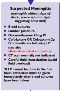
National (UK) guidelines on meningitis (community acquired meningitis in the immunocompetent host) available from meningitis.org. , including this box:

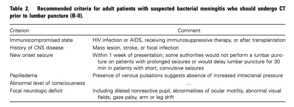
Practice Guidelines for the Management of Bacterial Meningitis These 2004 guidelines from the Infectious Diseases Society of America provide the following table listing the recommended criteria for adult patients with suspected bacterial meningitis who should undergo CT prior to lumbar puncture:

Clin Infect Dis. (2004) 39 (9): 1267-1284 Full text

4. This is potentially even more of an issue with paediatric patients

Fatal Lumbar Puncture: Fact Versus Fiction—An Approach to a Clinical Dilemma

An excellent summary of the above mentioned issues presented in a paediatric context, including the following:

On initial consideration a cranial CT would seem to be an appropriate and potentially useful diagnostic study for confirming the diagnosis of cerebral herniataion. The fallacy in this assessment has been emphasized by the finding that no clinically significant CT abnormalities are found that are not suspected on clinical assessments. Further, as previously noted, a normal CT examination may be found at about the time of a fatal herniation. Thus, the practical usefulness of a cranial CT in the majority of pediatric patients is limited to those rare patients whose increased ICP is secondary to mass lesions, not in the initial approach to acute meningitis.

Pediatrics. 2003 Sep;112(3 Pt 1):e174-6 Full Text

The last words should go to Dr Brad Spellberg, who in response to the IDSA’s guidelines wrote an excellent letter summarising much of the evidence at the time, confessed:

Why do we persist in using the CT scan for this purpose, despite the lack of supportive data? I am as guilty of this practice as anyone else, and the reason is simple: I am a chicken.

Clin Infect Dis. (2005) 40 (7): 1061 Full Text

Salicylate poisoning and pseudohyperchloraemia


Severe salicylate poisoning can cause metabolic acidosis from an accumulation of salicylic acid, lactic acid, and ketone bodies. A high anion gap acidosis is therefore the typical metabolic abnormality seen. A case series illustrates salicylate poisoning presenting with a normal gap (hyperchloraemic) acidosis – one patient had a chloride of 111 mmol/l and the other 123 mmol/l. This can occur when some analysers falsely read an elevated chloride in the presence of high concentrations of salicylate.


Severe salicylate poisoning is classically associated with an anion gap metabolic acidosis. However, high serum salicylate levels can cause false increase of laboratory chloride results on some analyzers. We present 2 cases of life-threatening salicylate poisoning with an apparently normal anion gap caused by an important laboratory interference. These cases highlight that the diagnosis of severe salicylism must be considered in all patients presenting with metabolic acidosis, even in the absence of an increased anion gap.

Falsely Normal Anion Gap in Severe Salicylate Poisoning Caused by Laboratory Interference
Ann Emerg Med. 2011 Sep;58(3):280-1

Easy rapid infusion set up

Kapoor and Singh's system from the Open Access article - click for explanation

Here’s a nice and simple set up for rapid iv infusions using simple cheap equipment

Full details at the Scandinavian Journal Site
Novel rapid infusion device for patients in emergency situations
Scand J Trauma Resusc Emerg Med. 2011 Jun 10;19:35 (Free Full Text)

Delta CVP with PEEP and fluid responsiveness

I’ve been (and remain) critical of the use of CVP to determine ‘filling status’ or more accurately volume-responsiveness, even using CVP trends; I’m generally in agreement with Dr Marik’s bold statement that “CVP should not be used to make clinical decisions regarding fluid management”1. However there might now appear to be a way of using CVP for this purpose.
Increasing PEEP in patients undergoing positive pressure ventilation can increase the CVP. It has been demonstrated in a small study of cardiac surgical patients2 that the degree to which a 10cmH2O increase in PEEP changes the CVP correlates with fluid responsiveness. The fluid responsiveness was determined by the change in cardiac output measured by thermodilution after a passive leg raise.
There are a number of limitations to this study that should prevent us from immediately extrapolating this method of determining fluid responsiveness to our ED / critical care patients, but the concept is interesting. This can be added to the growing pile of dynamic measures of circulatory filling.


Background Changes in central venous pressure (CVP) rather than absolute values may be used to guide fluid therapy in critically ill patients undergoing mechanical ventilation. We conducted a study comparing the changes in the CVP produced by an increase in PEEP and stroke volume variation (SVV) as indicators of fluid responsiveness. Fluid responsiveness was assessed by the changes in cardiac output (CO) produced by passive leg raising (PLR).

Methods In 20 fully mechanically ventilated patients after cardiac surgery, PEEP was increased +10 cm H2O for 5 min followed by PLR. CVP, SVV, and thermodilution CO were measured before, during, and directly after the PEEP challenge and 30° PLR. The CO increase >7% upon PLR was used to define responders.

Results Twenty patients were included; of whom, 10 responded to PLR. The increase in CO by PLR directly related (r=0.77, P<0.001) to the increase in CVP by PEEP. PLR responsiveness was predicted by the PEEP-induced increase in CVP [area under receiver-operating characteristic (AUROC) curve 0.99, P<0.001] and by baseline SVV (AUROC 0.90, P=0.003). The AUROC's for dCVP and SVV did not differ significantly (P=0.299).
Conclusions Our data in mechanically ventilated, cardiac surgery patients suggest that the newly defined parameter, PEEP-induced CVP changes, like SVV, appears to be a good parameter to predict fluid responsiveness.

1. Does central venous pressure predict fluid responsiveness? A systematic review of the literature and the tale of seven mares.
Chest. 2008 Jul;134(1):172-8
Full Text Link
2. Predicting cardiac output responses to passive leg raising by a PEEP-induced increase in central venous pressure, in cardiac surgery patients.
Br J Anaesth. 2011 Aug;107(2):251-7

Is there nothing ketamine can't do?

As well as the benefits of cardiovascular stability, maintenance of cerebral perfusion pressure, possibly lowering ICP and providing other neuroprotective benefits, ketamine may have other advantages. These are reviewed in a British Journal of Anaesthesia article from which I’ve selected those benefits of interest to practitioners of emergency medicine and critical care.


Additional Beneficial Effects of Ketamine

  • the dysphoric, or ’emergence’ reactions associated with ketamine may be reduced by pre-administration or co-administration of sedatives, such as benzodiazepines, propofol, dexmedetomidine, or droperidol.
  • ketamine potentiates opioid analgesia in multiple settings, reducing opioid total dose and in some groups of patients reducing postoperative desaturation
  • ketamine has possible anti-inflammatory effects demonstrated in some types of surgical patients
  • ketamine may prevent awareness, recall, or both during general anaesthesia

Ketamine: new uses for an old drug?
Br J Anaesth. 2011 Aug;107(2):123-6