
 High intensity focused ultrasound (HIFU) was hailed as the ‘surgery of the future’ a few years ago(1). As it’s now the future, where is it?
HIFU uses ultrasound to increase the heat within tissues at a specific area, causing local necrosis and cautery without injuring surrounding tissues. It is used to treat some cancers, but has shown promise in haemorrhage control. In animal studies it reduced or stopped bleeding in liver(2), spleen(3), and vascular injuries(4).
It has been proposed to offer a promising method for hemorrhage control in both civilivan and miltary trauma(5). Automated systems have been developed and tested that identify bleeding using Doppler ultrasound techniques that then allow targeting of the HIFU beam to the bleeding tissue(6). The United States Army has identified the need for a such systems and has designed a remotely operated robotic haemostatic system to save lives of soldiers. This was presented in 2006(7).
I would love to know where we are with this technology, and why nothing seems to have appeared about it in the literature for the last few years. If you have any information, please fill us in via the comments box.
1. High intensity focused ultrasound: surgery of the future?
Br J Radiol. 2003 Sep;76(909):590-9 Full text
2. Liver hemostasis using high-intensity focused ultrasound
Ultrasound Med Biol. 1997;23(9):1413-20
3. Control of splenic bleeding by using high intensity ultrasound
J Trauma. 1999 Sep;47(3):521-5
4. Hemostasis of punctured blood vessels using high-intensity focused ultrasound
Ultrasound Med Biol. 1998 Jul;24(6):903-10
5. Hemorrhage control using high intensity focused ultrasound
Int J Hyperthermia. 2007 Mar;23(2):203-11
6. Focused ultrasound: concept for automated transcutaneous control of hemorrhage in austere settings.
Aviat Space Environ Med. 2009 Apr;80(4):391-4
7. Remotely Operated Robotic High Intensity Focused Ultrasound (HIFU) Manipulator System for Critical Systems for Trauma and Transport (CSTAT)
Presented at the IEEE Ultrasonics Symposium, October 3-6, 2006, Vancouver, Canada – Full Text Here
Category Archives: All Updates
Targeted ICP reduction in TBI
A South American randomised controlled trial has demonstrated no improvement in mortality when traumatic brain injured patients had therapy targeted at keeping intracranial pressure below or equal to 20 mmHg as measured by an intraparenchymal monitor. The control group’s management was guided by neurologic examination and serial CT imaging(1).
Editorialist Dr Ropper summarises what we should do with this information well(2):
“[The authors]…do not advocate abandoning the treatment of elevated intracranial pressure any more than the authors of studies on wedge pressure reject the administration of fluid boluses in the treatment of shock”
BACKGROUND
Intracranial-pressure monitoring is considered the standard of care for severe traumatic brain injury and is used frequently, but the efficacy of treatment based on monitoring in improving the outcome has not been rigorously assessed.
METHODS
We conducted a multicenter, controlled trial in which 324 patients 13 years of age or older who had severe traumatic brain injury and were being treated in intensive care units (ICUs) in Bolivia or Ecuador were randomly assigned to one of two specific protocols: guidelines-based management in which a protocol for monitoring intraparenchymal intracranial pressure was used (pressure-monitoring group) or a protocol in which treatment was based on imaging and clinical examination (imaging–clinical examination group). The primary outcome was a composite of survival time, impaired consciousness, and functional status at 3 months and 6 months and neuropsychological status at 6 months; neuropsychological status was assessed by an examiner who was unaware of protocol assignment. This composite measure was based on performance across 21 measures of functional and cognitive status and calculated as a percentile (with 0 indicating the worst performance, and 100 the best performance).
RESULTS
There was no significant between-group difference in the primary outcome, a composite measure based on percentile performance across 21 measures of functional and cognitive status (score, 56 in the pressure-monitoring group vs. 53 in the imaging–clinical examination group; P=0.49). Six-month mortality was 39% in the pressure-monitoring group and 41% in the imaging–clinical examination group (P=0.60). The median length of stay in the ICU was similar in the two groups (12 days in the pressure-monitoring group and 9 days in the imaging–clinical examination group; P=0.25), although the number of days of brain-specific treatments (e.g., administration of hyperosmolar fluids and the use of hyperventilation) in the ICU was higher in the imaging–clinical examination group than in the pressure-monitoring group (4.8 vs. 3.4, P=0.002). The distribution of serious adverse events was similar in the two groups.
CONCLUSIONS
For patients with severe traumatic brain injury, care focused on maintaining monitored intracranial pressure at 20 mm Hg or less was not shown to be superior to care based on imaging and clinical examination
1. A Trial of Intracranial-Pressure Monitoring in Traumatic Brain Injury
N Eng J Med 367;26:2471-2381 Full Text
2. Brain in a Box
N Eng J Med DOI: 10.1056/NEJMe1212289 Full Text
Perimortem Caesarean Delivery: Late is Better than Not
“To date, approximately one-third of the women who die during pregnancy remain undelivered at the time of death”
Guidelines recommend cardiac arrest in pregnant women beyond 20 weeks gestation should be treated with perimortem caesarean delivery (PMCD) commenced within 4 minutes of arrest and completed within 5. These time intervals come from two papers, neither of which is current or used robust review methodology.
To address this, an up-to-date fairly comprehensive review was undertaken of published cases of maternal cardiac arrests occurring prior to delivery. The primary outcome measures were maternal and neonatal survival to hospital discharge and the relationship between PMCD and this outcome.
The Arrests
94 cases were included in the final analysis.Most pregnancies were singleton (90.4%, n = 85) with an average gestational age at the time of the arrest of 33 ± 7 weeks (median 35, range 10–42).
The most common causes of arrest were trauma, maternal cardiac problems, severe pre-eclampsia and amniotic fluid embolism, together comprising about 70% of arrests; two thirds occurred in hospital.
The Outcomes
Overall, return of spontaneous circulation (ROSC) was achieved more often than not (60.6%) and overall survival to hospital discharge was 54.3%
Only 57 cases (75%) reported the time from arrest to delivery; the average time was 16.6 ± 12.5 min (median 10, range 1–60), with only 4 cases making it under the advocated 4-min time limit.
Timing of PMCD and Maternal Survival
In cases undergoing PMCD the average time elapsing from arrest to PMCD was significantly different between surviving (27/57) and non-surviving (30/57) mothers [10.0 ± 7.2 min (median 9, range 1–37) and 22.6 ± 13.3 min (median 20, range 4–60) respectively (p < 0.001, 95%CI 6.9–18.2)].
Timing of PMCD and Neonatal Survival
Mean times to PMCD were 14±11min (median=10, range=1–47) and 22 ± 13 min (median = 20, range = 4–60) in neonatal survivors and non-survivors respectively (p=0.016)
In cases with PMCD which reported outcome, the overall neonatal survival rate was 63.6% (42/66).
“The 4-min time frame advocated for PMCD usually remains unmet yet neonatal survival is still likely if delivery occurs within 10 or even 15 min of arrest”
Both maternal & neonatal mortality were higher with prehospital arrest location.
Summary
The study may be limited by recall bias, under-reporting and publication bias, but provides a more comprehensive evidence base on which to base resuscitation recommendations. The authors provide a useful warning against becoming fixated with the recommended four minute window, which may lead teams to fail to attempt a potentially life-saving intervention:
“Fixation on specific time frames for PMCD may not be ideal. It may be more important to focus on event recognition and good overall performance…. It may be wise to advocate a short time frame for performance of PMCD in order to achieve better outcomes; however, blanket endorsement of an unrealistic time frame may well create a defeatist attitude when that time frame cannot be met.”
Maternal cardiac arrest and perimortem caesarean delivery: Evidence or expert-based?
Resuscitation. 2012 Oct;83(10):1191-200
[EXPAND Click for abstract]
AIM: To examine the outcomes of maternal cardiac arrest and the evidence for the 4-min time frame from arrest to perimortem caesarean delivery (PMCD) recommended in current resuscitation and obstetric guidelines.
DATA SOURCES AND METHODS: Review and data extraction from all reported maternal cardiac arrests occurring prior to delivery (1980-2010). Cases were included if they provided details regarding both the event and outcomes. Outcomes of arrest were assessed using survival, Cerebral Performance Category (CPC) and maternal/neonatal harm/benefit from PMCD. Outcome measures were maternal and neonatal survival.
RESULTS: Of 1594 manuscripts screened, 156 underwent full review. Data extracted from 80 relevant papers yielded 94 included cases. Maternal outcome: 54.3% (51/94) of mothers survived to hospital discharge, 78.4% (40/51) with a CPC of 1/2. PMCD was determined to have been beneficial to the mother in 31.7% of cases and was not harmful in any case. In-hospital arrest and PMCD within 10 min of arrest were associated with better maternal outcomes (ORs 5.17 and 7.42 respectively, p<0.05 both). Neonatal outcome: mean times from arrest to delivery were 14±11 min and 22±13 min in survivors and non-survivors respectively (receiver operating area under the curve 0.729). Neonatal survival was only associated with in-hospital maternal arrest (OR 13.0, p<0.001).
CONCLUSIONS: Treatment recommendations should include a low admission threshold to a highly monitored area for pregnant women with cardiorespiratory decompensation, good overall performance of resuscitation and delivery within 10 min of arrest. Cognitive dissonance may delay both situation recognition and the response to maternal collapse.
[/EXPAND]
London Calling – part 3
 Notes from Day 4 of the London Trauma Conference
The highlight for me was Mr Jonny Morrison speaking on Resuscitative Emergency Balloon Occlusion of the Aorta (REBOA). He is a British military surgeon currently out in Texas studying balloon occlusion of the aorta on pigs. Looking at trauma deaths, the next unexpected survivors will come from the uncontrollable haemorrhage group (truncal and junctional zones). This is by no means a new technique – described in the 1950’s during the Korean War – but like the early Star Wars chapters, needed to wait for technology to advance to make it feasible. It has the effect of cross clamping the aorta which provides afterload support, increases cerebral and coronary perfusion and provides proximal inflow control – without the mess of a resuscitative thoracotomy and greater access.
 The placement of the balloon is determined by the location of the injury (see photo) and falls into two zones. Zone 1 is the thoracic aorta and is used for truncal haemorrhage control, avoid Zone 2 where the celiac axis etc originates and Zone 3 is infrarenal, used for junctional bleeding and pelvic haemorrhage.
His studies have determined that for Zone 3 amenable bleeds balloon occlusion up to 60min is the optimal time. Any longer and the debt of the metabolic load is paid by increased inotropic support requirements. He also compared REBOA to the current standard treatment for junctional injuries, Celox™ gauze. If coagulation is normal then both treatments perform similarly, the benefit is seen in coagulopathic patients where REBOA outperforms the gauze.
Has REBOA been used on humans? Yes a case series of 13 – the technique improved the BP allowing time to get to definitive surgery (blogged here 2.5 years ago!).
The Zone 1 studies are looking at continuous vs intermittent balloon occlusion. The jury is still out as to which is better. With the intermittent occlusion (20min on, 1min off) there are inevitably some losses when the balloon is deflated, conversely the metabolic debt generated by continuous occlusion is too great in some also leading to deaths.
What was very clear is that for this technique to have an impact it must be delivered proactively and pre-hospital. The challenges that need to be overcome are access to the femoral artery and blind accurate placement.
Prof Karim Brohi brought the conference to a close with a summary of what we have learned about coagulation in trauma this year. Here are three things;
- FFP is good but as 43% deaths due to trauma in the UK are secondary to bleeding and occur in the first 3hr we are failing our patients by administering the treatment on average at 2.5hrs.
- Fibrinogen levels are low in coagulopathic trauma patients; we should give cryoprecipitate early and aim for Fib ≥2.0
- And finally whilst TEG is recommended to guide treatment and can provide results within 5 min, there are some aspects of coagulation it does not detect i.e. fibrinolysis was only detected in 8% of coagulopathic trauma patients – when measured in the plasma it was then detectable in 80%.
These are the highlights of the 2012 London Trauma Conference. I hope this whistle stop tour through these days has been informative and though provoking. I can assure you telephone hacking was not used to bring you this information and to my knowledge is correct.
This is Lou Chan, roving reporter for Resus ME! signing off.

‘London raises her head, shakes off the debris of the night from her hair, and takes stock of the damage done. The sign of a great fighter in the ring is can he get up from a fall after being knocked down… London does this every morning.’

London Calling – part 2
Notes from Days 2 & 3 of the London Trauma Conference
Day 2 of the LTC was really good. There were some cracking speakers who clearly had the ‘gift’ when it comes to entertaining the audience. No death by PowerPoint here (although it seems Keynote is now the presentation software of choice!). The theme of the day was prehospital care and major incidents.
The golden nuggets to take away include: (too many to list all of course)
- ‘Pull’ is the key to rapid extrication from cars if time critical from the Norweigan perspective. Dr Lars Wik of the Norweigen air ambulance presented their method of rapid extrication. Essentially they drag the car back on the road or away from what ever it has crashed into to control the environment and make space (360 style). They put a paramedic in the car whilst this is happening. They then make a cut in the A post near the roof, secure the rear of the car to a fire truck or fixed object with a chain and put another chain around the lower A post and steering wheel that is then winched tight. This has the effect of ‘reversing’ the crash and a few videos showed really fast access to the patient. The car seems to peel open. As they train specifically for it, there doesn’t seem to be any safety problems so far and its much quicker than their old method. I guess it doesnt matter really how you organise a rapid extrication method as long as it is trained for and everyone is on the same page.
- Dr Bob Winter presented his thoughts on hangings – to date no survivor of a non-judicial hanging has had a C-spine injury, so why do we collar them? Also there seems no point in cooling them. All imaging and concern for these patients should be based on the significant soft tissue injury that can be caused around the neck.
- Drownings – if the patient is totally submerged probably reasonable to search for 30mins in water that is >6 degrees or 90mins if <6 degrees. After that it becomes a body recovery (unless there is an air pocket or some exceptional circumstance). Patients that have drowned should have early ventilatory support if they show any signs of resp distress.
- Drs Julian Thompson and Mark Byers reassured us on a variety of safety issues at major incidents. It seems the risk to rescuers from secondary bombs at scene is low. Very few terrorist attacks world wide, ever, have had secondary devices so rescuers should be reassured (a bit). Greatest risk to the rescuer, like always, are the silly simple things that are a risk every day, like tripping over your own feet! With reference to chemical incidents, simple PPE seems to be sufficient for the vast majority of incidents, even fairly significant chemical ones, all this mucking about in full air tight suits is probably pointless and means patients cant be treated (at all). This led to the debate of how much risk should we, as rescue staff, accept? Clearly there are no absolute answers but minimising all risk to the rescuer is often at conflict with your ability to rescue. Where the balance should lie is a matter for organisations and individuals I guess.
 Sir Prof Keith Porter also gave us an update on the future of Prehospital emergency medicine as a recognised medical specialty. As those in the know, know, the specialty has been recognised by the GMC and the first draft of trainees are currently in post. More deaneries will be following suit soon to begin training but it is likely to take some time to build up large numbers of trained specialists. Importantly for those of us who already have completed our training there will be an option to sub specialise in PHEM but it will involve undertaking the FIMC exam. Great, more exams – see you there.

Day 3 – Major trauma
The focus of day 3 was that of damage control. Damage control surgery and damage control resucitation. We had indepth discussions about how to manage pelvic trauma and some of the finer points of trauma resuscitation.
Specific points raised were:
- Pelvic binders are great and can replace an ex fix if the abdomen needs opening to fix a spleen for example.
- You can catheterise patients with pelvic fractures (one gentle try).
- Most pelvic bleeds are venous which is why surgeons who can pack a pelvis is better than a radiologist who can mainly only treat arterial bleeds.
- Coagulopathy in trauma is not DIC and is probably caused by peripheral hypoperfusion.
- All the standard clotting tests that we use (INR etc) are useless and take too long to do. ROTEM or TEG is much better but still not perfect.
Also, as I am sure will please many – pressure isn’t flow so dont use pressors in trauma!
Chris Hill is an emergency and prehospital care physician based in the United Kingdom
London Calling!!
Notes from Day 1 of the London Trauma Conference
I’ve always fancied trying my hand at journalism so when this opportunity to cover the London Trauma Conference (LTC) presented itself how could I resist? The LTC is well established now running into its sixth year. So what little gems does it have left to offer?
The Air Ambulance Symposium opened the conference with strong representation from Norway.
Dr Marius Rehn presented a thought provoking talk on pre-hospital trauma triage. Pragmatically there will always be a proportion of patients that are mistriaged. So is under triage worse than over triage? It depends on whose point of view you take. If you’re the trauma victim then under triage is your greatest fear. But as clinicians we display loyalty bias (preferential consideration for our current patient over those we have no involvement with) which leads to over triage. The consequences are usually unseen as they manifest in other areas of the health system – studies have demonstrated a detrimental effect in cardiac patients arriving in units where a trauma patient is treated concurrently. Commonly under triaged are older patients that have low mechanism falls and children involved in RTC’s are over triaged. Triage protocols aren’t perfect but those based on physiology and anatomy are the best; even better still an experienced clinician (physicians better than paramedics) and in the future we should think about using lactate clearance.
I have never needed any convincing that ultrasound has a role in pre-hospital care. However Dr Nils Petter Oveland presented some of his research (due for publication next year) which reinforces this belief. He studied chest ultrasound for the detection of pneumothoraces. Plain radiography interpreted by a consultant radiologist can detect a 500ml pneumothorax; ultrasonography can detect a mere 50ml. Using pig models he demonstrated a linear relationship between the volume of the pneumothorax and the sternal – lung point distance (lung point = where the lung edge remains in contact with the pleura). Practically how can we use this? A small pneumothorax may be detected by ultrasound but have no clinical consequence. Prior to aero medical transfer the lung point can be marked and if clinical deterioration occurs en route repeat US can accurately determine an increase in pneumothorax volume and guide treatment. Genius!
 Prof Hans Morten Lossius provided a convincing argument for pre-hospital stroke thrombolysis. If you believe in this treatment, then it is more efficacious the sooner it is delivered (see photo). So why are we aiming for a thrombolysis time that is suboptimal? The thrombolysis times for a central Norwegian hospital were in the region of 3.5hrs, this reduced to 2.5hrs with rapid transportation. Approaching the problem from a different angle they trialled pre-hospital management with a mobile unit (CT scanner + neuroradiologist + neurologist) reducing time to thrombolysis to 72min (Lancet Neurology 2012, Walter). The next step is a multicentre RCT comparing standard treatment against a mobile CT + pre-hospital team with telemedical links to the Stroke centre……..
The Keynote address from Dr Gareth Davies took a look at the past and then a look to the future – the focus remained the same; providing the intervention patients need when they need it! Could this lead us into a future of Resuscitative Emergency Balloon Occlusion of the Aorta (REBOA) or Emergency Preservation Resuscitation (EPR) or emergency pre-hospital burr holes? Only time will tell.
Dr Steven Solid presented a double bill on patient safety. Admission to hospital is a high risk activity (as risky as bungee jumping!). Patient harm in aviation occurs 2 per 1000 flights. Only 25% were aviation related; mostly they are communication or equipment failures. He suggests medical line checks and team simulation training.
Dr Anne Weaver finished the first day with the story of her quest to get pre-hospital blood onto London HEMS to compliment the pre-hospital haemostatic resuscitation strategy they have for exsanguinating haemorrhage (tranexamic acid, prothrombin complex concentrate (for rapid warfarin reversal), POC INR machine, Buddy Lite™ blood warmers). Initial observations after the first six months are that ROSC is achieved more frequently in traumatic cardiac arrests although it’s too early to comment on mortality benefit. But this isn’t then end of the story – the next challenge is fresh frozen plasma.
Reassurance: difficult laryngoscopy in children remains rare
 I was taught a useful principle by a paediatric anaesthetist 10 years ago which has proven true in my experience and has contributed to keeping me calm when intubating sick kids. Unlike adults, in whom difficulty in intubation can often be unexpected, the vast majority of normal looking children are easy to intubate, and the ones who are difficult usually have obvious indicators such as dysmorphism.
This appears to be supported by recent evidence: in a large retrospective series of 11.219 anaesthesia patients, the overall incidence of difficult laryngoscopy [Cormack and Lehane (CML) grade III and IV] was only 1.35%, although was much higher in infants less than one year compared with older children. This low percentage is in the same ball park as two other paediatric studies. Besides younger age, their database suggested underweight, ASA III and IV physical status and, if obtainable, Mallampati III and IV findings as predictors for difficult laryngoscopy. The authors point out:
“…the oromaxillofacial surgery department with a high proportion of cleft palate interventions and pediatric cardiac surgery contributed substantially to the total number of difficult laryngoscopies. In patients undergoing pediatric cardiac surgery, a possible explanation for the higher incidence of CML III/IV findings might be that some congenital heart defects are associated with chromosomal anomalies like microdeletion 22q11.2 syndrome. This syndrome is also associated with extracardiac anomalies like cranio-facial dysmorphism“
Take home message: As a very rough rule of thumb to illustrate the difference between the ease/difficulty of laryngoscopy between adults and kids, I think it’s fair to say grade III or IV views occur in about 10% of adults but only about 1% of children.
Incidence and predictors of difficult laryngoscopy in 11.219 pediatric anesthesia procedures
Paediatr Anaesth. 2012 Aug;22(8):729-36
[EXPAND Click for abstract]
OBJECTIVE: Difficult laryngoscopy in pediatric patients undergoing anesthesia.
AIM: This retrospective analysis was conducted to investigate incidence and predictors of difficult laryngoscopy in a large cohort of pediatric patients receiving general anesthesia with endotracheal intubation.
BACKGROUND: Young age and craniofacial dysmorphy are predictors for the difficult pediatric airway and difficult laryngoscopy. For difficult laryngoscopy, other general predictors are not yet described.
METHODS: Retrospectively, from a 5-year period, data from 11.219 general anesthesia procedures in pediatric patients with endotracheal intubation using age-adapted Macintosh blades in a single center (university hospital) were analyzed statistically.
RESULTS: The overall incidence of difficult laryngoscopy [Cormack and Lehane (CML) grade III and IV] was 1.35%. In patients younger than 1 year, the incidence of CML III or IV was significantly higher than in the older patients (4.7% vs 0.7%). ASA Physical Status III and IV, a higher Mallampati Score (III and IV) and a low BMI were all associated (P < 0.05) with difficult laryngoscopy. Patients undergoing oromaxillofacial surgery and cardiac surgery showed a significantly higher rate of CML III/IV findings.
CONCLUSION: The general incidence of difficult laryngoscopy in pediatric anesthesia is lower than in adults. Our results show that the risk of difficult laryngoscopy is much higher in patients below 1 year of age, in underweight patients and in ASA III and IV patients. The underlying disease might also contribute to the risk. If the Mallampati score could be obtained, prediction of difficult laryngoscopy seems to be reliable. Our data support the existing recommendations for a specialized anesthesiological team to provide safe anesthesia for infants and neonates.
[/EXPAND]
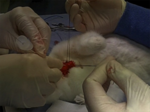
Transtracheal airways in kids. Well, pigs' kids anyway
Ever had to do a surgical airway in a child? Thought not. They’re pretty rare. Bill Heegaard MD from Henepin County Medical Center taught me a few approaches (with the help of an anaesthetised rabbit) which really got me thinking. It’s something I’d often trained for in my internal simulator, and I even keep the equipment for it in my house (listen out for an upcoming podcast on that). Research and experience has demonstrated that open surgical airway techniques are more reliable than transtracheal needle techniques in adults, but what about kids, in whom traditional teaching cautions against open techniques?
Australian investigators who were experienced airway proceduralists evaluated transtracheal needle techniques using a rabbit model (an excellent model for the infant airway). Their success rate was only 60% and they perforated the posterior tracheal wall in 42% of attempts. Of 13 attempts to insert a dedicated paediatric tracheotomy device, the Quicktrach Child, none were successful(1) (they did not use the Quicktrach Infant model as it is not available in Australia).
Danish investigators used fresh piglet cadavers weighing around 8 kg to assess two transtracheal cannulas, in which they achieved success rates of 65.6% and 68.8%(2). There was also a very high rate of posterior tracheal wall perforation. Using an open surgical tracheostomy technique, they were successful in 97% of attempts. These were also experienced operators, with a median anaesthetic experience of 12.5 years.
Their tracheotomy technique was nice and simple, and used just a scalpel, scissors, and surgical towel clips. Here’s their technique:
Simple tracheotomy procedure described by Holm-Knudsen et al
- Identify larynx and proximal trachea by palpation
- Vertical incision through the skin and subcutaneous tissue from the upper part of larynx to the sternal notch
- Grasp strap muscles with two towel forceps and separate in the midline
- Palpate and identify the trachea (palpate rather than look for tracheal rings, as in a live patient one would expect bleeding to obscure the view)
- Stabilise the trachea by grasping it with a towel forceps
- Insert sharp tip of the scissors between two tracheal rings and lift the trachea anteriorly to avoid damage to the posterior wall
- Cut vertically in the midline of the trachea with the scissors – they chose to use the scissors to cut the tracheal rings to facilitate tube insertion
- Insert the tracheal tube
Using ultrasound and CT to evaluate comparative airway dimensions, the authors concluded that the pig model is most useful for training emergency airway management in older children aged 5–10 years.
Why were they doing a tracheotomy rather than a cricothyroidotomy? Reasons given by the authors include:
- The infant cricothyroid membrane is very small
- Palpation of the thyroid notch may be hindered by the overlying hyoid bone
- The mandible may obstruct needle access to the cricothyroid membrane given the cephalad position in the neck of the infant larynx.
From an emergency medicine point of view, there are a couple of other reasons why we need to be able to access the trachea lower than the cricothyroid membrane. One is fractured larynx or other blunt or penetrating airway injury where there may be anatomical disruption at the cricothyroid level. The other situation is foreign body airway obstruction, when objects may lodge at the level of the cricoid ring which is functionally the narrowest part of the pediatric upper airway. Of course, alternative methods might be considered to remove the foreign body prior to tracheotomy, such as employing basic choking algorithms, and other techniques depending on whether you do or don’t have equipment.
Take home messages
- Transtracheal airways in kids are so rare, we can’t avoid extrapolating animal data
- Whichever infant or paediatric model is used, transtracheal needle techniques have a high rate of failure even by ‘experienced’ operators
- The small size and easy compressibility of the airway probably contributes to this failure rate, including the high rate of posterior wall puncture
- In keeping with adult audit data, open surgical techniques may have a higher success rate
- Tracheotomy may be necessary rather than cricothyroidotomy in infants and children depending on clinical scenario and accessibility of anatomy
- The stress and blood that is not simulated in cadaveric animal models will make open tracheotomy harder in a live patient, and so these success rates may not translate. However these factors do mean that whatever technique is used must be kept simple and should employ readily available and familiar equipment
- Something to maintain control and anterior position of the anterior trachea wall should be used during incision and intubation of the trachea. The study reported here used towel clips; sutures around the tracheal rings may also be used (see image below)
I recommend you add ‘paediatric tracheotomy’ to the list of procedures you might need to do (if it’s not already there). Identify what equipment you would use and run the simulation in your head and in your work environment.
Have fun.
1. The ‘Can’t Intubate Can’t Oxygenate’ scenario in Pediatric Anesthesia: a comparison of different devices for needle cricothyroidotomy
Paediatr Anaesth. 2012 Dec;22(12):1155-8
BACKGROUND: Little evidence exists to guide the management of the ‘Can’t Intubate, Can’t Oxygenate’ (CICO) scenario in pediatric anesthesia.
OBJECTIVES: To compare two intravenous cannulae for ease of use, success rate and complication rate in needle tracheotomy in a postmortem animal model of the infant airway, and trial a commercially available device using the same model.
METHODS: Two experienced proceduralists repeatedly attempted cannula tracheotomy in five postmortem rabbits, alternately using 18-gauge (18G) and 14-gauge (14G) BD Insyte(™) cannulae (BD, Franklin Lakes, NJ, USA). Attempts began at the first tracheal cartilage, with subsequent attempts progressively more caudad. Success was defined as intratracheal cannula placement. In each rabbit, an attempt was then made by each proceduralist to perform a cannula tracheotomy using the Quicktrach Child(™) device (VBM Medizintechnik GmbH, Sulz am Neckar, Germany).
RESULTS: The rabbit tracheas were of similar dimensions to a human infant. 60 attempts were made at cannula tracheotomy, yielding a 60% success rate. There was no significant difference in success rate, ease of use, or complication rate between cannulae of different gauge. Successful aspiration was highly predictive (positive predictive value 97%) and both sensitive (89%) and specific (96%) for tracheal cannulation. The posterior tracheal wall was perforated in 42% of tracheal punctures. None of 13 attempts using the Quicktrach Child(™) were successful.
CONCLUSION: Cannula tracheotomy in a model comparable to the infant airway is difficult and not without complication. Cannulae of 14- and 18-gauge appear to offer similar performance. Successful aspiration is the key predictor of appropriate cannula placement. The Quicktrach Child was not used successfully in this model. Further work is required to compare possible management strategies for the CICO scenario
2. Emergency airway access in children – transtracheal cannulas and tracheotomy assessed in a porcine model
Paediatr Anaesth. 2012 Dec;22(12):1159-65
OBJECTIVES: In the rare scenario when it is impossible to oxygenate or intubate a child, no evidence exists on what strategy to follow.
AIM: The aim of this study was to compare the time and success rate when using two different transtracheal needle techniques and also to measure the success rate and time when performing an emergency tracheotomy in a piglet cadaver model.
METHODS: In this randomized cross-over study, we included 32 anesthesiologists who each inserted two transtracheal cannulas (TTC) using a jet ventilation catheter and an intravenous catheter in a piglet model. Second, they performed an emergency tracheotomy. A maximum of 2 and 4 min were allowed for the procedures, respectively. The TTC procedures were recorded using a video scope.
RESULTS: Placement of a transtracheal cannula was successful in 65.6% and 68.8% of the attempts (P = 0.76), and the median duration of the attempts was 69 and 42 s (P = 0.32), using the jet ventilation catheter and the intravenous catheter, respectively. Complications were frequent in both groups, especially perforation of the posterior tracheal wall. Performing an emergency tracheotomy was successful in 97%, in a median of 88 s.
CONCLUSIONS: In a piglet model, we found no significant difference in success rates or time to insert a jet ventilation cannula or an intravenous catheter transtracheally, but the incidence of complications was high. In the same model, we found a 97% success rate for performing an emergency tracheotomy within 4 min with a low rate of complications.
Externally rotate leg for femoral vein access
Want to access the femoral vein? Externally rotate the leg at the hip and things might be a bit easier. This study was done in adult patients, with the knee straight and no abduction applied. External rotation is also helpful in kids, with abduction up to sixty degrees.
Objective: To determine if external rotation of the leg increases the size and accessibility of the femoral vein compared with a neutral position.
Methods: One hundred patients presenting to a tertiary teaching hospital were prospectively recruited. The right common femoral vein of each subject was scanned with a linear probe (5–10 MHz) inferior to the inguinal ligament, with the leg in a neutral position and then in the externally rotated position. The transverse diameter of the femoral vein, the accessible diameter of the vein (lying medial to the femoral artery) and the depth of the vein were measured.
Results: The mean diameter of the femoral vein in the externally rotated leg was greater than with the leg in the neutral position (15.4 mm vs 13.8 mm); the mean difference was 1.6 mm (95% CI 1.3–1.9). The mean accessible diameter of the femoral vein was larger with the leg externally rotated (13.8 mm vs 11.7 mm, mean difference 2.1 mm, 95% CI 1.8–2.5). The depth from the skin to the femoral vein was less with the leg in external rotation (20.9 mm vs 22.6 mm, mean difference 1.7 mm, 95% CI 1.2–2.2). The mean diameter and depth were greater in patients with overweight or obese body mass index (BMI) measurements in both leg positions. The increase in femoral vein diameter and accessibility with external rotation was observed in all BMI groups.
Conclusion: The total and accessible femoral vein diameter is increased and the surface depth of the vein is decreased by placing the leg in external rotation compared with the neutral position.
Simple external rotation of the leg increases the size and accessibility of the femoral vein
Emerg Med Australas. 2012 Aug;24(4):408-13
Hyperglycaemia & mortality in sepsis – lactate dependent?
I like this paper for introducing a new concept to me. For years the critical care community has recognised the link between hyperglycaemia and mortality, leading to early recommendations of intensive insulin regimens subsequently shown not to be of benefit. Now it appears that the association between hyperglycaemia and mortality may be less relevant in patients with a normal lactate.
In a study of adult nondiabetic critically ill patients, hyperglycaemia had a significant association with increased mortality risk using simple univariate analysis. When they adjusted for concurrent hyperlactataemia however, hyperglycaemia was not significantly associated with increased mortality risk.
The authors discuss several known or postulated aspects of interplay between lactate and glucose in sepsis:
- Hyperlactataemia appears to inhibit glucose uptake by muscle cells and decrease activity of the GLUT-4 transporters
- Hyperlactataemia has also been shown to increase insulin resistance directly
- Glucose and lactate levels tend to be elevated simultaneously in severe sepsis at baseline.
- Experimentally it has been estimated that 45% of infused (radiolabelled) lactate is either converted into glucose via gluconeogenesis or is transformed into glycogen via the Cori cycle, representing a higher proportion of glucose formation from lactate than in nonseptic controls.
- It is possible that elevated glucose and lactate levels in sepsis both may be measures of the same phenomenon: glucose accumulates due to the sympathomimetic response to a systemic infection with increased catecholamine levels leading to increased activity of the Na+K+-ATPase, resulting in accumulation of adenosine diphosphate (ADP). Increased levels of ADP in turn augment glycogenolysis.
- Mitochondrial metabolism cannot meet the increased cellular energy needs of sepsis, resulting in accumulation of ADP and leading to cytosolic glycolysis and lactate production, even in an aerobic environment.
The augmented glycolysis of sepsis (and during adrenergic therapy such as epinephrine/adrenaline or albuterol/salbutamol) is one of the causes of a raised lactate to consider when applying the LACTATES mnemonic I like to use.
Hyperlactatemia affects the association of hyperglycemia with mortality in nondiabetic adults with sepsis
Acad Emerg Med. 2012 Nov;19(11):1268-75
[EXPAND Click for abstract]
BACKGROUND: Admission hyperglycemia has been reported as a mortality risk factor for septic nondiabetic patients; however, hyperglycemia’s known association with hyperlactatemia was not addressed in these analyses.
OBJECTIVES: The objective was to determine whether the association of hyperglycemia with mortality remains significant when adjusted for concurrent hyperlactatemia.
METHODS: This was a post hoc, nested analysis of a retrospective cohort study performed at a single center. Providers had identified study subjects during their emergency department (ED) encounters; all data were collected from the electronic medical record (EMR). Nondiabetic adult ED patients hospitalized for suspected infection, two or more systemic inflammatory response syndrome (SIRS) criteria, and simultaneous lactate and glucose testing in the ED were enrolled. The setting was the ED of an urban teaching hospital from 2007 to 2009. To evaluate the association of hyperglycemia (glucose > 200 mg/dL) with hyperlactatemia (lactate ≥ 4.0 mmol/L), a logistic regression model was created. The outcome was a diagnosis of hyperlactatemia, and the primary variable of interest was hyperglycemia. A second model was created to determine if coexisting hyperlactatemia affects hyperglycemia’s association with mortality; the main outcome was 28-day mortality, and the primary risk variable was hyperglycemia with an interaction term for simultaneous hyperlactatemia. Both models were adjusted for demographics; comorbidities; presenting infectious source; and objective evidence of renal, respiratory, hematologic, or cardiovascular dysfunction.
RESULTS: A total of 1,236 ED patients were included, and the median age was 77 years (interquartile range [IQR] = 60 to 87 years). A total of 115 (9.3%) subjects were hyperglycemic, 162 (13%) were hyperlactatemic, and 214 (17%) died within 28 days of their initial ED visits. After adjustment, hyperglycemia was significantly associated with simultaneous hyperlactatemia (odds ratio [OR] = 4.14, 95% confidence interval [CI] = 2.65 to 6.45). Hyperglycemia and concurrent hyperlactatemia were associated with increased mortality risk (OR = 3.96, 95% CI = 2.01 to 7.79), but hyperglycemia in the absence of simultaneous hyperlactatemia was not (OR = 0.78, 95% CI = 0.39 to 1.57).
CONCLUSIONS: In this cohort of septic adult nondiabetic patients, mortality risk did not increase with hyperglycemia unless associated with simultaneous hyperlactatemia. The previously reported association of hyperglycemia with mortality in nondiabetic sepsis may be due to the association of hyperglycemia with hyperlactatemia.
[/EXPAND]