
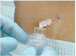
Here ultrasound was used to ascertain the best position for doing a lumbar puncture in kids, where the interspinous space was maximised:
BACKGROUND Lumbar punctures are commonly performed in the pediatric emergency department. There is no standard, recommended, optimal position for children who are undergoing the procedure.
OBJECTIVE To determine a position for lumbar punctures where the interspinous space is maximized, as measured by bedside ultrasound.
METHODS A prospective convenience sample of children under age 12 was performed. Using a portable ultrasound device, the L3-L4 or L4-L5 interspinous space was measured with the subject in 5 different positions. The primary outcome was the interspinous distance between 2 adjacent vertebrae. The interspinous space was measured with the subject sitting with and without hip flexion. In the lateral recumbent position, the interspinous space wasmeasured with the hips in a neutral position as well as in flexion, both with and without neck flexion. Data were analyzed by comparing pairwise differences.
RESULTS There were 28 subjects enrolled (13 girls and 15 boys) at a median age of 5 years. The sitting-flexed position provided a significantly increased interspinous space (P < .05). Flexion of the hips increased the interspinous space in both the sitting and lateral recumbent positions (P < .05). Flexion of the neck, did not significantly change the interspinous space (P = .998).
CONCLUSIONS The interspinous space of the lumbar spine was maximally increased with children in the sitting position with flexed hips; therefore we recommend this position for lumbar punctures. In the lateral recumbent position, neck flexion does not increase the interspinous space and may increase morbidity; therefore, it is recommended to hold patients at the level of the shoulders as to avoid neckflexion.
Positioning for lumbar puncture in children evaluated by bedside ultrasound
Pediatrics. 2010 May;125(5):e1149-53
A more recent study also used ultrasound in infants to investigate the anatomic necessity and advantage derived from a tight flexed lateral recumbent position, since hypoxia has been observed in that position:
Objectives: Hypoxia has been observed when infants undergo lumbar puncture in a tight flexed lateral recumbent position. This study used sonographic measurements of lumbar interspinous spaces to investigate the anatomic necessity and advantage derived from this tight flexed positioning in infants.
Methods: This was a brief, prospective, observational study of a convenience sample of patients. Twenty-one healthy infants under 1 month of age were scanned in two positions: prone in a spine-neutral position and lateral recumbent with their knees bent into their chest and their neck flexed. In each position, a 5- to 10-MHz linear array transducer was used to scan midline along the lumbar spinous processes in the sagittal plane. The distances between the spinous processes were measured near the ligamentum flavum using the ultrasound machine’s calipers. Pulse oximetry was monitored on all infants during flexed positioning.
Results: In the spine-neutral position, all studied interspinous spaces were much wider than a 22-gauge spinal needle (diameter 0.072 cm). The mean (±SD) interspinous spaces for L3-4, L4-5, and L5-S1 in a spine-neutral position were 0.42 (±0.07), 0.37 (±0.06), and 0.36 (±0.11) cm, respectively. Flexing the infants increased the mean lumbar interspinous spaces at L3-4, L4-5, and L5-S1 by 31, 51, and 44%, respectively.
Conclusions: This study verified that tight, lateral flexed positioning substantially enhances the space between the lumbar spinous processes and that a spine-neutral position also allows for a large enough anatomic interspinous space to perform lumbar puncture. However, further clinical research is required to establish the feasibility of lumbar puncture in a spine-neutral position.
Evaluating infant positioning for lumbar puncture using sonographic measurements
Acad Emerg Med. 2011 Feb;18(2):215-8
Category Archives: All Updates
Therapeutic hypothermia for newborns
More evidence that cooling the hypoxic neonatal brain improves outcomes….
OBJECTIVE Mild hypothermia after perinatal hypoxic-ischemic encephalopathy (HIE) reduces neurologic sequelae without significant adverse effects, but studies are needed to determine the most-efficacious methods.
METHODS In the neo.nEURO.network trial, term neonates with clinical and electrophysiological evidence of HIE were assigned randomly to either a control group, with a rectal temperature of 37°C (range: 36.5–37.5°C), or a hypothermia group, cooled and maintained at a rectal temperature of 33.5°C (range: 33–34°C) with a cooling blanket for 72 hours, followed by slow rewarming. All infants received morphine (0.1 mg/kg) every 4 hours or an equivalent dose of fentanyl. Neurodevelopmental outcomes were assessed at the age of 18 to 21 months. The primary outcome was death or severe disability.
RESULTS A total of 129 newborn infants were enrolled, and 111 infants were evaluated at 18 to 21 months (53 in the hypothermia group and 58 in the normothermia group). The rates of death or severe disability were 51% in the hypothermia group and 83% in the normothermia group (P = .001; odds ratio: 0.21 [95% confidence interval [CI]: 0.09–0.54]; number needed to treat: 4 [95% CI: 3–9]). Hypothermia also had a statistically significant protective effect in the group with severe HIE (n = 77; P = .005; odds ratio: 0.17 [95% CI: 0.05–0.57]). Rates of adverse events during the intervention were similar in the 2 groups except for fewer clinical seizures in the hypothermia group.
CONCLUSION Systemic hypothermia in the neo.nEURO.network trial showed a strong neuroprotective effect and was effective in the severe HIE group.
Systemic Hypothermia After Neonatal Encephalopathy: Outcomes of neo.nEURO.network RCT
Pediatrics. 2010 Oct;126(4):e771-8
Update Dec 2014:
An RCT to determine if longer duration cooling (120 hours), deeper cooling (32.0°C), or both are superior to cooling at 33.5°C for 72 hours in neonates who are full-term with moderate or severe hypoxic ischemic encephalopathy.
Longer cooling, deeper cooling, or both compared with hypothermia at 33.5°C for 72 hours did not reduce NICU death. Small study.
Effect of depth and duration of cooling on deaths in the NICU among neonates with hypoxic ischemic encephalopathy: a randomized clinical trial
JAMA. 2014 Dec 24;312(24):2629-39
2J or 4J/kg in Paediatric Defibrillation?
Should we shock with 2J/kg or 4J/kg in Paediatric Defibrillation? The answer seems to be ‘we still don’t know’. Don’t worry – just follow the guidelines (reproduced for you at the bottom)
OBJECTIVE To examine the effectiveness of initial defibrillation attempts. We hypothesized that (1) an initial shock dose of 2 ± 10 J/kg would be less effective for terminating fibrillation than suggested in published historical data and (2) a 4 J/kg shock dose would be more effective.
PATIENTS AND METHODS This was a National Registry of Cardiopulmonary Resuscitation prospective, multisite, observational study of in-hospital pediatric (aged 18 years) ventricular fibrillation or pulseless ventricular tachycardia cardiac arrests from 2000–2008. Termination of ventricular fibrillation or pulseless ventricular tachycardia and event survival after initial shocks of 2 J/kg were compared with historic controls and a 4 J/kg shock dose.

RESULTS Of 266 children with 285 events, 173 of 285 (61%) survived the event and 61 of 266 (23%) survived to discharge. Termination of fibrillation after initial shock was achieved for 152 of 285 (53%) events. Termination of fibrillation with 2 ± 10 J/kg was much less frequent than that seen among historic control subjects (56% vs 91%; P < .001), but not different than 4 J/kg. Compared with 2 J/kg, an initial shock dose of 4 J/kg was associated with lower rates of return of spontaneous circulation (odds ratio: 0.41 [95% confidence interval: 0.21–0.81]) and event survival (odds ratio: 0.42 [95% confidence interval: 0.18–0.98]).
CONCLUSIONS The currently recommended 2 J/kg initial shock dose for in-hospital cardiac arrest was substantially less effective than previously published. A higher initial shock dose (4 J/kg) was not associated with superior termination of ventricular fibrillation or pulseless ventricular tachycardia or improved survival rates. The optimal pediatric defibrillation dose remains unknown.
Effect of defibrillation energy dose during in-hospital pediatric cardiac arrest
Pediatrics. 2011 Jan;127(1):e16-23
Here’s what the guidelines say:
Many AEDs have high specificity in recognizing pediatric shockable rhythms, and some are equipped to decrease (or attenuate) the delivered energy to make them suitable for infants and children <8 years of age. For infants a manual defibrillator is preferred when a shockable rhythm is identified by a trained healthcare provider (Class IIb, LOE C). The recommended first energy dose for defibrillation is 2 J/kg. If a second dose is required, it should be doubled to 4 J/kg. If a manual defibrillator is not available, an AED equipped with a pediatric attenuator is preferred for infants. An AED with a pediatric attenuator is also preferred for children <8 year of age. If neither is available, an AED without a dose attenuator may be used (Class IIb, LOE C). AEDs that deliver relatively high energy doses have been successfully used in infants with minimal myocardial damage and good neurological outcomes
Pediatric Basic Life Support: 2010 American Heart Association Guidelines for Cardiopulmonary Resuscitation and Emergency Cardiovascular Care
Full text document
Out of hospital monitoring in kids
I don’t have full text access to the Journal Pediatrics, so I’m not sure what I make of this small randomised trial comparing two types of blood pressure monitoring during paediatric transport:
BACKGROUND The “golden-hour” concept has led to emphasis on speed of patient delivery during pediatric interfacility transport. Timely intervention, in addition to enhanced monitoring during transport, is the key to improved outcomes in critically ill patients. Taking the ICU to the patient may be more beneficial than rapid delivery to a tertiary care center.

METHODS The Improved Monitoring During Pediatric Interfacility Transport trial was the first randomized controlled trial in the out-of-hospital pediatric transport environment. It was designed to determine the impact of improved blood pressure monitoring during pediatric interfacility transport and the effect on clinical outcomes in patients with systemic inflammatory response syndrome and moderate-to-severe head trauma. Patients in the control group had their blood pressure monitored intermittently with an oscillometric device; those in the intervention group had their blood pressure monitored every 12 to 15 cardiac contractions with a near-continuous, noninvasive device.
RESULTS Between May 2006 and June 2007, 1995, consecutive transport patients were screened, and 94 were enrolled (48 control, 46 intervention). Patients in the intervention group received more intravenous fluid (19.8 ± 22.2 vs 9.9 ± 9.9 mL/kg; P = .01), had a shorter hospital stay (6.8 ± 7.8 vs 10.9 ± 13.4 days; P = .04), and had less organ dysfunction (18 of 206 vs 32 of 202 PICU days; P = .03).
CONCLUSIONS Improved monitoring during pediatric transport has the potential to improve outcomes of critically ill children. Clinical trials, including randomized controlled trials, can be accomplished during pediatric transport. Future studies should evaluate optimal equipment, protocols, procedures, and interventions during pediatric transport, aimed at improving the clinical and functional outcomes of critically ill patients.
Enhanced Monitoring Improves Pediatric Transport Outcomes: A Randomized Controlled Trial
Pediatrics. 2011 Jan;127(1):42-8
Pre-hospital / HEMS podcast
I was lucky enough to be interviewed by the amazing Scott Weingart, an emergency medicine intensivist who runs the spectacular EMcrit podcast. We covered some stuff on pre-hospital airway management, physicians in pre-hospital care, and I had a rant about ‘scoop and run’ versus ‘stay and play’. Worryingly, Scott is keeping back some audio footage for a later podcast, probably containing an even bigger rant about things like ATLS.
Click the image to be taken to the EMcrit site where you can listen to the podcast.

Plasma:red cell ratios
In some circles, ‘wuntwuntwun’ is in danger of becoming the new dogma of trauma fluid replacement (ie. 1 unit of plasma and 1 unit of platelets for every unit of red cells). Since it takes longer to thaw some plasma than it does to throw in some O negative packed red cells, some really sick patients may be dead before they get the plasma, biasing comparisons that show a reduced mortality in patients who were still alive to receive plasma. This ‘survivor bias’ has been suggested as a reason that high plasma:red cell ratios are associated with mortality reduction, although this has been challenged.
The survivor bias explanation receives some new support by the following (small) study from Journal of Trauma:
BACKGROUND: In light of recent data, controversy surrounds the apparent 30-day survival benefit of patients achieving a fresh frozen plasma (FFP) to packed red blood cell (PRBC) ratio of at least 1:2 in the face of massive transfusions (MT) (≥10 units of PRBC within 24 hours of admission). We hypothesized that initial studies suffer from survival bias because they do not consider early deaths secondary to uncontrolled exsanguinating hemorrhage. To help resolve this controversy, we evaluated the temporal relationship between blood product administration and mortality in civilian trauma patients receiving MT.

METHODS: Patients requiring MT over a 22-month period were identified from the resuscitation registry of a Level I trauma center. Shock severity at admission and timing of shock-trauma admission, blood product administration, and death were determined. Patients were divided into high- and low-ratio groups (≥1:2 and<1:2 FFP:PRBC, respectively) and compared. Kaplan-Meier analysis and log-rank test was used to examine 24-hour survival.
RESULTS: One hundred three patients (63% blunt) were identified (66 high-ratio and 37 low-ratio). Those patients who achieved a high-ratio in 24 hours had improved survival. However, severity of shock was less in the high-group (base excess: -8.0 vs. -11.2, p=0.028; lactate: 6.3 vs. 8.4, p=0.03). Seventy-five patients received MT within 6 hours. Of these, 29 received a high-ratio in 6 hours. Again, severity of shock was less in the high-ratio group (base excess: -7.6 vs. -12.7, p=0.008; lactate: 6.7 vs. 9.4, p=0.02). For these patients, 6-hour mortality was less in the high-group (10% vs. 48%, p<0.002). After accounting for early deaths, groups were similar from 6 hours to 24 hours.
CONCLUSIONS: Improved survival was observed in patients receiving a higher plasma ratio over the first 24 hours. However, temporal analysis of mortality using shorter time periods revealed those who achieve early high-ratio are in less shock and less likely to die early from uncontrolled hemorrhage compared with those who never achieve a high-ratio. Thus, the proposed survival advantage of a high-ratio may be because of selection of those not likely to die in the first place; that is, patients die with a low-ratio not because of a low-ratio.
The authors state “The current study underscores the need for well-designed prospective studies to address the important question of which ratio results in improved survival and stresses the importance of timing of blood product administration as this may impact survival.”
Improved survival after hemostatic resuscitation: does the emperor have no clothes?
J Trauma. 2011 Jan;70(1):97-102
Helicopters between hospitals
More National Trauma Databank analysis coming out in favour of helicopter transport: this time looking at interhospital transfer:
Background: Helicopter transport (HT) is frequently used for interfacility transfer of injured patients to a trauma center. The benefits of HT over ground transport (GT) in this setting are unclear. By using a national sample, the objective of this study was to assess whether HT impacted outcomes following interfacility transfer of trauma patients.

Methods: Patients transferred by HT or GT in 2007 were identified using the National Trauma Databank (version 8). Injury severity, resource utilization, and survival to discharge were compared. Stepwise logistic regression was used to determine whether transport modality was a predictor of survival after adjusting for covariates. Regression analysis was repeated in subgroups with Injury Severity Score (ISS) ≤15 and ISS >15.
Results: There were 74,779 patients transported by helicopter (20%) or ground (80%). Mean ISS was higher in patients transported by helicopter (17 ± 11 vs. 12 ± 9; p < 0.01) as was the proportion with ISS >15 (49% vs. 28%; odds ratio [OR], 2.53; 95% confidence interval [CI], 2.43-2.63). Patients transported by helicopter had higher rates of intensive care unit admission (54% vs. 29%; OR, 2.86; 95% CI, 2.75-2.96), had shorter transport time (61 ± 55 minutes vs. 98 ± 71 minutes; p < 0.01), and had shorter overall prehospital time (135 ± 86 minutes vs. 202 ± 132 minutes; p < 0.01). HT was not a predictor of survival overall or in patients with ISS ≤15. In patients with ISS >15, HT was a predictor of survival (OR, 1.09; 95% CI, 1.02-1.17; p = 0.01).
Conclusions: Patients transported by helicopter were more severely injured and required more hospital resources than patients transported by ground. HT offered shorter transport and overall prehospital times. For patients with ISS >15, HT was a predictor of survival. These findings should be considered when developing interfacility transfer policies for patients with severe injuries.
Helicopters Improve Survival in Seriously Injured Patients Requiring Interfacility Transfer for Definitive Care
J Trauma. 2011 Feb;70(2):310-4.
Neck movement in spite of collar
A cadaveric study using an artificially created unstable cervical spine injury has shown marked displacement of the vertebrae when cervical collars were applied, and when the bodies were moved in a way that simulated normal transfer and log-rolling. There was no comparison with a no-collar situation, so we can’t say from this that collars are necessarily bad, just that they’re no good in this cadaveric model. I like this statement by the authors:
“A variety of collars, backboards, and other equipment and techniques are being used in an attempt to achieve spine stabilization, largely without any validation of efficacy when used in the presence of a severe cervical injury. Randomized, prospective clinical trial designs are challenging in this domain theless, basic cadaver studies can provide valuable insight into potential clinical efficacy.”

Even more musical to my ears is the editorial commentary by neurosurgery professor Richard L. Saunders, MD:
“…the more compelling question is whether there is a place for collars in emergent protection of the injured cervical spine or are they simply a gimcrack***?
The incidence of second injuries to the spinal cord in the extraction of accident victims under the best of EMT performance is not known and would be difficult to determine. However, in an effort to minimize that incidence, paramedical gospel is the application of a cervical collar, maintaining the neck in in-line and in a neutral position. By definition, this gospel implies the deliberate movement of the neck to apply an orthotic known to be nonprotective. Furthermore, the neutral and in-line admonition implies that the patient’s neck position can be safely adjusted to “look better” without a shred of evidence that this might be a safer strategy than avoiding any unnecessary neck movement whatsoever….
…In a conclusion common to many small study reports, the authors recommend that more work should be done in this area. In my opinion that might be best in refinements of extraction methods with an eye to only that neck movement necessary to resuscitation, collar be damned.”
Motion Within the Unstable Cervical Spine During Patient Maneuvering: The Neck Pivot-Shift Phenomenon
J Trauma. 2011 Jan;70(1):247-50
*** I confess never to have encountered this word before. According to the freedictionary.com, a gimcrack is ‘A cheap and showy object of little or no use; a gewgaw‘. Now, WTF is a gewgaw?!?!
Testing of Low-Risk Chest Pain Patients
A summary of the literature on low risk chest pain, including history, physical exam, ECG, biomarkers, and investigations such as exercise tolerance testing, myocardial perfusion imaging, and other investigations, is provided in the American Heart Association’s recently published scientific statement.
The document contains a number of useful statistics on the limitations of clinical assessment in ruling out coronary artery disease, such as these:
“…the Multicenter Chest Pain Study found that 22% of patients presenting with symptoms described as sharp or stabbing pain (13% with pleuritic pain and 7% with pain reproduced on palpation) were eventually diagnosed with ACS.”
Testing of Low-Risk Patients Presenting to the Emergency Department With Chest Pain
A Scientific Statement From the American Heart Association
Circulation. 2010;122:1756-1776 – Full Text
CPR on your own? Stay at the head end
In this manikin study, single-rescuer bag-mask ventilation (BMV) with chest compressions was tried in three different positions. Staying at the head end to deliver effective BMV, with ‘over-the-head’ chest compressions from that position, was best.

Background The 2005 guidelines for cardiopulmonary resuscitation (CPR) do not include a statement on performance of basic life support by a single healthcare professional using a bagevalveemask device. Three positions are possible: chest compressions and ventilations from over the head of the casualty (over-the-head CPR), from the side of the casualty (lateral CPR), and chest compressions from the side and ventilations from over the head of the casualty (alternating CPR). The aim of this study was to compare CPR quality of these three positions.
Methods 102 healthcare professionals were randomised to a crossover design and performed a 2-min CPR test on a manikin for each position.
Results The hands-off time over a 2-min interval was not significantly different between over-the-head (median 31 s) and lateral (31 s) CPR, but these compared favourably with alternating CPR (36 s). Over-the-head CPR resulted in significantly more chest compressions (155) compared with lateral (152) and alternating CPR (149); the number of correct chest compressions did not differ significantly (119 vs 122 vs 109). Alternating CPR resulted in significantly less inflations (eight) compared with over-the-head (ten) and lateral CPR (ten). Lateral CPR led to significantly less correct inflations (three) compared with over-the-head (five) and alternating CPR (four).
Conclusions In the case of a single healthcare professional using a bagevalveemask device, the quality of over-the-head CPR is at least equivalent to lateral, and superior to alternating CPR. Because of the potential difficulties in bagevalveemask ventilation in the lateral position, the authors recommend over-the-head CPR.
Comparison of the over-the-head, lateral and alternating positions during cardiopulmonary resuscitation performed by a single rescuer with a bag valve mask device
Emerg Med J. 2010 Oct 14. [Epub ahead of print]