Category Archives: Resus

Life-saving medicine

Reversing new oral anticoagulants

A small study on normal volunteers examined reversal of the new oral anticoagulants, Rivaroxaban and Dabigatran.
Rivaroxaban is a Factor Xa inhibitor and Dabigatran is a direct thrombin inhibitor.

Image from "Australian Prescriber" website. Click for Original

We should note that this was a study on the reversal of effects on various coagulation tests, not on reversal of bleeding, which is what we would be interested in for our ED/critical care patients.
Nevertheless, it’s helpful to note that prothrombin complex concentrate appeared to reverse the effects of Rivaroxaban, but not of Dabigatran.


Background Rivaroxaban and dabigatran are new oral anticoagulants that specifically inhibit factor Xa and thrombin, respectively. Clinical studies on the prevention and treatment of venous and arterial thromboembolism show promising results. A major disadvantage of these anticoagulants is the absence of an antidote in case of serious bleeding or when an emergency intervention needs immediate correction of coagulation. This study evaluated the potential of prothrombin complex concentrate (PCC) to reverse the anticoagulant effect of these drugs.

Methods and Results In a randomized, double-blind, placebo-controlled study, 12 healthy male volunteers received rivaroxaban 20 mg twice daily (n=6) or dabigatran 150 mg twice daily (n=6) for 2½ days, followed by either a single bolus of 50 IU/kg PCC (Cofact) or a similar volume of saline. After a washout period, this procedure was repeated with the other anticoagulant treatment. Rivaroxaban induced a significant prolongation of the prothrombin time (15.8±1.3 versus 12.3±0.7 seconds at baseline; P<0.001) that was immediately and completely reversed by PCC (12.8±1.0; P<0.001). The endogenous thrombin potential was inhibited by rivaroxaban (51±22%; baseline, 92±22%; P=0.002) and normalized with PCC (114±26%; P<0.001), whereas saline had no effect. Dabigatran increased the activated partial thromboplastin time, ecarin clotting time (ECT), and thrombin time. Administration of PCC did not restore these coagulation tests.
Conclusion Prothrombin complex concentrate immediately and completely reverses the anticoagulant effect of rivaroxaban in healthy subjects but has no influence on the anticoagulant action of dabigatran at the PCC dose used in this study.

Reversal of Rivaroxaban and Dabigatran by Prothrombin Complex Concentrate
Circulation. 2011 Oct 4;124(14):1573-9

So what do we do about bleeding patients who are taking Dabigatran? If you haven’t seen it already, take a look at this video from HQMEDED.com made by my heroes at Hennepin County Medical Centre:

Bleeding in the Patient on Dabigatran from hqmeded.com on Vimeo.

They have an algorithm for the patient who is bleeding on dabigatran therapy that you can download a PDF of here

Myoclonus no longer a show-stopper

In comatose survivors of cardiac arrest, myoclonus is considered a grave prognostic sign. The American Academy of Neurology stated in 20061 that:
After cardiac arrest, the following clinical findings accurately predict poor outcome;

  • myoclonus status epilepticus within the first 24 hours in patients with primary circulatory arrest
  • absence of pupillary responses within days 1 to 3 after CPR
  • absent corneal reflexes within days 1 to 3 after CPR
  • and absent or extensor motor responses after 3 days.

However in the age of targeted temperature management the presence and/or timing of these signs needs to be re-evaluated. It has been suggested that therapeutic hypothermia and sedation required for induced cooling might delay recovery of motor reactions up to 5–6 days after cardiac arrest. Now a series of three survivors of cardiac arrest who had massive myoclonus in the first four hours after return of spontaneous circulation (ROSC) is reported2, all of whom were treated with TTM and experienced good neurologic outcomes.


Early myoclonus in comatose survivors of cardiac arrest, even when it is not myoclonic status epilepticus (MSE), is considered a sign of severe global brain ischemia and has been associated with high rates of mortality and poor neurologic outcomes. We report on three survivors of primary circulatory cardiac arrests who had good neurologic outcomes (two patients with a CPC score=1 and one patient with a CPC score=2) after mild therapeutic hypothermia, despite exhibiting massive myoclonus within the first four hours after return of spontaneous circulation. The concept that early myoclonus heralds a uniformly poor prognosis may need to be reconsidered in the era of post-cardiac arrest mild therapeutic hypothermia.

1. Practice Parameter: Prediction of outcome in comatose survivors after cardiopulmonary resuscitation (an evidence-based review): Report of the Quality Standards Subcommittee of the American Academy of Neurology
Neurology. 2006 Jul 25;67(2):203-10 Full Text
2. Neurologic Recovery After Therapeutic Hypothermia in Patients with Post-Cardiac Arrest Myoclonus
Resuscitation published on line 03 October 2011

Offensive medicine: CT before LP

I’m getting worn down by clinicians – often other specialists – who insist that CT imaging of the brain is mandatory prior to lumbar puncture in all patients. There is surely a subgroup of patients (especially young ones) in whom the benefit:harm balance of CT comes out in favour of NOT doing the imaging. In these cases, getting the scan is not ‘defensive medicine’ but ‘offensive medicine’ – offending the principle of primum non nocere. During ED shifts I have recently had to perform online searches in order to furnish colleagues and patients’ medically qualified relatives with printouts of the literature on this. This page is here to save me having to repeat those searches. Regarding the practice of performing a routine head CT prior to lumbar puncture to rule out risk of herniation:

  • Mass effect on CT does not predict herniation
  • Lack of mass effect on CT does not rule out raised ICP or herniation
  • Herniation has occurred in patients who did not undergoing lumbar puncture because of CT findings
  • Clinical predictors of raised ICP are more reliable than CT findings
  • CT may delay diagnosis and treatment of meningitis
  • Even in patients in whom LP may be considered contraindicated (cerebral abscess, mass effect on CT), complications from LP were rare in several studies

Best practice, it would seem, is the following

  • If you think CT will show a cause for the headache, do a CT
  • If a CT is indicated for other reasons (depressed conscious level, focal neurology), do a CT
  • If a GCS 15 patient is to undergo LP for suspected (or to rule out) meningitis, and they have a normal neurological exam (including fundi), and are not elderly or immunosuppressed, there is no need to do a CT first.
  • If you’re seriously worried about meningitis and are intent on getting a CT prior to LP, don’t let the imaging delay antimicrobial therapy.

Here are some useful references:

1. The CT doesn’t help

CT head before lumbar puncture in suspected meningitis BestBET evidence summary: In cases of suspected meningitis it is very unlikely that patients without clinical risk factors (immunocompromise/ history of CNS disease/seizures) or positive neurological findings will have a contraindication to lumbar puncture on their CT scan If CT scan is deemed to be necessary, administration of antibiotics should not be delayed. BestBETS website

Computed Tomography of the Head before Lumbar Puncture in Adults with Suspected Meningitis Much cited NEJM paper from 2001 which concludes: “In adults with suspected meningitis, clinical features can be used to identify those who are unlikely to have abnormal findings on CT of the headN Engl J Med. 2001 Dec 13;345(24):1727-33 Full Text

Cranial CT before Lumbar Puncture in Suspected Meningitis Correspondence in 2002 NEJM including study of 75 patients with pneumococcal meningitis: CT cannot rule out risk of herniation Cranial CT before Lumbar Puncture in Suspected Meningitis N Engl J Med. 2002 Apr 18;346(16):1248-51 Full Text

2. The CT may harm

Cancer risk from CT Paucis verbis card, from the wonderful Academic Life in EM

3. Guidelines say CT is not always needed

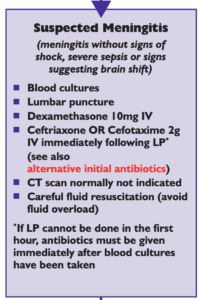
National (UK) guidelines on meningitis (community acquired meningitis in the immunocompetent host) available from meningitis.org. , including this box:

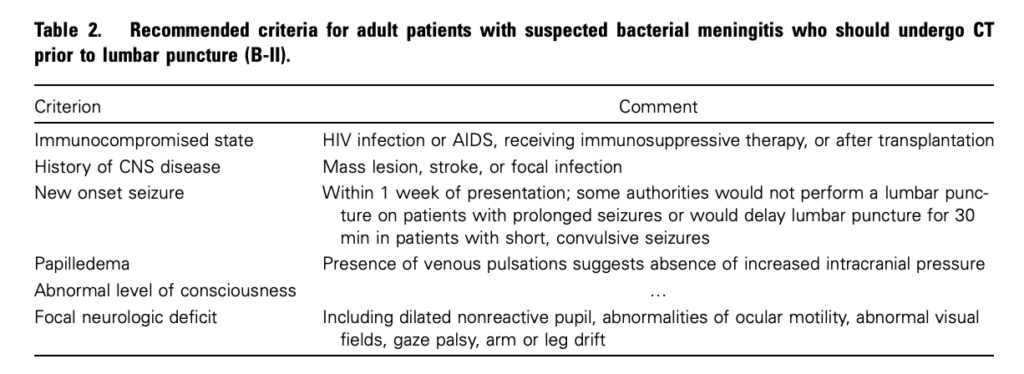
Practice Guidelines for the Management of Bacterial Meningitis These 2004 guidelines from the Infectious Diseases Society of America provide the following table listing the recommended criteria for adult patients with suspected bacterial meningitis who should undergo CT prior to lumbar puncture:

Clin Infect Dis. (2004) 39 (9): 1267-1284 Full text

4. This is potentially even more of an issue with paediatric patients

Fatal Lumbar Puncture: Fact Versus Fiction—An Approach to a Clinical Dilemma

An excellent summary of the above mentioned issues presented in a paediatric context, including the following:

On initial consideration a cranial CT would seem to be an appropriate and potentially useful diagnostic study for confirming the diagnosis of cerebral herniataion. The fallacy in this assessment has been emphasized by the finding that no clinically significant CT abnormalities are found that are not suspected on clinical assessments. Further, as previously noted, a normal CT examination may be found at about the time of a fatal herniation. Thus, the practical usefulness of a cranial CT in the majority of pediatric patients is limited to those rare patients whose increased ICP is secondary to mass lesions, not in the initial approach to acute meningitis.

Pediatrics. 2003 Sep;112(3 Pt 1):e174-6 Full Text

The last words should go to Dr Brad Spellberg, who in response to the IDSA’s guidelines wrote an excellent letter summarising much of the evidence at the time, confessed:

Why do we persist in using the CT scan for this purpose, despite the lack of supportive data? I am as guilty of this practice as anyone else, and the reason is simple: I am a chicken.

Clin Infect Dis. (2005) 40 (7): 1061 Full Text

Salicylate poisoning and pseudohyperchloraemia


Severe salicylate poisoning can cause metabolic acidosis from an accumulation of salicylic acid, lactic acid, and ketone bodies. A high anion gap acidosis is therefore the typical metabolic abnormality seen. A case series illustrates salicylate poisoning presenting with a normal gap (hyperchloraemic) acidosis – one patient had a chloride of 111 mmol/l and the other 123 mmol/l. This can occur when some analysers falsely read an elevated chloride in the presence of high concentrations of salicylate.


Severe salicylate poisoning is classically associated with an anion gap metabolic acidosis. However, high serum salicylate levels can cause false increase of laboratory chloride results on some analyzers. We present 2 cases of life-threatening salicylate poisoning with an apparently normal anion gap caused by an important laboratory interference. These cases highlight that the diagnosis of severe salicylism must be considered in all patients presenting with metabolic acidosis, even in the absence of an increased anion gap.

Falsely Normal Anion Gap in Severe Salicylate Poisoning Caused by Laboratory Interference
Ann Emerg Med. 2011 Sep;58(3):280-1

Oxygen prevented hypoxia. Yep.


Why wouldn’t you give oxygen prophylactically to someone undergoing procedural sedation? One argument is that this will delay the detection of respiratory depression since a pre-oxygenated patient can be hyponoeic/apnoeic for longer prior to desaturation. This is not an issue for those of us who use non-invasive capnography during sedation.
In this randomized trial of oxygen vs air during ED propofol procedural sedation there was less hypoxia when high-flow supplemental oxygen was added. The authors made the following observations:

  • There was no difference between groups in the incidence of respiratory depression, confirming previous research that supplemental oxygen does not exacerbate respiratory depression
  • 5 patients in the compressed air group developed hypoxia without preceding respiratory depression, so capnography cannot be completely relied on in this setting.

They summarise:
“…assuming that capnography is in place to monitor ventilatory function, our results strongly support the routine use of high-flow oxygen during ED propofol sedation”


STUDY OBJECTIVE: We determine whether high-flow oxygen reduces the incidence of hypoxia by 20% in adults receiving propofol for emergency department (ED) sedation compared with room air.

METHODS: We randomized adults to receive 100% oxygen or compressed air at 15 L/minute by nonrebreather mask for 5 minutes before and during propofol procedural sedation. We administered 1.0 mg/kg of propofol, followed by 0.5 mg/kg boluses until the patient was adequately sedated. Physicians and patients were blinded to the gas used. Hypoxia was defined a priori as an oxygen saturation less than 93%; respiratory depression was defined as an end tidal CO(2) greater than 50 mm Hg, a 10% absolute change from baseline, or loss of waveform.

RESULTS: We noted significantly less hypoxia in the 59 patients receiving high-flow oxygen compared with the 58 receiving compressed air (19% versus 41%; P=.007; difference 23%; 95% confidence interval 6% to 38%). Respiratory depression was similar between groups (51% versus 48%; difference 2%; 95% confidence interval -15% to 22%). We observed 2 adverse events in the high-flow group (1 hypotension, 1 bradycardia) and 2 in the compressed air group (1 assisted ventilation, 1 hypotension).

CONCLUSION: High-flow oxygen reduces the frequency of hypoxia during ED propofol sedation in adults.

The Utility of High-Flow Oxygen During Emergency Department Procedural Sedation and Analgesia With Propofol: A Randomized, Controlled Trial
Ann Emerg Med. 2011 Oct;58(4):360-364

Easy rapid infusion set up

Kapoor and Singh's system from the Open Access article - click for explanation

Here’s a nice and simple set up for rapid iv infusions using simple cheap equipment

Full details at the Scandinavian Journal Site
Novel rapid infusion device for patients in emergency situations
Scand J Trauma Resusc Emerg Med. 2011 Jun 10;19:35 (Free Full Text)

Delta CVP with PEEP and fluid responsiveness

I’ve been (and remain) critical of the use of CVP to determine ‘filling status’ or more accurately volume-responsiveness, even using CVP trends; I’m generally in agreement with Dr Marik’s bold statement that “CVP should not be used to make clinical decisions regarding fluid management”1. However there might now appear to be a way of using CVP for this purpose.
Increasing PEEP in patients undergoing positive pressure ventilation can increase the CVP. It has been demonstrated in a small study of cardiac surgical patients2 that the degree to which a 10cmH2O increase in PEEP changes the CVP correlates with fluid responsiveness. The fluid responsiveness was determined by the change in cardiac output measured by thermodilution after a passive leg raise.
There are a number of limitations to this study that should prevent us from immediately extrapolating this method of determining fluid responsiveness to our ED / critical care patients, but the concept is interesting. This can be added to the growing pile of dynamic measures of circulatory filling.


Background Changes in central venous pressure (CVP) rather than absolute values may be used to guide fluid therapy in critically ill patients undergoing mechanical ventilation. We conducted a study comparing the changes in the CVP produced by an increase in PEEP and stroke volume variation (SVV) as indicators of fluid responsiveness. Fluid responsiveness was assessed by the changes in cardiac output (CO) produced by passive leg raising (PLR).

Methods In 20 fully mechanically ventilated patients after cardiac surgery, PEEP was increased +10 cm H2O for 5 min followed by PLR. CVP, SVV, and thermodilution CO were measured before, during, and directly after the PEEP challenge and 30° PLR. The CO increase >7% upon PLR was used to define responders.

Results Twenty patients were included; of whom, 10 responded to PLR. The increase in CO by PLR directly related (r=0.77, P<0.001) to the increase in CVP by PEEP. PLR responsiveness was predicted by the PEEP-induced increase in CVP [area under receiver-operating characteristic (AUROC) curve 0.99, P<0.001] and by baseline SVV (AUROC 0.90, P=0.003). The AUROC's for dCVP and SVV did not differ significantly (P=0.299).
Conclusions Our data in mechanically ventilated, cardiac surgery patients suggest that the newly defined parameter, PEEP-induced CVP changes, like SVV, appears to be a good parameter to predict fluid responsiveness.

1. Does central venous pressure predict fluid responsiveness? A systematic review of the literature and the tale of seven mares.
Chest. 2008 Jul;134(1):172-8
Full Text Link
2. Predicting cardiac output responses to passive leg raising by a PEEP-induced increase in central venous pressure, in cardiac surgery patients.
Br J Anaesth. 2011 Aug;107(2):251-7

Is there nothing ketamine can't do?

As well as the benefits of cardiovascular stability, maintenance of cerebral perfusion pressure, possibly lowering ICP and providing other neuroprotective benefits, ketamine may have other advantages. These are reviewed in a British Journal of Anaesthesia article from which I’ve selected those benefits of interest to practitioners of emergency medicine and critical care.


Additional Beneficial Effects of Ketamine

  • the dysphoric, or ’emergence’ reactions associated with ketamine may be reduced by pre-administration or co-administration of sedatives, such as benzodiazepines, propofol, dexmedetomidine, or droperidol.
  • ketamine potentiates opioid analgesia in multiple settings, reducing opioid total dose and in some groups of patients reducing postoperative desaturation
  • ketamine has possible anti-inflammatory effects demonstrated in some types of surgical patients
  • ketamine may prevent awareness, recall, or both during general anaesthesia

Ketamine: new uses for an old drug?
Br J Anaesth. 2011 Aug;107(2):123-6

Lateral trauma position

Image from sjtrem.com - click for original

The tradition of transporting trauma patients to hospital in a supine position may not be the safest approach in obtunded patients with unprotected airways. The ‘solution’ of having them on an extrication board (backboard / long spine board) to enable rolling them to one side in the event of vomiting may not be practicable for limited crew numbers.
The Norwegians have been including the option of the lateral trauma position in their pre-hospital trauma life support training for some years now.
A questionnaire study demonstrates that this method has successfully been adopted by Norwegian EMS systems.
The method of application is described as:

  • Check airways (look, listen, feel).
  • Apply chin lift/jaw thrust, suction if needed.
  • Apply stiff neck collar.
  • If the patient is unresponsive, but has spontaneous respiration: Roll patient to lateral/recovery position while maintaining head/neck position.
  • Roll to side that leaves the patient facing outwards in ambulance coupé.
  • Transfer to ambulance stretcher (Scoop-stretcher, log-roll onto stretcher mattress, or use multiple helpers, lifting by patient’s clothing).
  • Support head, secure with three belts (across legs, over hip, over shoulder)
  • Manual support of head, supply oxygen, observation, suction, BVM (big valve mask) ventilation when needed.

Different options for supporting the head in the lateral position, according to questionnaire responders, include:

  • putting padding under the head, such as a pillow or similar item (81%)
  • a combination of padding and putting the head on the lower arm (7%)
  • rest the head on the lower arm alone (10%)
  • rest the head on the ground (<1%)


BACKGROUND: Trauma patients are customarily transported in the supine position to protect the spine. The Airway, Breathing, Circulation, Disability, and Exposure (ABCDE) principles clearly give priority to airways. In Norway, the lateral trauma position (LTP) was introduced in 2005. We investigated the implementation and current use of LTP in Norwegian Emergency Medical Services (EMS).

METHODS: All ground and air EMS bases in Norway were included. Interviews were performed with ground and air EMS supervisors. Questionnaires were distributed to ground EMS personnel.

RESULTS: Of 206 ground EMS supervisors, 201 answered; 75% reported that LTP is used. In services using LTP, written protocols were present in 67% and 73% had provided training in LTP use. Questionnaires were distributed to 3,025 ground EMS personnel. We received 1,395 (46%) valid questionnaires. LTP was known to 89% of respondents, but only 59% stated that they use it. Of the respondents using LTP, 77% reported access to written protocols. Flexing of the top knee was reported by 78%, 20% flexed the bottom knee, 81% used under head padding. Of 24 air EMS supervisors, 23 participated. LTP is used by 52% of the services, one of these has a written protocol and three arrange training.

CONCLUSIONS: LTP is implemented and used in the majority of Norwegian EMS, despite little evidence as to its possible benefits and harms. How the patient is positioned in the LTP differs. More research on LTP is needed to confirm that its use is based on evidence that it is safe and effective.

The lateral trauma position: What do we know about it and how do we use it? A cross-sectional survey of all Norwegian emergency medical services
Scand J Trauma Resusc Emerg Med. 2011 Aug 4;19:45
Open Access Full Text

Capillary refill time

A review of capillary refill time (CRT) reveals some interesting details about this test:

  • CRT is affected by age – the upper limit of normal for neonates is 3 seconds.
  • It increases with age – one study recommended the upper limit of normal for adult women should be increased to 2.9 seconds and for the elderly to 4.5 seconds.
  • It is affected by multiple external factors (especially ambient temperature).
  • Although it is claimed to have some predictive value in the assessment of dehydration and serious infection in children, studies vary in where and for how long pressure should be applied, and there is poor interobserver reliability.

The latest (5th Edition) of the Advanced Paediatric Life Support Manual states:
Poor capillary refill and differential pulse volumes are neither sensitive nor specific indicators of shock in infants and children, but are useful clinical signs when used in conjunction with the other signs described
In my view, it is best used as a monitor of trends (in accordance with skin temperature and other markers of perfusion), rather than by placing emphasis on the exact number of seconds of a single reading. See below for a video of my perfectly happy and healthy son demonstrating a CRT of over six seconds in a cool room during an English Summer’s day.
The authors of the review caution:
Operating rooms are cold, patients are often draped, which limits access, and because most anesthetics are potent vasodilators, the use of CRT to guide practice is not justified. The possibility of a false-positive or false-negative assessment is simply too great.


Capillary refill time (CRT) is widely used by health care workers as part of the rapid, structured cardiopulmonary assessment of critically ill patients. Measurement involves the visual inspection of blood returning to distal capillaries after they have been emptied by pressure. It is hypothesized that CRT is a simple measure of alterations in peripheral perfusion. Evidence for the use of CRT in anesthesia is lacking and further research is required, but understanding may be gained from evidence in other fields. In this report, we examine this evidence and factors affecting CRT measurement. Novel approaches to the assessment of CRT are under investigation. In the future, CRT measurement may be achieved using new technologies such as digital videography or modified oxygen saturation probes; these new methods would remove the limitations associated with clinical CRT measurement and may even be able to provide an automated CRT measurement.

Capillary Refill Time: Is It Still a Useful Clinical Sign?
Anesth Analg. 2011 Jul;113(1):120-3
The Capillary Refill Video