Newly trained BLS providers were more likely to spend two minutes counting 5 cycles of 30:2 compression:ventilation than to correctly identify when two minutes had elapsed.

Using the 30 : 2 compression–ventilation ratio: five cycles is easier to follow than 2 min of cardiopulmonary resuscitation
Eur J Emerg Med. 2009 Dec;16(6):339-41
Category Archives: Resus
Life-saving medicine
Abnormal head CT in altered mental status
In a study of 674 patients with altered mental status who received a CT scan of the brain, logistic regression analysis identified a series of clinical factors that were associated with an abnormal CT result.
Factors with an adjusted odds ratio between 1 and 2.5 included GCS less than 15, focal weakness, diastolic blood pressure greater than 80mmHg and antiplatelet use.
Four variables were associated with an adjusted odds ratio of 2.5 or above. These included presence of headache, dilated pupils (either unilateral or bilateral), upgoing plantar response and anticoagulant use.
Identifying risk factors for an abnormal computed tomographic scan of the head among patients with altered mental status in the Emergency Department
Eur J Emerg Med. 2009 Sep 21. [Epub ahead of print]
EGDT sepsis bundle challenged
An article in American Journal of Emergency Medicine by two intensivists challenges the science behind Rivers’ early goal-directed therapy (EGDT) protocol for severe sepsis / septic shock. In a nutshell:
- Rivers’ study was small (n = 263), nonblinded, industry-supported and single-center
- early fluids and antibiotics are a sound idea, but other components of EGDT are flawed
- targeting a CVP is meaningless and could result in hypovolaemia or pulmonary oedema; dynamic markers of preload responsiveness such as pulse pressure variation or IVC diameter variation are better guides to fluid resuscitation
- ScvO2 may be normal or elevated in septic shock patients; the low average ScvO2 in Rivers’ study has not been reproduced in subsequent studies.
- packed cells have significant side effects and their non-deformability, pro-inflammatory and pro-thrombotic effects may impair microvascular perfusion and paradoxically worsen tissue oxygen delivery
- dobutamine can potentially further worsen the haemodynamic status of patients with hypovolaemia, vasodilation, or a hyperdynamic circulation, which cannot be differentiated using CVP and ScvO2
Early goal-directed therapy: on terminal life support?
Am J Emerg Med. 2010 Feb;28(2):243-5
I like this paper, mainly because I have been uncomfortable with the chasing of arbitrary targets for some time. My own practice is to try to improve markers of organ hypoperfusion (such as lactate, urine output, mental status, and skin perfusion as well as blood pressure) by early antibiotics, fluid resuscitation guided by clinical and sonographic (IVC) signs, and vasoactive drugs guided by clinical and sonographic (basic echo) findings. I place a central venous catheter for access for the vasoactive drugs, rather than to get a CVP reading. I do measure ScvO2 with a central venous blood gas, but have rarely seen one below 70% even in severely shocked patients – I’m far more interested in clearing the lactate, as are these guys.
Biphasic shocks for AF and Atrial flutter
Based on a study of 453 consecutive patients undergoing their first transthoracic electrical cardioversion for atrial tachyarrhythmias, recommendations were developed to aim at delivering the lowest possible total cumulative energy with ≤2 consecutive shocks using the specific truncated exponential biphasic waveform incorporated in Medtronic Physio-Control devices: they recommend an initial energy setting of 50 J in patients with atrial flutter or atrial tachycardia, of 100 J in patients with atrial fibrillation (AF) of 2 or less days in duration, and of 150 J with AF of more than 2 days in duration. If the initial shock fails to restore sinus rhythm, a rescue shock of 250 J for AFL/AT or of 360 J for AF should be applied to secure the highest possible probability of successful cardioversion for each patient.
Optimization of initial energy for cardioversion of atrial tachyarrhythmias with biphasic shocks
Am J Emerg Med. 2010 Feb;28(2):159-65
Bad news for etomidate from CORTICUS
In an a priori substudy of the CORTICUS multi-centre, randomised, double-blind, placebo-controlled trial of hydrocortisone in septic shock, the use and timing of etomidate administration was examined in relation to outcome.
Of 499 analysable patients, 96 (19.2%) received etomidate within the 72 h prior to inclusion. The proportion of non-responders to ACTH was significantly higher in patients who were given etomidate than in other patients (61.0 vs. 44.6%, P = 0.004). Etomidate therapy was associated with a higher 28-day mortality in univariate analysis (P = 0.02) and after correction for severity of illness (42.7 vs. 30.5%; P=0.06 and P=0.03) in two multi-variant models. Hydrocortisone administration did not change the mortality of patients receiving etomidate (45 vs. 40%).
Some of the previous attacks on etomidate have not been founded on the most rigorous evidence. However this study adds further to the difficulty in justifying etomidate’s use when a perfectly acceptable alternative (ketamine) exists for rapid sequence induction in the haemodynamically unstable septic patient.
The effects of etomidate on adrenal responsiveness and mortality in patients with septic shock.
Intensive Care Med. 2009 Nov;35(11):1868-76
Whole-body CT during trauma resuscitation
German trauma patients are more likely to survive if they have a whole body CT rather than selective scans. Or that’s what this paper would have you believe IF you’re happy with the retrospective comparison, multivariate adjustments, and potential confounders. Still, if it helps you get your radiologists to play ball, the reference is…
Effect of whole-body CT during trauma resuscitation on survival: a retrospective, multicentre studyLancet. 2009 Apr 25;373(9673):1455-61
Ketamine and procedural success
There is a myth that increased muscular tone caused by ketamine leads to an increased failure rate of joint manipulations when this agent is used for procedural sedation in the ED. This is neither borne out by the published evidence nor our own experience of a series of cases, which have been presented by Louisa Chan at a former (UK) College of Emergency Medicine Conference. At the Australasian College of Emergency Medicine Annual Scientific Conference in Melbourne these data were presented by A/Professor Taylor’s team in Victoria, which provide evidence that procedural failure rate is in fact lower with ketamine than with other commonly used sedatives. Here is the abstract reproduced with the kind permission of A/Prof Taylor:
Failure to successfully complete a procedure following emergency department sedation
DMcD Taylor1,2 for the Emergency Department Sedation Study Investigators
1Austin Health; 2University of Melbourne, Melbourne, Australia
Aims: To determine the nature and incidence of, and factors contributing to, failure to successfully complete a procedure fol- lowing sedation in the ED
Methods: Eleven Australian ED enrolled consecutive adult and paediatric patients between January 2006 and December 2008. Patients were included if a sedative drug was administered for an ED procedure. Data collection was prospective and employed a specifically designed form.
Results: Two thousand six hundred and twenty three patients were enrolled (60.3% male, mean age 39.2 years). Failure to successfully complete the procedure occurred in 148 (5.6%) cases. Most failures occurred with attempted reductions of fractured/dislocated shoulders (35 cases), hips (32), ankles (21) and elbows (14). However, failure rates were highest among fractured/dislocated hips (18.5%), digits (13.7%), femurs (11.1%), mandibles (10.2%) and elbows (9.3%). Failure rates for residents/registrars (5.9%), consultants (5.6%) and nurse practitioners (5.9%) did not differ (P = 0.92). Overall, failure rates for the various drugs (used alone or in combina- tion) did not differ (P = 0.07). However, ketamine (used alone or in combination) was associated with a much lower failure rate (2.9%) than all other sedation drugs used (midazolam 5.8%, propofol 6.5%, fentanyl 6.9%, nitrous oxide 7.1%, and morphine 7.8%).
Conclusion: Procedural failure is uncommon although some pro- cedures are at higher risk, especially dislocated hip reduction. Failure rates do not appear to be affected by the designation of the operator or the sedative drug used. However, ketamine use is associated with lower failure rates. For those procedures at higher risk of failure, the provision of optimal conditions (spe- cialist unit assistance, venue, drug selection) may minimise failure rates.
Emergency Medicine Australasia 2010;22(S1):A52-3
Cardiocerebral resuscitation
An emergency medical service introduced a cardiocerebral resuscitation protocol and compared outcomes with a standard ACLS protocol.
Cardiocerebral resuscitation (CCR) was defined as:
- initiation of 200 immediate, uninterrupted chest compressions at a rate of 100 compressions ⁄ min
- analyzing the rhythm and delivering a single defibrillator shock, if indicated
- 200 more chest compressions before the first pulse check or rhythm reanalysis
- epinephrine (1 mg intravenous or intraosseous) as soon as possible or with each 200 compression cycle
- endotracheal intubation delayed until after three cycles of chest compressions
Data was analysed from a registry including data on 3515 patients from 62 EMS agencies, some of which instituted CCR (in a total of 1024 patients). Outcome predictors were identified using logistic regression analysis and
Individuals who received CCR had better outcomes across age groups. The increase in survival for the subgroup with a witnessed Vfib was most prominent on those <40 years of age (3.7% for standard ALS patients vs. 19% for CCR patients, odds ratio [OR] = 5.94, 95% confidence interval [CI] = 1.82 to 19.26). Neurologic outcomes were also better in the patients who received CCR (OR = 6.64, 95% CI = 1.31 to 32.8). Within the subgroup that received CCR, the factors most predictive of improved survival included witnessed arrest, initial rhythm of Vfib⁄Vtach, agonal respirations upon arrival, EMS response time, and age. Neurologic outcome was not adversely affected by age.
Cardiocerebral Resuscitation Is Associated With Improved Survival and Neurologic Outcome from Out-of-hospital Cardiac Arrest in Elders
Academic Emergency Medicine 2010;17(3):269 – 275
End tidal CO2 and procedural sedation
One hundred and thirty-two adults underwent propofol sedation in the emergency department and were randomised into a group in which treating physicians had access to the capnography and a blinded group in which they did not. All patients received supplemental oxygen (3 L/minute) and opioids greater than 30 minutes before. Propofol was dosed at 1.0 mg/kg, followed by 0.5 mg/kg as needed.
Hypoxia (defined as SpO2 less than 93%) was observed in 17 of 68 (25%) subjects with capnography and 27 of 64 (42%) with blinded capnography (p=.035; difference 17%; 95% confidence interval 1.3% to 33%). Capnography identified all cases of hypoxia before onset (sensitivity 100%; specificity 64%), with the median time from capnographic evidence of respiratory depression to hypoxia 60 seconds (range 5 to 240 seconds).
The journal comments: ‘this study provides compelling evidence that capnography can aid in the detection of respiratory depression and reduce hypoxia during procedural sedation.’
However in an accompanying article outlining a pro-con debate for introducing capnography as standard practice in ED procedural sedation, the point is made that the safety benefit purported in this and similar studies is decreased hypoxemia, according to thresholds ranging from 90% to 95%, lasting from 5 to 15 seconds. In the clinical context, many of these events are self-limiting or resolve with minimal interventions such as airway repositioning or supplemental oxygen, and other more clinically relevant outcomes are rarely examined (perhaps due to the rarity of genuinely adverse events in ED procedural sedation by emergency physicians).
Does end tidal CO2 monitoring during emergency department procedural sedation and analgesia with propofol decrease the incidence of hypoxic events? A randomized, controlled trial
Ann Emerg Med. 2010 Mar;55(3):258-64
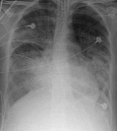
Higher PEEP in ARDS
The current mortality of 35% associated with acute lung injury (ALI) is roughly three times higher than that associated with ST-segment elevation myocardial infarction. Protective ventilation strategies limiting tidal volumes and plateau pressures improve outcome, but the optimial level of PEEP is debated. In patients with ALI and its more severe form acute respiratory distress syndrome (ARDS), higher levels of PEEP may prevent atelectasis, recruit already collapsed alveolar units, and reduce pulmonary damage by avoiding the cyclical opening and collapse of alveoli.

In a systematic review and meta-analysis of individual-patient data, researchers investigated the association between higher vs lower PEEP levels and patient-important outcomes among adults with acute lung injury or ARDS who receive ventilation with low tidal volumes.
Randomized trials eligible for this review compared higher with lower levels of PEEP (mean difference of at least 3 cm H2O between groups) in critically ill adults with ALI or ARDS. Eligible trials incorporated a target tidal volume of less than 8 mL/kg of predicted body weight in both the experimental and the control ventilation strategies and provided patient follow-up to death or for at least 20 days.

Three trials, including 2299 patients, met the eligibility criteria: the Assessment of Low Tidal Volume and Elevated End-Expiratory Pressure to Obviate Lung Injury (ALVEOLI) trial, the Lung Open Ventilation to Decrease Mortality in the Acute Respiratory Distress Syndrome (LOVS) study, and the Expiratory Pressure Study (EXPRESS).
There were 374 hospital deaths in 1136 patients (32.9%) assigned to treatment with higher PEEP and 409 hospital deaths in 1163 patients (35.2%) assigned to lower PEEP (adjusted relative risk [RR], 0.94; 95% confidence interval [CI], 0.86-1.04; P = .25). Treatment effects varied with the presence or absence of ARDS (as opposed to ALI). In patients with ARDS (n = 1892), there were 324 hospital deaths (34.1%) in the higher PEEP group and 368 (39.1%) in the lower PEEP group (adjusted RR, 0.90; 95% CI, 0.81-1.00; P = .049). Rates of pneumothorax and vasopressor use were similar.
The authors conclude that treatment with higher vs lower levels of PEEP was not associated with improved hospital survival overall when ALI/ARDS were considered together, but higher levels were associated with improved survival among the pre-defined subgroup of patients with ARDS.
Higher vs lower positive end-expiratory pressure in patients with acute lung injury and acute respiratory distress syndrome: systematic review and meta-analysis
JAMA. 2010 Mar 3;303(9):865-73
