Tag Archives: airway

Advanced airways and worse outcomes in cardiac arrest

A new study demonstrates an association between advanced prehospital airway management and worse clinical outcomes in patients with cardiac arrest. Done in Japan, the numbers of patients included are staggering: this nationwide population-based cohort study included 658 829 adult patients. They found that CPR with advanced airway management (use of tracheal tubes and even supraglottic airways) was a significant predictor of poor neurological outcome compared with conventional bag-valve-mask ventilation.
Association of Prehospital Advanced Airway Management With Neurologic Outcome and Survival in Patients With Out-of-Hospital Cardiac Arrest
JAMA 2013;309(3):257-66
[EXPAND Click to read abstract]


Importance It is unclear whether advanced airway management such as endotracheal intubation or use of supraglottic airway devices in the prehospital setting improves outcomes following out-of-hospital cardiac arrest (OHCA) compared with conventional bag-valve-mask ventilation.

Objective To test the hypothesis that prehospital advanced airway management is associated with favorable outcome after adult OHCA.

Design, Setting, and Participants Prospective, nationwide, population-based study (All-Japan Utstein Registry) involving 649 654 consecutive adult patients in Japan who had an OHCA and in whom resuscitation was attempted by emergency responders with subsequent transport to medical institutions from January 2005 through December 2010.

Main Outcome Measures Favorable neurological outcome 1 month after an OHCA, defined as cerebral performance category 1 or 2.

Results Of the eligible 649 359 patients with OHCA, 367 837 (57%) underwent bag-valve-mask ventilation and 281 522 (43%) advanced airway management, including 41 972 (6%) with endotracheal intubation and 239 550 (37%) with use of supraglottic airways. In the full cohort, the advanced airway group incurred a lower rate of favorable neurological outcome compared with the bag-valve-mask group (1.1% vs 2.9%; odds ratio [OR], 0.38; 95% CI, 0.36-0.39). In multivariable logistic regression, advanced airway management had an OR for favorable neurological outcome of 0.38 (95% CI, 0.37-0.40) after adjusting for age, sex, etiology of arrest, first documented rhythm, witnessed status, type of bystander cardiopulmonary resuscitation, use of public access automated external defibrillator, epinephrine administration, and time intervals. Similarly, the odds of neurologically favorable survival were significantly lower both for endotracheal intubation (adjusted OR, 0.41; 95% CI, 0.37-0.45) and for supraglottic airways (adjusted OR, 0.38; 95% CI, 0.36-0.40). In a propensity score–matched cohort (357 228 patients), the adjusted odds of neurologically favorable survival were significantly lower both for endotracheal intubation (adjusted OR, 0.45; 95% CI, 0.37-0.55) and for use of supraglottic airways (adjusted OR, 0.36; 95% CI, 0.33-0.39). Both endotracheal intubation and use of supraglottic airways were similarly associated with decreased odds of neurologically favorable survival.

Conclusion and Relevance Among adult patients with OHCA, any type of advanced airway management was independently associated with decreased odds of neurologically favorable survival compared with conventional bag-valve-mask ventilation.

[/EXPAND]

Lifting the Fogg on ED Intubaton

Fellow retrieval specialist and Royal North Shore Hospital emergency physician Dr Toby Fogg and coauthors have published their audit of intubations in an Australian Emergency Department(1). More important than the results themselves is that the process of monitoring ones practice inevitably leads to improvements. For example, at Toby’s institution an intubation checklist has been introduced since the audit began. Other Australasian EDs are encouraged to participate using the free resources at airwayregistry.org.au.
Recently we have also seen the publication of Korean registry data on paediatric intubations performed in 13 academic EDs over 5 years(2), in which first pass success rates (overall 67.6%) were higher with emergency physicians compared with paediatricians. Interestingly, a rapid sequence intubation technique was only used in 22.4% of intubations, which was more likely to be used by emergency physicians and was associated with a greater likelihood of first pass success.
This relatively low first pass success rate is reminiscent of the American study published in September(3) which raised some eyebrows with its 52% first pass intubation success rates in a paediatric ED, and which also showed that attending-level providers were 10 times more likely to be successful on the first attempt than all trainees combined. Possible reasons for such a low first pass success rate compared with adult registry data include the rigorous video analysis method used, or perhaps more likely that paediatric emergency subspecialists are exposed to fewer critical procedures, resuscitations, and intubations than their general emergency medicine counterparts(4).
As a specialty we must continue to seek to do better, and I salute all these brave authors who are telling it like it is. Particularly with children, whose airways are relatively easy, we have to develop the training, preparation, supervision, monitoring and feedback to aim for as high a success rate as possible.

Study authors Toby Fogg and Nick Annesley demonstrate the 'Happiness Triad'

1. Prospective observational study of the practice of endotracheal intubation in the emergency department of a tertiary hospital in Sydney, Australia
Emerg Med Australas. 2012 Dec;24(6):617-24
[EXPAND Click to read abstract]


OBJECTIVE: To describe the practice of endotracheal intubation in the ED of a tertiary hospital in Australia, with particular emphasis on the indication, staff seniority, technique, number of attempts required and the rate of complications.

METHODS: A prospective observational study.

RESULTS: Two hundred and ninety-five intubations occurred in 18 months. Trauma was the indication for intubation in 30.5% (95% CI 25.3-36.0) and medical conditions in 69.5% (95% CI 64.0-74.5). Emergency physicians were team leaders in 69.5% (95% CI 64.0-74.5), whereas ED registrars or senior Resident Medical Officers made the first attempt at intubation in 88.1% (95% CI 83.9-91.3). Difficult laryngoscopy occurred in 24.0% (95% CI 19.5-29.3) of first attempts, whereas first pass success occurred in 83.4% (95% CI 78.7-87.2). A difficult intubation occurred in 3.4% (95% CI 1.9-6.1) and all patients were intubated orally in five or less attempts. A bougie was used in 30.9% (95% CI 25.8-36.5) of first attempts, whereas a stylet in 37.5% (95% CI 32.1-43.3). Complications occurred in 29.0% (95% CI 23.5-34.1) of the patients, with desaturation the commonest in 15.7% (95% CI 11.9-20.5). Cardiac arrest occurred in 2.2% (95% CI 0.9-4.4) after intubation. No surgical airways were undertaken.

CONCLUSION: Although the majority of results are comparable with overseas data, the rates of difficult laryngoscopy and desaturation are higher than previously reported. We feel that this data has highlighted the need for practice improvement within our department and we would encourage all those who undertake emergent airway management to audit their own practice of this high-risk procedure.

[/EXPAND]
2. The factors associated with successful paediatric endotracheal intubation on the first attempt in emergency departments: a 13-emergency-department registry study
Resuscitation. 2012 Nov;83(11):1363-8
[EXPAND Click to read abstract]


BACKGROUND: We investigated which factors are associated with successful paediatric endotracheal intubation (ETI) on the first attempt in emergency department (EDs) from multicentre emergency airway registry data.

METHODS: We created a multicentre registry of intubations at 13 EDs and performed surveillance over 5 years. Each intubator filled out a data form after an intubation. We defined “paediatric patients” as patients younger than 10 years of age. We assessed the specialty and level of training of intubator, the method, the equipment, and the associated adverse events. We analysed the intubation success rates on the first attempt (first-pass success, FPS) based on these variables.

RESULTS: A total of 430 ETIs were performed on 281 children seen in the ED. The overall FPS rate was 67.6%, but emergency medicine (EM) physicians showed a significantly greater success rate of 74.4%. In the logistic regression analysis, the intubator’s specialty was the only independent predictive factor for paediatric FPS. In the subgroup analysis, the EM physicians used the rapid sequence intubation/intubation (RSI) method and Macintosh laryngoscope more frequently than physicians of other specialties. ETI-related adverse events occurred in 21 (7.2%) out of the 281 cases. The most common adverse event in the FPS group was mainstem bronchus intubation, and vomiting was the most common event in the non-FPS group. The incidence of adverse events was lower in the FPS group than in the non-FPS group, but this difference was not statistically significant.

CONCLUSIONS: The intubator’s specialty was the major factor associated with FPS in emergency department paediatric ETI, The overall ETI FPS rate among paediatric patients was 67.6%, but the EM physicians had a FPS rate of 74.4%. A well structured airway skill training program, and more actively using the RSI method are important and this could explain this differences.

[/EXPAND]
3.Rapid sequence intubation for pediatric emergency patients: higher frequency of failed attempts and adverse effects found by video review.
Ann Emerg Med. 2012 Sep;60(3):251-9
[EXPAND Click to read abstract]


STUDY OBJECTIVE: Using video review, we seek to determine the frequencies of first-attempt success and adverse effects during rapid sequence intubation (RSI) in a large, tertiary care, pediatric emergency department (ED).

METHODS: We conducted a retrospective study of children undergoing RSI in the ED of a pediatric institution. Data were collected from preexisting video and written records of care provided. The primary outcome was successful tracheal intubation on the first attempt at laryngoscopy. The secondary outcome was the occurrence of any adverse effect during RSI, including episodes of physiologic deterioration. We collected time data from the RSI process by using video review. We explored the association between physician type and first-attempt success.

RESULTS: We obtained complete records for 114 of 123 (93%) children who underwent RSI in the ED during 12 months. Median age was 2.4 years, and 89 (78%) were medical resuscitations. Of the 114 subjects, 59 (52%) were tracheally intubated on the first attempt. Seventy subjects (61%) had 1 or more adverse effects during RSI; 38 (33%) experienced oxyhemoglobin desaturation and 2 required cardiopulmonary resuscitation after physiologic deterioration. Fewer adverse effects were documented in the written records than were observed on video review. The median time from induction through final endotracheal tube placement was 3 minutes. After adjusting for patient characteristics and illness severity, attending-level providers were 10 times more likely to be successful on the first attempt than all trainees combined.

CONCLUSION: Video review of RSI revealed that first-attempt failure and adverse effects were much more common than previously reported for children in an ED.

[/EXPAND]
4. A is for airway: a pediatric emergency department challenge.
Ann Emerg Med. 2012 Sep;60(3):261-3

Reassurance: difficult laryngoscopy in children remains rare

I was taught a useful principle by a paediatric anaesthetist 10 years ago which has proven true in my experience and has contributed to keeping me calm when intubating sick kids. Unlike adults, in whom difficulty in intubation can often be unexpected, the vast majority of normal looking children are easy to intubate, and the ones who are difficult usually have obvious indicators such as dysmorphism.
This appears to be supported by recent evidence: in a large retrospective series of 11.219 anaesthesia patients, the overall incidence of difficult laryngoscopy [Cormack and Lehane (CML) grade III and IV] was only 1.35%, although was much higher in infants less than one year compared with older children. This low percentage is in the same ball park as two other paediatric studies. Besides younger age, their database suggested underweight, ASA III and IV physical status and, if obtainable, Mallampati III and IV findings as predictors for difficult laryngoscopy. The authors point out:

…the oromaxillofacial surgery department with a high proportion of cleft palate interventions and pediatric cardiac surgery contributed substantially to the total number of difficult laryngoscopies. In patients undergoing pediatric cardiac surgery, a possible explanation for the higher incidence of CML III/IV findings might be that some congenital heart defects are associated with chromosomal anomalies like microdeletion 22q11.2 syndrome. This syndrome is also associated with extracardiac anomalies like cranio-facial dysmorphism

Take home message: As a very rough rule of thumb to illustrate the difference between the ease/difficulty of laryngoscopy between adults and kids, I think it’s fair to say grade III or IV views occur in about 10% of adults but only about 1% of children.

Incidence and predictors of difficult laryngoscopy in 11.219 pediatric anesthesia procedures
Paediatr Anaesth. 2012 Aug;22(8):729-36
[EXPAND Click for abstract]


OBJECTIVE: Difficult laryngoscopy in pediatric patients undergoing anesthesia.

AIM: This retrospective analysis was conducted to investigate incidence and predictors of difficult laryngoscopy in a large cohort of pediatric patients receiving general anesthesia with endotracheal intubation.

BACKGROUND: Young age and craniofacial dysmorphy are predictors for the difficult pediatric airway and difficult laryngoscopy. For difficult laryngoscopy, other general predictors are not yet described.

METHODS: Retrospectively, from a 5-year period, data from 11.219 general anesthesia procedures in pediatric patients with endotracheal intubation using age-adapted Macintosh blades in a single center (university hospital) were analyzed statistically.

RESULTS: The overall incidence of difficult laryngoscopy [Cormack and Lehane (CML) grade III and IV] was 1.35%. In patients younger than 1 year, the incidence of CML III or IV was significantly higher than in the older patients (4.7% vs 0.7%). ASA Physical Status III and IV, a higher Mallampati Score (III and IV) and a low BMI were all associated (P < 0.05) with difficult laryngoscopy. Patients undergoing oromaxillofacial surgery and cardiac surgery showed a significantly higher rate of CML III/IV findings.

CONCLUSION: The general incidence of difficult laryngoscopy in pediatric anesthesia is lower than in adults. Our results show that the risk of difficult laryngoscopy is much higher in patients below 1 year of age, in underweight patients and in ASA III and IV patients. The underlying disease might also contribute to the risk. If the Mallampati score could be obtained, prediction of difficult laryngoscopy seems to be reliable. Our data support the existing recommendations for a specialized anesthesiological team to provide safe anesthesia for infants and neonates.

[/EXPAND]

Transtracheal airways in kids. Well, pigs' kids anyway

Ever had to do a surgical airway in a child? Thought not. They’re pretty rare. Bill Heegaard MD from Henepin County Medical Center taught me a few approaches (with the help of an anaesthetised rabbit) which really got me thinking. It’s something I’d often trained for in my internal simulator, and I even keep the equipment for it in my house (listen out for an upcoming podcast on that). Research and experience has demonstrated that open surgical airway techniques are more reliable than transtracheal needle techniques in adults, but what about kids, in whom traditional teaching cautions against open techniques?

Australian investigators who were experienced airway proceduralists evaluated transtracheal needle techniques using a rabbit model (an excellent model for the infant airway). Their success rate was only 60% and they perforated the posterior tracheal wall in 42% of attempts. Of 13 attempts to insert a dedicated paediatric tracheotomy device, the Quicktrach Child, none were successful(1) (they did not use the Quicktrach Infant model as it is not available in Australia).

Danish investigators used fresh piglet cadavers weighing around 8 kg to assess two transtracheal cannulas, in which they achieved success rates of 65.6% and 68.8%(2). There was also a very high rate of posterior tracheal wall perforation. Using an open surgical tracheostomy technique, they were successful in 97% of attempts. These were also experienced operators, with a median anaesthetic experience of 12.5 years.
Their tracheotomy technique was nice and simple, and used just a scalpel, scissors, and surgical towel clips. Here’s their technique:

Simple tracheotomy procedure described by Holm-Knudsen et al

  1. Identify larynx and proximal trachea by palpation
  2. Vertical incision through the skin and subcutaneous tissue from the upper part of larynx to the sternal notch
  3. Grasp strap muscles with two towel forceps and separate in the midline
  4. Palpate and identify the trachea (palpate rather than look for tracheal rings, as in a live patient one would expect bleeding to obscure the view)
  5. Stabilise the trachea by grasping it with a towel forceps
  6. Insert sharp tip of the scissors between two tracheal rings and lift the trachea anteriorly to avoid damage to the posterior wall
  7. Cut vertically in the midline of the trachea with the scissors – they chose to use the scissors to cut the tracheal rings to facilitate tube insertion
  8. Insert the tracheal tube

Using ultrasound and CT to evaluate comparative airway dimensions, the authors concluded that the pig model is most useful for training emergency airway management in older children aged 5–10 years.
Why were they doing a tracheotomy rather than a cricothyroidotomy? Reasons given by the authors include:

  • The infant cricothyroid membrane is very small
  • Palpation of the thyroid notch may be hindered by the overlying hyoid bone
  • The mandible may obstruct needle access to the cricothyroid membrane given the cephalad position in the neck of the infant larynx.

From an emergency medicine point of view, there are a couple of other reasons why we need to be able to access the trachea lower than the cricothyroid membrane. One is fractured larynx or other blunt or penetrating airway injury where there may be anatomical disruption at the cricothyroid level. The other situation is foreign body airway obstruction, when objects may lodge at the level of the cricoid ring which is functionally the narrowest part of the pediatric upper airway. Of course, alternative methods might be considered to remove the foreign body prior to tracheotomy, such as employing basic choking algorithms, and other techniques depending on whether you do or don’t have equipment.

Take home messages

  • Transtracheal airways in kids are so rare, we can’t avoid extrapolating animal data
  • Whichever infant or paediatric model is used, transtracheal needle techniques have a high rate of failure even by ‘experienced’ operators
  • The small size and easy compressibility of the airway probably contributes to this failure rate, including the high rate of posterior wall puncture
  • In keeping with adult audit data, open surgical techniques may have a higher success rate
  • Tracheotomy may be necessary rather than cricothyroidotomy in infants and children depending on clinical scenario and accessibility of anatomy
  • The stress and blood that is not simulated in cadaveric animal models will make open tracheotomy harder in a live patient, and so these success rates may not translate. However these factors do mean that whatever technique is used must be kept simple and should employ readily available and familiar equipment
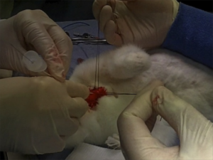
  • Something to maintain control and anterior position of the anterior trachea wall should be used during incision and intubation of the trachea. The study reported here used towel clips; sutures around the tracheal rings may also be used (see image below)
I recommend you add ‘paediatric tracheotomy’ to the list of procedures you might need to do (if it’s not already there). Identify what equipment you would use and run the simulation in your head and in your work environment.
Have fun.

1. The ‘Can’t Intubate Can’t Oxygenate’ scenario in Pediatric Anesthesia: a comparison of different devices for needle cricothyroidotomy
Paediatr Anaesth. 2012 Dec;22(12):1155-8

BACKGROUND: Little evidence exists to guide the management of the ‘Can’t Intubate, Can’t Oxygenate’ (CICO) scenario in pediatric anesthesia.

OBJECTIVES: To compare two intravenous cannulae for ease of use, success rate and complication rate in needle tracheotomy in a postmortem animal model of the infant airway, and trial a commercially available device using the same model.

METHODS: Two experienced proceduralists repeatedly attempted cannula tracheotomy in five postmortem rabbits, alternately using 18-gauge (18G) and 14-gauge (14G) BD Insyte(™) cannulae (BD, Franklin Lakes, NJ, USA). Attempts began at the first tracheal cartilage, with subsequent attempts progressively more caudad. Success was defined as intratracheal cannula placement. In each rabbit, an attempt was then made by each proceduralist to perform a cannula tracheotomy using the Quicktrach Child(™) device (VBM Medizintechnik GmbH, Sulz am Neckar, Germany).

RESULTS: The rabbit tracheas were of similar dimensions to a human infant. 60 attempts were made at cannula tracheotomy, yielding a 60% success rate. There was no significant difference in success rate, ease of use, or complication rate between cannulae of different gauge. Successful aspiration was highly predictive (positive predictive value 97%) and both sensitive (89%) and specific (96%) for tracheal cannulation. The posterior tracheal wall was perforated in 42% of tracheal punctures. None of 13 attempts using the Quicktrach Child(™) were successful.

CONCLUSION: Cannula tracheotomy in a model comparable to the infant airway is difficult and not without complication. Cannulae of 14- and 18-gauge appear to offer similar performance. Successful aspiration is the key predictor of appropriate cannula placement. The Quicktrach Child was not used successfully in this model. Further work is required to compare possible management strategies for the CICO scenario

2. Emergency airway access in children – transtracheal cannulas and tracheotomy assessed in a porcine model
Paediatr Anaesth. 2012 Dec;22(12):1159-65

OBJECTIVES: In the rare scenario when it is impossible to oxygenate or intubate a child, no evidence exists on what strategy to follow.

AIM: The aim of this study was to compare the time and success rate when using two different transtracheal needle techniques and also to measure the success rate and time when performing an emergency tracheotomy in a piglet cadaver model.

METHODS: In this randomized cross-over study, we included 32 anesthesiologists who each inserted two transtracheal cannulas (TTC) using a jet ventilation catheter and an intravenous catheter in a piglet model. Second, they performed an emergency tracheotomy. A maximum of 2 and 4 min were allowed for the procedures, respectively. The TTC procedures were recorded using a video scope.

RESULTS: Placement of a transtracheal cannula was successful in 65.6% and 68.8% of the attempts (P = 0.76), and the median duration of the attempts was 69 and 42 s (P = 0.32), using the jet ventilation catheter and the intravenous catheter, respectively. Complications were frequent in both groups, especially perforation of the posterior tracheal wall. Performing an emergency tracheotomy was successful in 97%, in a median of 88 s.

CONCLUSIONS: In a piglet model, we found no significant difference in success rates or time to insert a jet ventilation cannula or an intravenous catheter transtracheally, but the incidence of complications was high. In the same model, we found a 97% success rate for performing an emergency tracheotomy within 4 min with a low rate of complications.

Don't bronchodilators work in infants?

Inpatient paediatric teams can be scornful when bronchodilators are given by ED staff to wheezing infants, correctly referring to the lack of evidence of clinical benefit(1). There is however a persisting meme out there I’ve heard on a number of occasions that ‘young infants don’t have the receptors so inhaled beta agonists will never work.’ I’d love to know where this comes from.
Apparently, beta 2-receptors are present from the 16th gestational week(2). Pulmonary function testing of ventilated, very-low-birth-weight babies has shown that some consistently responded to beta-agonists whereas others did not(3). A newly published study reports that a quarter of mechanically ventilated infants with bronchiolitis were responders to inhaled albuterol, defined as a reduction in respiratory system resistance more than 30% below baseline(4).
In summary: beta-agonist bronchodilators have not been shown to improve clinical outcomes in wheezing infants. However some infants with some wheezing disorders will show a response in terms of pulmonary function. The receptors are there, and in life-threatening presentations bronchodilators should certainly be considered.
1. Short acting beta agonists for recurrent wheeze in children under 2 years of age
Cochrane Database Syst Rev. 2002;(3):CD002873
[EXPAND Click to read abstract]


BACKGROUND: Wheeze is a common symptom in infancy and is a common cause for both primary care consultations and hospital admission. Beta2-adrenoceptor agonists (b2-agonists) are the most frequently used as bronchodilator but their efficacy is questionable.
OBJECTIVES: To determine the effectiveness of b2-agonist for the treatment of infants with recurrent and persistent wheeze.
SEARCH STRATEGY: Relevant trials were identified using the Cochrane Airways Group database (CENTRAL), Medline and Pubmed. The database search used the following terms: Wheeze or asthma and Infant or Child and Short acting beta-agonist or Salbutamol (variants), Albuterol, Terbutaline (variants), Orciprenaline, Fenoterol

SELECTION CRITERIA: Randomised controlled trials comparing the effect of b2-agonist against placebo in children under 2 years of age who had had two or more previous episodes of wheeze, not related to another form of chronic lung disease.
DATA COLLECTION AND ANALYSIS: Eight studies met the criteria for inclusion in this meta-analysis. The studies investigated patients in three settings: at home (3 studies), in hospital (2 studies) and in the pulmonary function laboratory (3 studies). The main outcome measure was change in respiratory rate except for community based studies where symptom scores were used.

MAIN RESULTS: The studies were markedly heterogeneous and between study comparisons were limited. Improvement in respiratory rate, symptom score and oxygen saturation were noted in one study in the emergency department following two salbutamol nebulisers but this had no impact on hospital admission. There was a reduction in bronchial reactivity following salbutamol. There was no significant benefit from taking regular inhaled salbutamol on symptom scores recorded at home.

REVIEWER’S CONCLUSIONS: There is no clear benefit of using b2-agonists in the management of recurrent wheeze in the first two years of life although there is conflicting evidence. At present, further studies should only be performed if the patient group can be clearly defined and there is a suitable outcome parameter capable of measuring a response.

[/EXPAND]
2. The beta-2-agonists in asthma in infants and young children
Arch Pediatr. 2002 Aug;9 Suppl 3:384s-389s
[EXPAND Click to read abstract]


Beta 2-agonists, by inducing a fast and long relaxation of the bronchial smooth muscle, are considered as the more potent bronchodilators. beta 2-receptors are present from the 16th gestational week, explaining a possible bronchial response in the youngest children. beta 2-agonists do not induce any bronchodilator response in healthy children. Short-acting beta 2-agonists (salbutamol or albuterol, terbutaline) are indicated for asthma attacks, as needed in chronic asthma, and for prevention of symptoms during effort. They are safe and secure. The more efficient route of administration in preschool children is pressurized metered-dose inhaler used with a spacer device. Therefore, whatever the route of inhalation chosen (inhalation, injection, or continuous nebulization in acute asthma attack), more specified indications and doses are needed in young children. Long-acting beta 2-agonists (formoterol, salmeterol) are not authorized in France in children under 4 to 5 years of age depending on the drug used. Because of new oral formulations and recent considerations about their use in asthma attack, instead of short-acting beta 2-agonists, their indication in preschool asthmatic children might be reconsidered.

[/EXPAND]
3. Use of a beta-agonist in ventilated, very-low-birth-weight babies: a longitudinal evaluation
Dev Pharmacol Ther. 1990;15(2):61-7
[EXPAND Click to read abstract]


To determine if there is a specific postnatal (PNA) or postconceptional age (PCA) at which ventilated preterm infants respond to beta-agonists, we evaluated 15 infants with a mean gestational age of 26.5 +/- 1.5 weeks and mean birth weight of 0.89 +/- 0.23 kg who required mechanical ventilation at 10 days of age. Weekly pulmonary function testing (PFT) was performed before and 1 h after administration of albuterol. Taking the group as a whole, as well as individual babies, regression analysis showed no relationship between positive response and either PNA or PCA. Evaluation of individual infants, however, showed that some consistently responded to beta-agonists whereas others did not. We recommend individual PFT to identify those infants who will benefit from use of beta-agonists.

[/EXPAND]
4. Pulmonary mechanics following albuterol therapy in mechanically ventilated infants with bronchiolitis
J Asthma. 2012 Sep;49(7):688-96
[EXPAND Click to read abstract]


BACKGROUND AND AIMS: Bronchiolitis is a common cause of critical illness in infants. Inhaled β(2)-agonist bronchodilators are frequently used as part of treatment, despite unproven effectiveness. The purpose of this study was to describe the physiologic response to these medications in infants intubated and mechanically ventilated for bronchiolitis.

MATERIALS AND METHODS: We conducted a prospective trial of albuterol treatment in infants intubated and mechanically ventilated for bronchiolitis. Before and for 30 minutes following inhaled albuterol treatment, sequential assessments of pulmonary mechanics were determined using the interrupter technique on repeated consecutive breaths.

RESULTS: Fifty-four infants were enrolled. The median age was 44 days (25-75%; interquartile range (IQR) 29-74 days), mean hospital length of stay (LOS) was 18.3 ± 13.3 days, mean ICU LOS was 11.3 ± 6.4 days, and mean duration of mechanical ventilation was 8.5 ± 3.5 days. Fifty percent (n = 27) of the infants were male, 81% (n = 44) had public insurance, 80% (n = 41) were Caucasian, and 39% (n = 21) were Hispanic. Fourteen of the 54 (26%) had reduction in respiratory system resistance (Rrs) that was more than 30% below baseline, and were defined as responders to albuterol. Response to albuterol was not associated with demographic factors or hospitalization outcomes such as LOS or duration of mechanical ventilation. However, increased Rrs, prematurity, and non-Hispanic ethnicity were associated with increased LOS.

CONCLUSIONS: In this population of mechanically ventilated infants with bronchiolitis, relatively few had a reduction in pulmonary resistance in response to inhaled albuterol therapy. This response was not associated with improvements in outcomes.

[/EXPAND]

From BURP to BILP: backwards internal laryngeal pressure

A burns patient whose tracheal tube was accidentally dislodged and ended up placed in the oesophagus on day 2 of his ICU stay continued to spontaneously ventilate and maintain saturations on a midazolam infusion. The oesophageal tube was left in during laryngoscopy (after propofol but no muscle relaxant due to anticipated difficult airway) which revealed a cormack-lehane grade 3 view. The operator’s hand which was holding a bougie rested on the oesophageal tube, which displaced it backwards. This resulted in backwards displacement of the larynx and improved the glottic view to 2b, facilitating intubation.
The discovery of this ‘backwards internal laryngeal pressure’ manoeuvre led the authors to make the recommendation that during difficult intubation an inadvertently placed oesophageal tube should be left in place to allow a BILP manouevre, but removed if it impedes the passage of the tracheal tube.
I love anything that might improve success rates of critical procedures and this one could conceivably come in handy. I can just see Minh Le Cong inventing a transoesophageal posterior laryngal retractor for under 50 bucks…
The use of “Internal Laryngeal Pressure” to improve the laryngeal view following inadvertent oesophageal intubation in a patient with difficult airway
Anaesth Intensive Care. 2012 Jul;40(4):736-7

Size matters when you're sick

A nice example of a difference between elective anaesthesia and critical care practice when it comes to airway management is the selection of appropriate tracheal tube size when intubating, which is highlighted in a recent Anaesthesia article.
In recent years progressively smaller tubes have been used in anaesthesia in pursuit of decreased tracheal injury, sore throat, and hoarseness and increased ease of placement.
Patients likely to remain intubated for some time due to critical illness, however, may benefit from larger diameter tubes for the following reasons:

  • Accumulation of biofilm debris, which increases with duration of intubation – this can significantly decrease the luminal internal diameter, but is less likely to be significant with larger tubes.
  • Work of breathing during weaning: spontaneous breathing trials prior to extubation require patients to breathe through tracheal tubes. Volunteer studies have demonstrated that work of breathing increases as tube diameter decreases.
  • Bronchoscopes and suction catheters: the standard adult ICU fibreoptic bronchoscope has a diameter of 5.7 mm with a 2-mm suction channel to enable adequate suction, which limits the tracheal tube to those larger than 7.5–8.0 mm, and even with an 8.0-mm tube, the bronchoscope occupies more than 50% of the tube diameter, which can lead to ventilation issues during bronchoscopy.

The authors conclude by recommending:


‘If admission to ICU is contemplated then the time-honoured ‘8.0 for females, 9.0 for males’ is a reasonable rule of thumb, unless circumstances dictate otherwise, e.g. in difficult airways or particularly small patients.’

Size matters: choosing the right tracheal tube
Anaesthesia. 2012 Aug;67(8):815-9

Laryngospasm after Ketamine

A case is reported in Prehospital Emergency Care1 in which an agitated patient (due to mania and alcohol intoxication) received 5 mg/kg (500 mg) of ketamine intramuscularly by an EMS crew which dissociated him within a few minutes. He subsequently developed episodes of laryngospasm in the emergency department which were unrelieved by head tilt, chin lift and simple airway adjuncts but responded to bag-mask ventilation (BMV). The patient was intubated because the laryngospasm recurred, although it had again responded to BMV.
The authors make the point that because of the response of laryngospasm to simple manoeuvres, and because in the prehospital environment a patient will not be left without an EMS provider present, ‘restricting ketamine to EMS units capable of rapid-sequence intubation therefore seems unnecessary.
This is one for EMS directors to consider seriously. Personally, I think practicing prehospital care without access to ketamine is like having a hand tied behind my back. Ketamine opens up a world of possibilities in controlling combative patients, optimising scene safety, providing sedation for painful procedures including extrication, and enabling severe pain to be controlled definitively.
I’ve been using ketamine regularly for prehospital analgesia and emergency department procedural sedation in both adults and kids for more than a decade. I’ve seen significant laryngospasm 5 times (twice in kids). On one of those occasions, a 3 year old child desaturated to around 50% twice during two episodes of laryngospasm. We weren’t slow to pick it up – that was just her showing us how quickly kids can desaturate which continued while we went through a stepwise approach until BMV resolved it. It was however an eye opener for the registrar (senior resident) assisting me, who became extremely respectful of ketamine after that. Our ED sedation policy (that I wrote) required that suxamethonium was ready and available and that an appropriate dose had been calculated before anyone got ketamine. Paralysis may extremely rarely be required, but when it’s needed you need to be ready.

The best monitor for laryngospasm – noninvasive capnography

Laryngospasm is rare but most regular prescribers of ketamine will have seen it; the literature says it occurs in about 1-2% of sedations, although anecdotally I think it’s a bit less frequent. Importantly for those weighing the risks of allowing non-RSI competent prescribers, the requirement for intubation is exceptionally rare (2 of 11,589 reported cases in one review). Anyone interested should read this excellent review of ketamine-related adverse effects provided by Chris Nickson at Life in The Fast Lane. Chris reminds us of the Larson manouevre, which is digital pressure in the notch behind and below the ear, described by Larson2 as follows:

The technique involves placing the middle finger of each hand in what I term the laryngospasm notch. This notch is behind the lobule of the pinna of each ear. It is bounded anteriorly by the ascending ramus of the mandible adjacent to the condyle, posteriorly by the mastoid process of the temporal bone, and cephalad by the base of the skull. The therapist presses very firmly inward toward the base of the skull with both fingers, while at the same time lifting the mandible at a right angle to the plane of the body (i.e., forward displacement of the mandible or “jaw thrust”). Properly performed, it will convert laryngospasm within one or two breaths to laryngeal stridor and in another few breaths to unobstructed respirations.

I use this point most often to provide painful stimuli when assessing GCS in a patient, particular those I think may be feigning unconsciousness (I’ve done this for a number of years since learning how painful it can be when I was shown it by a jujitsu instructor). Dr Larson says he was taught the technique by Dr Guadagni, so perhaps we should be calling it the ‘Guadagni manouevre’. The lack of published evidence has led to some appropriate skepticism3, but as it can be combined with a jaw thrust it needn’t delay more aggressive interventions should they become necessary, it may work, and it’s likely to be harmless.
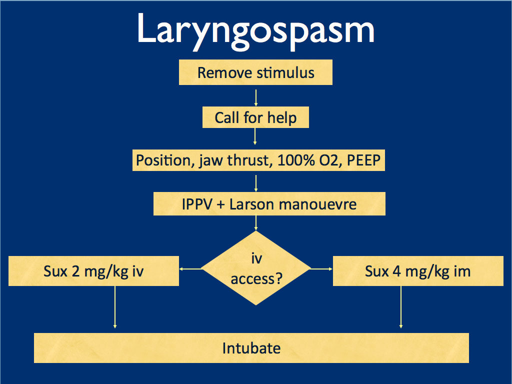
I presented the following suggested algorithm for management of laryngospasm during ketamine procedural sedation at a regional emergency medicine ‘Fellows Forum’ meeting in November 2007 in the UK. Since many paediatric procedural sedations were done using intramuscular (im) ketamine, it gives guidance based on whether or not vascular access has been obtained:

Some things I considered were:

    • Neuromuscular blockade (NMB) isn’t always necessary – laryngospasm may be managed with other sedatives such as propofol. However, titrating further sedatives in a desaturating child in my view is inferior to definitive airway management and laryngeal relaxation with suxamethonium and a tube.
    • Laryngospasm may be managed with much smaller doses of suxamethonium than are required for intubation – as little as 0.1 mg/kg may be effective. However, I think once we go down the NMB route we’re committed to intubation and therefore we should use a dose guaranteed to be effective in achieving intubating conditions.
    • In the child without vascular access, I considered intraosseous and intralingual sux. However, intramuscular suxamethonium is likely to have a relaxant effect on the laryngeal muscles within 30-45 seconds, which has to be compared with time taken to insert and confirm intraosseous needle placement. I do not think the traditionally recommended intralingual injection has any role to play in modern airway management.
  • At the time I wrote this most paediatric resuscitation bays in my area in the United Kingdom had breathing circuits capable of delivering PEEP – usually the Ayr’s T-Piece (specifically the Mapleson F system), which is why PEEP was included early in in the algorithm prior to BMV.
I have since modified it for two reasons: firstly, we might as well do the Larson manoeuvre during the jaw thrust; secondly, many Australasian and US EDs will only be able to deliver PEEP with a PEEP valve attached to a BVM, so PEEP has been moved to the BVM stage.
I would love to hear what people are doing in their prehospital and inhospital practice. Should ketamine only be administered by providers who can offer RSI? Do you have a laryngospasm protocol? If so, I’d love to see it. If not, feel free to use or adapt my unvalidated one at your own risk.

ABSTRACT An advanced life support emergency medical services (EMS) unit was dispatched with law enforcement to a report of a male patient with a possible overdose and psychiatric emergency. Police restrained the patient and cleared EMS into the scene. The patient was identified as having excited delirium, and ketamine was administered intramuscularly. Sedation was achieved and the patient was transported to the closest hospital. While in the emergency department, the patient developed laryngospasm and hypoxia. The airway obstruction was overcome with bag–valve–mask ventilation. Several minutes later, a second episode of laryngospasm occurred, which again responded to positive-pressure ventilation. At this point the airway was secured with an endotracheal tube. The patient was uneventfully extubated several hours later. This is the first report of laryngospam and hypoxia associated with prehospital administration of intramuscular ketamine to a patient with excited delirium.

1. Laryngospasm and Hypoxia After Intramuscular Administration of Ketamine to a Patient in Excited Delirium
Prehosp Emerg Care. 2012 Jul;16(3):412-4
2. Laryngospasm-The best treatment
Anesthesiology 1998; 89:1293-4
3. Management of Laryngospasm
http://www.respond2articles.com/ANA/forums/thread/1096.aspx

Intubation of the critically ill in Scotland

Hi folks! Cliff has given me the helm of his blogsite for this week whilst he is teaching prehospital and critical care ultrasound with the Americans at Castlefest 2012
He invited me to write an article on this latest paper in British Journal of Anaesthesia on Scottish ICU audit of emergency tracheal intubation. For those who don’t know, Cliff has a proud Scottish heritage and this paper is a useful audit of his home land’s performance of this critical care intervention. I have done airway audits and this one is quite a reasonable 4 month effort albeit not every ICU in Scotland participated, which is not unusual for those wanting to do these kind of audits. Airway management gets a bit personal and some find review of their emergency airway performance to be confronting. It should not be. Now it’s a fine distinction but its important to be clear on this. A FAILED AIRWAY DOES NOT MEAN YOU ARE A FAILURE!! FAILED OXYGENATION IS ANOTHER STORY….
There are always recurring themes from audits like these and I will highlight a few.
The first and foremost, is the absolutely essential role of capnography for tracheal tube confirmation and monitoring of airway patency and ventilator status. My FDEAR aeromedical intubation audit showed this was an issue of patient safety that should be improved.
This Scottish ICU study revealed that capnography was used in only 54% of emergency intubations despite the vast majority being in hospital locations where such monitoring is available! This is a recurring theme amongst emergency airway audits and coroners reports like this one.
Paradoxically this Scottish audit had a high number of intubating doctors with greater than 24 months of anaesthetic training and one hypothesis I have is that as doctors become more confident in emergency intubations, perhaps less reliance is felt required on monitoring like capnography? In human factors research into anaesthetic related crises, we call this the invulnerability or superman complex : “If I say the tube has gone in, I must be right!”
Secondly, the length of anaesthetic training of the intubating doctor appeared related to overall airway success rates and a low complication rate. There was only one surgical airway required over the 4 month period and 794 recorded intubations. The authors discuss though the potential problems that may face up and coming critical care doctors in the United Kingdom who may not be exposed to terms of anaesthetic training of up to 2 years. My own personal view is that it does not and should not matter where you get your emergency airway training but it should be structured and specific to the work that you are going to do. Learning to do epidural anaesthesia in laboring women might not be so helpful for the bilateral pneumonia swine flu patient with a BMI of 50! And certainly no point learning to use airway equipment that you will rarely or never have available where you normally work!
Thirdly and I find this fascinating having heard talks and debates on this topic by Dr Scott Weingart and Dr Paul Mayo, but in this Scottish paper of bloody sick patients needing intubation, 8% were performed without paralytics at all and overall intubation success and number of attempts were not significantly different compared to the paralytic assisted group. My view is that overall in critically ill patients , paralytics are your friend as these folks need the airway secured, one way or another. However this paper and Dr Mayo’s work certainly demonstrate that sedation only intubation is successful and is a reasonable alternative.
Finally, 61% of these emergency intubations utilized propofol and there was an association with post intubation hypotension (systolic <70mmHg). Ketamine use was low at 3% and I think this just reflects the greater anaesthetic training of the doctors in the study. I am aware Cliff has done a previous podcast rant on Propofol assasins
I don’t want to rant and am not as good at it as Cliff. BUT Choose your poison carefully! This paper reminds us what we all know. The milk of amnesia has issues! Ask the Jackson family!
Anyway that’s enough for this paper. I gotta pick myself off the floor again after listening to Cliff’s propofol rant..
– Dr Minh Le Cong, Royal Flying Doctor Service, Australia

BACKGROUND: Complications associated with tracheal intubation may occur in up to 40% of critically ill patients. Since practice in emergency airway management varies between intensive care units (ICUs) and countries, complication rates may also differ. We undertook a prospective, observational study of tracheal intubation performed by critical care doctors in Scotland to identify practice, complications, and training.
METHODS: For 4 months, we collected data on any intubation performed by doctors working in critical care throughout Scotland except those in patients having elective surgery and those carried out before admission to hospital. We used a standardized data form to collect information on pre-induction physical state and organ support, the doctor carrying out the intubation, the techniques and drugs used, and complications noted.
RESULTS: Data from 794 intubations were analysed. Seventy per cent occurred in ICU and 18% occurred in emergency departments. The first-time intubation success rate was 91%, no patient required more than three attempts at intubation, and one patient required surgical tracheostomy. Severe hypoxaemia ( <80%) occurred in 22%, severe hypotension (systolic arterial pressure <80 mm Hg) in 20%, and oesophageal intubation in 2%. Three-quarters of intubations were performed by doctors with more than 24 months formal anaesthetic training and all but one doctor with <6 months training had senior supervision.
CONCLUSIONS: Tracheal intubation by critical care doctors in Scotland has a higher first-time success rate than described in previous reports of critical care intubation, and technical complications are few. Doctors carrying out intubation had undergone longer formal training in anaesthesia than described previously, and junior trainees are routinely supervised. Despite these good results, further work is necessary to reduce physiological complications and patient morbidity.

Tracheal intubation in the critically ill: a multi-centre national study of practice and complications
Br J Anaesth. 2012 May;108(5):792-9

Extubation guidelines

Tracheal extubation is a high risk procedure in anaesthesia and critical care. Until now most guidelines have focused on intubation, with little to guide the process of extubation. Complications may relate to the following issues:

  • Exaggerated reflexes – laryngospasm (which can lead to both hypoxia and negative pressure pulmonary oedema) and bronchospasm
  • Reduced airway reflexes
  • Dysfunctional laryngeal reflexes
  • Depletion of oxygen stores at extubation
  • Airway injury
  • Physiological compromise in other systems
  • Human factors

The goal is to ensure uninterrupted oxygen delivery to the patient’s lungs, avoid airway stimulation, and have a back-up plan, that would permit ventilation and re-intubation with minimum difficulty and delay should extubation fail.
The Difficult Airway Society has now published guidelines for the management of tracheal extubation, describing four steps:

Step 1: plan extubation.

Step 2: prepare for extubation.

Step 3: perform extubation.

Step 4: post-extubation care: recovery and follow-up.

During step 3, emphasis is on pre-oxygenation, positioning, and suction. This is followed by simultaneous deflation of the tracheal tube cuff and removal of the tube at the peak of a sustained inflation. This generates a passive exhalation, which may assist in the expulsion of secretions and possibly reduce the incidence of laryngospasm and breathholding.
The guideline refers to low-risk and at-risk extubations. ‘Low-risk’ (routine) extubation is characterised by the expectation that reintubation could be managed without difficulty, if required. ‘At-risk’ means the presence of general and/or airway risk factors that suggest that a patient may not be able to maintain his/her own airway after removal of the tracheal tube. ‘At-risk’ extubation is characterised by the concern that airway management may not be straightforward should reintubation be required.
These guidelines are written for the peri-operative patient but the text contains some interesting points that are pertinent to the ED or ICU patient. Some simple algorithms are presented:


Difficult Airway Society Guidelines for the management of tracheal extubation
Anaesthesia. 2012 Mar;67(3):318-40 Free full text

More guidelines from the Difficult Airway Society