Tag Archives: ECMO

Louisa in London – Prehospital Lessons from LTC2015

The London Trauma Conference remains up there on my list of ‘must go’ conferences to attend. It marks the end of the year, fills me with hope and inspires me for the future. Unfortunately this year I was torn between the conference and the demands of clinical directorship so I could only get to the “Air Ambulance & Prehospital Care Day”. At least this way I’m saved from the dilemma of which sessions to attend!
So what were the highlights of the Prehospital Day? For me, they were Prehospital ECMO,’Picking Up the Pieces’, and the REBOA update.
Prehospital ECMO
Professor Pierre Carli gave us an update on prehospital ECMO. Professor Carli (not to be confused with the equally awesome Professor Carley) is the medical director of Service d’Aide Médicale Urgente (SAMU) in Paris. They’ve been doing prehospital ECMO in Paris since 2011 and the data analysed over three years reveals a 10% survival to hospital discharge rate. We know from the work in Asia that successful outcome following traditional cardiac arrest management and ECPR is related to the speed of the intervention. Transposing the time to intervention from his 2011 – 2013 data onto the survival curve that Chen et al produced explains why the success rate is limited:
LTC2015
The revised 2015 process aims to reduce the duration of CPR, reduce time to ECMO and therefore improve survival to discharge rates. They are doing this by dispatching the ECMO team earlier.
The eligibility criteria for ECPR is also changing; patients >18 and <75years, refractory cardiac arrest (defined as failure of ROSC after 20min of CPR), no flow for < 5 minutes with shockable rhythm or signs of life or hypothermia or intoxication, EtCO2 > 10mmHg at time of inclusion and no major comorbidity.
Already there appears to be an improvement with 16 patients treated using the revised protocol with 5 survivors (31%) – although we must be wary of the small numbers.
A concern that was expressed by the French Department of Health was the fear of a reduction in organ donation with the introduction of ECPR – it turns out that rates have remained stable. In fact the condition of non heart beating donated organs is better when ECMO has been instigated; the long term effects on organ donation are being assessed.
I’m without doubt that prehospital ECMO/ED ECMO is the future although currently in the UK our hospital systems aren’t ready for this. If you want to learn more then look at the ED ECMO site or book on one of the many emerging courses on ED ECMO including the one that is run by Dr Simon Finney at the London Trauma Conference, or if you want to go further afield you could try San Diego (although places are fully booked on the next course).
Picking Up the Pieces
The Keynote speaker was Professor Sir Simon Wessely. He is a psychiatrist with a specialist interest in military psychology and his brief was to describe to us the public response to traumatic incidents. He has worked with the military and in civilian situations. After the 7/7 London bombings the population of London was surveyed: those most likely to be affected were of lower social class, of Muslim faith, those that had a relative that was injured, those unsure of the safety of others, those with no previous experience of terrorism and those experiencing difficulty in contacting others by mobile phone. Obviously there are many factors that we cannot influence however on the basis of the last risk factor our response to incidents has changed – the active discouragement to make phone calls has been changed to a recommendation of making short calls to friends and relatives.
The previous practice of offering immediate psychological debriefing to those involved in incidents was discounted by Prof Wessely – his research demonstrated that this intervention was not only not required but could actually result in harm: only a minority have ongoing psychological distress that can benefit from formal psychological input, which should occur later.
The approach that should be taken is to allow that individual to utilise their own social networks (family, friends, and colleagues) and to accept that in some cases the individual may not want or need to talk. This has led to the development of the Trauma Risk Management (TRIM) system which provides individuals within organisations that are exposed to traumatic events the skills required to identify those at risk of developing psychological problems and to recognise the signs and symptoms of those in difficulty. To a certain extent we naturally do this for our peers – I have spent many a night sitting in the ‘Good Samaritan’ pub with colleagues from the Royal London Hospital and London’s Air Ambulance – but having a more formal system is probably of benefit to enable those who have ongoing difficulties to access additional support.
REBOA update
Finally, the REBOA update – Resuscitative Endovascular Balloon Occlusion of the Aorta. One year on, Dr Sammy Sadek informed us that there are now more courses teaching the REBOA technique than there are (prehospital) patients that have received it. Over the last year only seven patients have qualified for this intervention in London, far fewer than they had anticipated. Another three patients died before REBOA could be instigated. All patients had a positive cardiovascular response. Four of the seven died from causes other than exsanguination. Is it worth all the effort and resource to deliver this intervention when such a select group will benefit?
Obviously there was much more covered in the day, this is just a taste. If you’ve never been to the London Trauma Conference then I definitely would recommend it and even if you have been before there are so many breakout sessions now there is always something for everyone.
More on the London Trauma Conference:

Merry Christmas and see you next year!
Louisa Chan

London Cardiac Arrest Symposium 2014

The focus of the entire day is cardiac arrest and this is the second day of the London Cardiac Arrest Symposium.

Professor Niklas Nielsen kicked off with a presentation of his Targeted Temperature Management trial.  It seems that even now there is uncertainty in the interpretation of this latest study. I take heart from the knowledge that Prof Nielsen has changed the practice of his institution to reflect the findings of his study – I have certainly changed my practice. But we need to remain aware that there is more work to be done to answer the multiple questions that remain and the need for further RCTs is recognised.

The management of Cardiac arrest after avalanche is not a clinical scenario that I imagine I’ll ever find myself in. The management is well documented in the ICAR MEDCOM guidelines 2012. Dr Peter Paal reminded us that you’re not dead until you’re rewarmed and dead unless: with asystole, CPR may be terminated (or withheld) if a patient is lethally injured or completely frozen, the airway is blocked and duration of burial >35 min, serum potassium >12 mmol L(-1), risk to the rescuers is unacceptably high or a valid do-not-resuscitate order exists.

The age old question about prognostication after cardiac arrest was tackled by Prof Mauro Oddo. He covered the evidence for clinical examination, SSPE, EEG, and neurone specific enolase. Bottom line, all of these modalities are useful but none are specific enough to be used as a stand alone test so multiple modalities are required.

SAMU is leading the way with prehospital ECMO. They have mastered the art of cannulation (in the Louvre no less!) but there haven’t enough cases to demonstrate a mortality benefit. The commencement of ECMO prehospital reduces low flow time and theoretically should improve outcomes. This is begging for a RCT.

The experience of the Italians with in hospital ECMO shoes a better survival rate for in-hospital rather than out of hospital cardiac arrests, explained Dr Tomasso Mauri. They treat patients with a no flow time of <6min and low flow rate of <45min and had a 31% ICU survival rate. If you want to learn more about ED ECMO go to http://edecmo.org.

VA-ECMO

The Douglas Chamberlain lecture this year was Selective aortic arch perfusion presented by Prof James Manning. He spoke about the use of this technique in cardiac arrest and also in trauma (where it is known to you as Zone 1 REBOA).

image-1

In cardiac arrest the aim is to improve coronary perfusion, to preserve perfusion to the heart and the brain, offer a route of rapid temperature control and offer a direct route of administration of adrenaline. Coronary perfusion is seen to be supra normal after SAAP. And the suggested place for SAAP is prior to ECMO.

image-5

It’s more familiar ground talking about SAAP in trauma. This Zone 1 occlusion preserves cerebral and cardiac perfusion while blood loss is limited and rapid fluid resuscitation can occur.

image-3

You can hear Prof Manning on SAAP over at EMCrit (of course!). 

It’s been another great conference. Put the dates for next year’s London Trauma & Cardiac Arrest Conferences in your diary: 8th-10th December 2015!

Happy Holidays & Keep Well

Louisa Chan


London Trauma Conference Day 2

London Trauma Conference 2013 – Day 2  by Dr Louisa Chan
So I find myself torn today: do I join the the main track with a Major incident theme or the Cardiac Masterclass? I never liked the thought of missing out on anything so I went to a bit of both.
 
Cardiac Masterclass
A lot of people probably think that managing cardiac arrest isn’t challenging and a bit dull because the patient is dead. But the Cardiac Masterclass would inspire you to think of a bright future for cardiac arrest management.
Mark Whitbread reminded us of how important dispatch is in the chain of survival. How much focus do we put on improving bystander CPR rates? Dispatcher assisted CPR has been shown to improve outcomes and needs to be skilfully done.
Ajay Jain pushes for all OHCA patients to be taken to a Cardiac Arrest centre for PCI. Why? Because the results he has from his centre for PCI in OHCA patients results in 77% (101/132) patients surviving to hosp discharge, 65% neurologically intact.
He also tells us that the ECG post arrest is a very poor predictor of PCI findings (although STEMI predicts a positive result) so they all should have PCI.
Lyon-survivors
 
More data from TOPCAT shows us that non survivors of OHCA are easy to cool.
 
LTC-mice
 
 
And maybe we should be cooling DURING cardiac arrest to minimise the reperfusion injury.
 
 
For persistent VF Prof Redwood says revascularisation is the key; when that doesn’t work then reducing LV volume may help so aspiration or an Impella may work. Failing that – ECMO.
 
Major Incidents
Major Incidents by their nature do not happen every day, so experience in these incidents is limited. The challenge then is how can we learn from incidents?
A standardised reporting system for a major incident database would be a good idea – www.majorincidentreporting.org – is where you will find the standard report form and open access database.
And then all I can suggest is that you need to come to the LTC and listen to the accounts of those who have been there. We heard about the Tokyo Sarin attack, Mumbai, and a very compelling story of multiple drownings from Steen Barnung.
Lessons from Tokyo – Sarin attack:

It will happen again
It will be chaos
Crowds cannot be controlled
Comms will fail
Clinical diagnosis – need a senior clinician
Treatment must be immediately available – 3min to absorb sarin
Decontamination – get naked, 90% decon with clothes removal.
Stream casualties
Empower the man on the ground.

 
Gadgets
LTC-MSUThe great thing about the London Trauma Conference is that it’s not just about the content of the tracks, there’s the networking and the opportunity to see new pieces of equipment.
The Norwegians won on the equipment front with their Mobile Stroke Unit. It’s due to go on line in 2014.
So TTFN and more from me on Day 3 of #LTC2013

Prehospital ECLS – it's happening

Patients with refractory (>30 mins) cardiac arrest underwent prehospital cannulation for extracorporeal life support in a French feasibility study. A physician-paramedic team responded by car in Paris to cardiac arrest cases that met inclusion criteria. Mechanical CPR devices (Autopulse or LUCAS) were applied during cannulation. Femoral venoarterial ECMO was instituted using a Maquet Cardiohelp system. Blood products and inotropes, echocardiography, and hypothermia were included in the prehospital management package.
Seven patients were treated, with a mean age of 42 (+/- SD of 16, no median given). ECLS was started an average 57 min (±21) after the onset of ACLS. One patient survived to discharge neurologically intact. Two brain dead patients became organ donors. The survivor had hypertrophic cardiomyopathy with refractory ventricular fibrillation.
Safety and feasibility of prehospital extra corporeal life support implementation by non-surgeons for out-of-hospital refractory cardiac arrest
Resuscitation. 2013 Nov;84(11):1525-9
[EXPAND Abstract]


BACKGROUND: Extra corporeal life support (ECLS) has been recently introduced in the treatment of refractory cardiac arrest (CA). Several studies have assessed the use of ECLS in refractory CA once the patients reach hospital. The time between CA and the implementation of ECLS is a major prognostic factor for survival. The main predictive factor for survival is ECLS access time. Pre hospital ECLS implementation could reduce access time. We therefore decided to assess the feasibility and safety of prehospital ECLS implementation (PH-ECLS) in a pilot study.

METHODS AND RESULTS: From January 2011 to January 2012, PH-ECLS implementation for refractory CA was performed in 7 patients by a PH-ECLS team including emergency and/or intensivist physicians and paramedics. Patients were included prospectively and consecutively if the following criteria were met: they had a witnessed CA; CPR was initiated within the first 5min of CA and/or there were signs of life during CPR; an PH-ECLS team was available and absence of severe comorbidities. ECLS flow was established in all patients. ECLS was started 22min (±6) after the incision, and 57min (±21) after the onset of advanced cardiovascular life support (ACLS). In one patient, ECLS was stopped for 10min due to an accidental decannulation. One patient survived without sequelae. Three patients developed brain death.

CONCLUSIONS: This pilot study suggests that PH-ECLS performed by non-surgeons is safe and feasible. Further studies are needed to confirm the time saved by this strategy and its potential effect on survival.

[/EXPAND]

Avoiding intubation in ARDS with awake ECMO

ECMOillusiconA letter in Intensive Care Medicine by Hoeper and colleagues from Hannover describes a small case series of six ARDS patients with severe hypoxaemia who went straight from non-invasive ventilation to awake veno-venous ECMO. All had single organ failure and four were immunocompromised, the latter factor influencing the decision to try to avoid invasive mechanical ventilation. Four of the six patients survived to hospital discharge. A larger multicentre study is being planned.

Clinical illustration courtesy of Dr Brian Burns

Extracorporeal membrane oxygenation instead of invasive mechanical ventilation in patients with acute respiratory distress syndrome
Intensive Care Med. 2013 Nov;39(11):2056-2057 (no abstract)

ECMO for paediatric cardiac arrest

The Taiwanese are at it again with their extracorporeal life support. This time, they report their outcomes in children who received ECMO for in-hospital cardiac arrest. Interestingly, the patients with pure cardiac causes of cardiac arrest had a survival rate similar to patients with non-cardiac causes.


PURPOSE: The study aims to describe 11 years of experience with extracorporeal cardiopulmonary resuscitation (ECPR) for in-hospital paediatric cardiac arrest in a university affiliated tertiary care hospital.

METHODS: Paediatric patients who received extracorporeal membrane oxygenation (ECMO) during active extracorporeal cardiopulmonary resuscitation (ECPR) at our centre from 1999 to 2009 were included in this retrospective study. The results from three different cohorts (1999-2001, 2002-2005 and 2006-2009) were compared. Survival rates and neurological outcomes were analysed. Favourable neurological outcome was defined as paediatric cerebral performance categories (PCPC) 1, 2 and 3.

RESULTS: We identified 54 ECPR events. The survival rate to hospital discharge was 46% (25/54), and 21 (84%) of the survivors had favourable neurological outcomes. The duration of CPR was 39±17 min in the survivors and 52±45 min in the non-survivors (p=NS). The patients with pure cardiac causes of cardiac arrest had a survival rate similar to patients with non-cardiac causes (47% (18/38) vs. 44% (7/16), p=NS). The non-survivors had higher serum lactate levels prior to ECPR (13.4±6.4 vs. 8.8±5.1 mmol/L, p<0.01) and more renal failure after ECPR (66% (19/29) vs. 20% (5/25), p<0.01). The patients resuscitated between 2006 and 2009 had shorter durations of CPR (34±13 vs. 78±76 min, p=0.032) and higher rates of survival (55% (16/29) vs. 0% (0/8), p=0.017) than those resuscitated between 1999 and 2002.

CONCLUSIONS: In our single-centre experience with ECPR for paediatric in-hospital cardiac arrest, the duration of CPR has become shorter and outcomes have improved in recent years. Higher pre-ECPR lactate levels and the presence of post-ECPR renal failure were associated with increased mortality. The presence of non-cardiac causes of cardiac arrest did not preclude successful ECPR outcomes. The duration of CPR was not significantly associated with poor outcomes in this study.

Eleven years of experience with extracorporeal cardiopulmonary resuscitation for paediatric patients with in-hospital cardiac arrest
Resuscitation. 2012 Jun;83(6):710-4

Surgical treatment for acute massive pulmonary embolism

A recent paper reminds us that surgery is an option in the management of massive pulmonary embolism(1), to be considered in the patient for whom thrombolysis has failed or is contraindicated. Good outcomes were produced when surgery was performed in a centre capable of cardiopulmonary bypass (6% 30-day postoperative mortality), but is surgery an option when these facilities are unavailable?

The “venous inflow occlusion” technique involves clamping the venae cavae prior to removing clot directly from the pulmonary artery and its branches after median sternotomy, and can be performed in any hospital with surgical facilities. Under normothermic conditions, speed is of the essence once cardiac arrest occurs, since the irreversible anoxic cerebral injury will occur after just a few minutes.
Clarke and Abrams wrote in the Lancet in 1972(2):

Our use of venous inflow-occlusion has given results which compare well with those obtained with extracorporeal circulation. 50% of our patients survived. All patients who had emboli removed without an episode of ventricular asystole survived surgery. Late deaths in 3 patients were from causes unrelated to pulmonary embolism, and from a further massive pulmonary embolus a week later. The technique has been applied with equal success in a major hospital fully equipped for cardiac surgery and in hospitals where resident and nursing staff had no experience of either thoracic or cardiac surgery. The simplicity and speed of the method has enabled the obstructed right ventricle to be relieved within thirty minutes of the onset of symptoms. The interval between induction of anaesthesia and the skin incision should be kept as short as possible, and drugs to maintain the blood pressure should be given. The period between skin incision and the restoration of the circulation has, with practice, been reduced to ten minutes.

But this clearly still requires surgical expertise and facilities. Emergency physicians can open the chest to deal with penetrating trauma. Could an ED thoracotomy facilitate clot removal from the pulmonary artery?

In 1969, a lady in her 50s arrested on the ward after an operation to remove a mass via a left lateral thoractomy. Pulmonary embolism was suspected and her thoracotomy wound was re-opened and the pulmonary artery incised, resulting in the removal of large amounts of clot. Return of spontaneous circulation resulted after a brief period of internal cardiac massage. Her case was written up decades later, in 1998(3):

The patient recovered rapidly and left the hospital on the 21st day without signs of cerebral damage. This patient is now 86 years old, mentally normal, living alone, and doing her own housekeeping. She remembers the hospital stay and the past years as worth living

Some patients may be considered too high risk for surgery and in some centres Extracorporeal Membrane Oxygenation (ECMO) is an option. It has been used both as life support pending surgery(4), or as an alternative to surgery to allow heparinisation to be used(5,6).

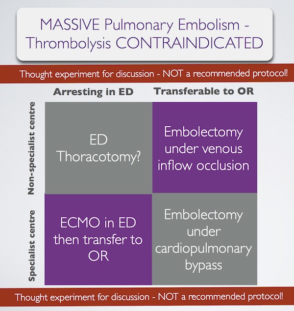
In summary, some patients with massive pulmonary embolism may benefit from surgery (contraindication to ‘lysis or failed ‘lysis). Getting them to surgery alive, or operating on them during cardiac arrest, is a challenge. Ideally they would undergo embolectomy under cardopulmonary bypass in the operating room, or could be placed on ECMO in the ED prior to going to the OR. If they present to a centre without these facilities, then the venous inflow occlusion technique could be used in the OR without bypass. Just rarely a patient may present in extremis with PE to an ED without these options. If that patient has major contraindications to thrombolysis, would an ED thoracotomy be something you would entertain?

I have done several thoracotomies for penetrating trauma but never for PE. I do not pretend to know how, and cannot find a case report of ED thoracotomy for pulmonary embolism in the literature. I’m therefore NOT recommending it. However, I would love to know people’s views on its feasibility. A possible approach could be summarised as:

Massive pulmonary embolism fascinates me, because it’s seen in the ‘talk and die’ patient. It is a single, treatable pathology that if diagnosed and treated appropriately truly makes the difference between life and death. When medicine presents us with an opportunity ‘on a plate’ like that to save a life, we need to be prepared. I have had great saves with this diagnosis and sadly have seen disastrous failures to act. When the time comes, we need to ask: ‘have we explored all options?’.

1. Surgical treatment of acute pulmonary embolism–a 12-year retrospective analysis.
Scand Cardiovasc J. 2012 Jun;46(3):172-6. Epub 2012 Mar 27.

OBJECTIVES: Surgical embolectomy for acute pulmonary embolism (PE) is considered to be a high risk procedure and therefore a last treatment option. We wanted to evaluate the procedures role in modern treatment of acute PE.
DESIGN: All data on patients treated with surgical embolectomy for acute PE were retrieved from our clinical database. The mortality was extracted from the Danish mortality register.
RESULTS: From October 1998 to July 2010, 33 patients underwent surgical embolectomy. All procedures were done through a median sternotomy and extracorporeal circulation. Twenty-six patients were diagnosed with a high risk PE and 7 with an intermediate risk PE and intracardial pathology. Six patients had been insufficiently treated with thrombolysis. Thirteen patients had contraindication for thrombolysis. Six patients were brought to the operating theatre in cardiogenic shock, 8 needed ventilator support, and 1 was in cardiac arrest. The postoperative 30-day mortality was 6% and during the 12-year follow-up the cumulative survival was 80% with 4 late deaths.
CONCLUSION: Surgical pulmonary embolectomy can be performed with low mortality although the treated patients belong to the most compromised part of the PE population. The results support surgical embolectomy as a vital part of the treatment algorithm for acute PE.

2. Pulmonary embolectomy with venous inflow-occlusion.
The Lancet 1972;1(7754):767–769

Massive pulmonary emboli have been removed surgically from 26 patients. The technique of normothermic circulatory arrest by venous inflow-occlusion was used in 25 patients. 13 patients survived. There were 10 operative deaths and 3 hospital deaths. Diagnosis was based upon clinical findings supplemented by electrocardiography and a plain radiograph of the chest. Surgery was offered to patients having a pulmonary embolus sufficiently massive to produce sustained hypotension. All patients whose hearts stopped beating before the embolectomy died. 6 successful operations were performed in hospitals without facilities for cardiac surgery. The method is recommended for its simplicity.

3. Left Anterior Thoracotomy for Pulmonary Embolectomy With 29-Year Follow-up
The Annals of Thoracic Surgery 1998, 66(4):1420-1421

Pulmonary embolectomy is usually performed in cardiopulmonary bypass. In acute situations too much time can be lost in setting up and connecting the pump oxygenator; this delay can cause cerebral damage in a patient with circulatory arrest. In such a situation left anterior thoracotomy can provide an ideal approach. An emergency thoracotomy can be performed in a few seconds. The lung automatically retracts. The phrenic nerve, pulmonary artery, and pericardium are clearly seen, and they outline the area for embolectomy. A case in which such an approach was successfully used is described.

4.ECMO treatment saved life of a young woman with acute pulmonary embolism
Lakartidningen 2004, 101(44):3420-3421

A 42-year old obese female using contraceptive medication was admitted to the emergency room because of sudden onset of dyspnoea and hypoxia. Computed tomography showed massive pulmonary emboli. Despite initial treatment with thrombolysis her condition deteriorated further and she was referred for acute surgery to our clinic. Before putting the patient to sleep extracorporeal circulation was instituted with access from the groin. After anaesthesia a median sternotomy was performed. With the heart beating, the main pulmonary artery was incised and a 9 cm long thrombus was removed. Immediate weaning from the heart-lung machine was not possible, mainly because of bleeding to the airways. The right atrium and the aorta was therefore cannulated and an extracorporeal circulation membrane oxygenator (ECMO) was used for three days. The patient required several re-entries for bleeding and a tracheotomy during the postoperative course. She was fully recovered three months after the operation.

5. Extracorporeal membrane oxygenator for pulmonary embolism.
The Annals of Thoracic Surgery 1997, 64(3):883-884 Free full text

6. Peripheral Extracorporeal Membrane Oxygenation: Comprehensive Therapy for High-Risk Massive Pulmonary Embolism
Ann Thorac Surg 2012;94:104–8

Background: Although commonly reserved as a last line of defense, experienced centers have reported excellent results with pulmonary embolectomy for massive and submassive pulmonary embolism (PE). We present a contemporary surgical series for PE that demonstrates the utility of peripheral extracorporeal membrane oxygenation (pECMO) for high-risk surgical candidates.
Methods: Between June 2005 and April 2011, 29 patients were treated for massive or submassive pulmonary embolism, with surgical embolectomy performed in 26. Four high-risk patients were placed on pECMO, established by percutaneously cannulating the right atrium through a femoral vein and perfusing by a Dacron graft anastomosed to the axillary artery. A small, extracorporeal, rotary assist device was used, interposing a compact oxygenator in the circuit, and maintaining anticoagulation with heparin.
Results: Extracorporeal membrane oxygenation was weaned in 3 of 4 patients after 5.3 days (5, 5, and 6), with normalization of right ventricular dysfunction and pulmonary artery pressure (44.0 ± 2.0 to 24.5 ± 5.5 mm Hg) by ECHO. Follow-up computed tomographies showed several peripheral, nearly resorbed emboli in 1 case and complete resolution in 2 others. The fourth patient, not improving after 10 days, underwent surgery where an embolic liposarcoma was extracted. For all 29 cases, hospital and 30-day mortality was 0% and all patients were discharged, with average postoperative length of stay of 15 days for embolectomy and 17 days for pECMO.
Conclusions: Heparin therapy with pECMO support is a rapid, effective option for patients who might benefit from pulmonary embolectomy but are at high risk for surgery.

Extracorporeal cardiopulmonary resuscitation

You have a patient in cardiac arrest who has had excellent resuscitation from the point of collapse, and who has treatable underlying pathology (eg. PE or STEMI). However you’re unable to get return of spontaneous circulation so you call it. Someone just died for whom the technology exists to save them. Extracorporeal life support (ECLS) supports heart and lung function by externally providing circulatory flow and gas exchange until the patient’s underlying cause of arrest is treated or recovers.
ECLS requires an extracorporeal membrane oxygenation (ECMO) circuit to be placed during the cardiac arrest resuscitation. This may sound like extreme stuff, but there have been some amazing saves with this technology, and large numbers of in-hospital and out-of-hospital arrest patients have been treated in Japan, Korea, and Taiwan. ECMO has even been commenced in the field by prehospital emergency physicians.
An inspiring EMCrit podcast with Dr Joe Bellezzo described how this technology is applied at Sharp Memorial Hospital in San Diego. Bellezzo and colleagues have now published a series of their out-of-hospital arrest cases who received ECLS initiated by emergency physicians(1).
Coming back to the Japanese, a multicentre prospective cohort study of ECLS for out-of hospital cardiac arrest (the ‘SAVE-J’ study) selected patients with VF or pulseless VT in whom no ROSC was achieved with standard resuscitative measures. Their striking results mirror other ECLS studies and were published in abstract form in November 2011(2).
To me, the overwhelming take home messages from what I’ve seen and read on this are:


1. ECLS can provide dramatic saves with neurologically intact survival in cardiac arrest cases that otherwise would be dead.

2. The critical factor for successful clinical outcomes and avoidance of wasted resources and clinical futility is case selection. The underlying cause of arrest needs to be reversible (eg. myocarditis) or treatable (eg. STEMI) and good resuscitation needs to have been in place prior to ECLS.

3. In the right hospital with the right resuscitation team, it can be done.

1. Emergency physician-initiated extracorporeal cardiopulmonary resuscitation
Resuscitation. 2012 Aug;83(8):966-70
[EXPAND Click for abstract]


CONTEXT: Extracorporeal cardiopulmonary resuscitation (ECPR) refers to emergent percutaneous veno-arterial cardiopulmonary bypass to stabilize and provide temporary support of patients who suffer cardiopulmonary arrest. Initiation of ECPR by emergency physicians with meaningful long-term patient survival has not been demonstrated.

OBJECTIVE: To determine whether emergency physicians could successfully incorporate ECPR into the resuscitation of patients who present to the emergency department (ED) with cardiopulmonary collapse refractory to traditional resuscitative efforts.

DESIGN: A three-stage algorithm was developed for ED ECPR in patients meeting inclusion/exclusion criteria. We report a case series describing our experience with this algorithm over a 1-year period.

RESULTS: 42 patients presented to our ED with cardiopulmonary collapse over the 1-year study period. Of these, 18 patients met inclusion/exclusion criteria for the algorithm. 8 patients were admitted to the hospital after successful ED ECPR and 5 of those patients survived to hospital discharge neurologically intact. 10 patients were not started on bypass support because either their clinical conditions improved or resuscitative efforts were terminated.

CONCLUSION: Emergency physicians can successfully incorporate ED ECPR in the resuscitation of patients who suffer acute cardiopulmonary collapse. More studies are necessary to determine the true efficacy of this therapy.

[/EXPAND]
2. Multicenter Non-Randomized Prospective Cohort Study of Extracorporeal Cardiopulmonary Resuscitation for Out-of Hospital Cardiac Arrest: Study of Advanced Life Support for Ventricular Fibrillation with Extracorporeal Circulation in Japan (SAVE-J)
Circulation 2011; 124: A18132
[EXPAND Click for abstract]


Background: This study is aimed to examine the efficacy of extracorporeal cardiopulmonary resuscitation (ECPR) for patients in out-of hospital cardiac arrest (OHCA) with ventricular fibrillation (VF) or pulseless ventricular tachycardia (VT).

Method: The design of this study is a multicenter non-randomized prospective cohort study. Hypothesis is that the outcome of OHCA with VF or pulseless VT is similar between ECPR and conventional advanced life support (ALS). During from Oct. 2008 to Dec. 2010, forty six tertiary emergency hospitals were participated in this study. Patient inclusion criteria were 1) VF or pulseless VT on scene, 2) cardiac arrest on arrival at hospital, 3) within 45 minutes from a call to an arrival of hospital, and 4) non-ROSC by conventional ALS during 15 minutes after an arrival at hospital. Exclusion criteria were 1) age: 75 yr, 2) poor activities of daily livings, 3) non-cardiac verified cardiac arrest, and 4) hypothermia. According to the inclusion criteria, ECPR was adopted for OHCA in 26 hospitals (ECPR group) and conventional ALS was planned in 20 hospitals (non-ECPR group). Both groups (Intention-to-treat) were analyzed about the proportion of patients with favorable outcome (CPC1 or 2) assessed with the Glasgow-Pittsburgh Cerebral Performance and Overall Performance Categories at 1 month by chi square test and Fisher exact probability test.

Results: One hundred and eighty patients of ECPR group and 134 patients of non-ECPR group were enrolled. There was no difference between the background of ECPR group and non-ECPR group; Average age (56.0 VS 56.9), Witnessed (72.8% VS 75.4%), Lay-rescuer CPR (49.4% VS 45.5%), Acute coronary syndrome (65.6% VS 61.4%), Minutes from collapse to emergency department (26.8 VS 30.0). The favorable outcome rate in ECPR group (12.4%, 22 patients) was statistically higher than the rate in non-ECPR group (1.6%, two patients) (p<0.001).

Conclusion: Extracorporeal cardiopulmonary resuscitation may improve the outcome of out-of hospital cardiac arrest with VF or pulseless VT without ROSC by conventional ALS during 15 minutes after an arrival at hospital.

[/EXPAND]

Circulatory support in cardiogenic shock

An editorial1 reviewing options for circulatory support in patients with cardiogenic shock argues that traditional inotrope therapy may be replaced by newer alternatives that have a less detrimental effect on myocardial oxygen demand.
Newer inotropic agents include levosimendan, istaroxime, and omecamtiv mecarbil. Mechanical therapies include intra-aortic balloon pumps (IABP), ventricular assist devices (VAD), and extracorporeal membrane oxygenation (ECMO).

Intra-aortic balloon pump in the resus room

Levosimendan is an inodilator, with the following characteristics:

  • stabilises the myocardial calcium-troponin C complex
  • activates adenosine triphosphate (ATP)-sensitive potassium channels in vascular smooth muscle and cardiac mitochondria,
  • acts as a traditional phosphodiesterase inhibitor at higher doses
  • improved cardiac output and a reduction in filling pressures compared with dobutamine
  • may also improve diastolic function by increasing relaxation rate
  • modulates the neuroendocrine response to heart failure by reducing brain natriuretic peptide levels
  • has anti-apoptotic and anti-inflammatory effects
  • renal function may also improve
  • is associated with a similar risk of ventricular arrhythmias to dobutamine
  • increases risk of new onset atrial fibrillation
  • has conflicting literature surrounding mortality
  • has shown a lack of consistent outcome benefits in studies
  • may be useful in postmyocardial infarction cardiac dysfunction and septic shock through increasing coronary flow and attenuating inflammatory activation, respectively2.

Istaroxime, a novel inotrope with positive lusitropic (cardiac relaxant) effects3:

  • is an inhibitor of the sodium-potassium-ATPase (resulting, like digoxin, in elevated intracellular calcium) with additional stimulatory effects on the sarcoplasmic reticulum calcium pump (SERCA)
  • provides a dose-dependant increase in cardiac output without significant change in heart rate or arrhythmia
  • in one study reducesd pulmonary capillary wedge pressure, increased systolic blood pressure, and reduced heart rate and left ventricular end-diastolic volume
  • requires further clinical evaluation.

Omecamtiv mecarbil is a cardiac myosin activator. This new drug:

  • improves myocardial contraction by increasing the hydrolysis of ATP by myosin ATPase
  • this produces the power stroke between actin and myosin and subsequent shortening of sarcomere length
  • in phase-2a studies in patients with systolic heart failure it demonstrated improved stroke volume without an increase in heart rate, although cardiac ischaemia emerged at high plasma concentrations4,5.

1. Do inotropes really have a future?
Anaesthesia. 2011 Nov;66(11):972-6.
2. Inotropes in cardiac patients: update 2011
Curr Opin Crit Care. 2010 Oct;16(5):432-41
[EXPAND Abstract]


PURPOSE OF REVIEW: ICU patients frequently develop low output syndromes due to cardiac dysfunction, myocardial injury, and inflammatory activation. Conventional inotropic agents seem to be useful in restoring hemodynamic parameters and improving peripheral organ perfusion, but can increase short-term and long-term mortality in these patients. Novel inotropes may be promising in the management of ICU patients, having no serious adverse effects. This review summarizes all the current knowledge about the use of conventional and new inotropic agents in various clinical entities of critically ill patients.

RECENT FINDINGS: In recent European Society of Cardiology guidelines, inotropic agents are administered in patients with low output syndrome due to impaired cardiac contractility, and signs and symptoms of congestion. The most recommended inotropes in this condition are levosimendan and dobutamine (both class of recommendation: IIa, level of evidence: B). Recent data indicate that levosimendan may be useful in postmyocardial infarction cardiac dysfunction and septic shock through increasing coronary flow and attenuating inflammatory activation, respectively. Furthermore, calcium sensitizing by levosimendan can be effectively used for weaning of mechanical ventilation in postcardiac surgery patients and has also cardioprotective effect as expressed by the absence of troponin release in this patient population. Finally, new agents, such as istaroxime and cardiac myosin activators may be safe and improve central hemodynamics in experimental models of heart failure and heart failure patients in phase II clinical trials; however, large-scale randomized clinical trials are required.

SUMMARY: In an acute cardiac care setting, short-term use of inotropic agents is crucial for the restoration of arterial blood pressure and peripheral tissue perfusion, as well as weaning of cardiosurgery. New promising agents should be tested in randomized clinical trials.

[/EXPAND]
3. Combining SERCA2a activation and Na-K ATPase inhibition: a promising new approach to managing acute heart failure syndromes with low cardiac output.
Discov Med. 2011 Aug;12(63):141-51 Free Full Text
[EXPAND Abstract]


Heart failure (HF) patients are a medically complex and heterogeneous population with multiple cardiac and non-cardiac comorbidities. Although there are a multitude of etiologic substrates and initiating and amplifying mechanisms contributing to disease progression, these pathophysiologic processes ultimately all lead to impaired myocardial function. The myocardium must both pump oxygenated, nutrient-rich blood throughout the body (systolic function) and receive deoxygenated, nutrient-poor blood returning from the periphery (diastolic function). At the molecular level, it is well-established that Ca2+ plays a central role in excitation-contracting coupling with action potentials stimulating the opening of L-type Ca2+ in the plasma membrane and ryanodine receptor 2 (RyR2) in the sarcoplasmic reticulum (SR) membrane during systole and the Na-Ca2+ exchanger and SERCA2a returning Ca2+ to the extracellular space and SR, respectively, during diastole. However, there is increasing recognition that impaired Ca2+ cycling may contribute to myocardial dysfunction. Preclinical studies and clinical trials indicate that combining SERCA2a activation and Na-K ATPase inhibition may increase contractility (inotropy) and facilitate active relaxation (lusitropy), improving both systolic and diastolic functions. Istaroxime, a novel luso-inotrope that activates SERCA2a and inhibits the Na-K ATPase, is currently in phase II clinical development and has been shown to improve systolic and diastolic functions and central hemodynamics, increase systolic but not diastolic blood pressure, and decrease substantially heart rate. Irrespective of its clinical utility, the development of istaroxime has evolved our understanding of the clinical importance of inhibiting the Na-K ATPase in order to obtain a clinically significant effect from SERCA2a activation in the setting of myocardial failure.

[/EXPAND]
4. Dose-dependent augmentation of cardiac systolic function with the selective cardiac myosin activator, omecamtiv mecarbil: a first-in-man study
Lancet. 2011 Aug 20;378(9792):667-75
[EXPAND Abstract]


BACKGROUND: Decreased systolic function is central to the pathogenesis of heart failure in millions of patients worldwide, but mechanism-related adverse effects restrict existing inotropic treatments. This study tested the hypothesis that omecamtiv mecarbil, a selective cardiac myosin activator, will augment cardiac function in human beings.

METHODS: In this dose-escalating, crossover study, 34 healthy men received a 6-h double-blind intravenous infusion of omecamtiv mecarbil or placebo once a week for 4 weeks. Each sequence consisted of three ascending omecamtiv mecarbil doses (ranging from 0·005 to 1·0 mg/kg per h) with a placebo infusion randomised into the sequence. Vital signs, blood samples, electrocardiographs (ECGs), and echocardiograms were obtained before, during, and after each infusion. The primary aim was to establish maximum tolerated dose (the highest infusion rate tolerated by at least eight participants) and plasma concentrations of omecamtiv mecarbil; secondary aims were evaluation of pharmacodynamic and pharmacokinetic characteristics, safety, and tolerability. This study is registered at ClinicalTrials.gov, number NCT01380223.

FINDINGS: The maximum tolerated dose of omecamtiv mecarbil was 0·5 mg/kg per h. Omecamtiv mecarbil infusion resulted in dose-related and concentration-related increases in systolic ejection time (mean increase from baseline at maximum tolerated dose, 85 [SD 5] ms), the most sensitive indicator of drug effect (r(2)=0·99 by dose), associated with increases in stroke volume (15 [2] mL), fractional shortening (8% [1]), and ejection fraction (7% [1]; all p<0·0001). Omecamtiv mecarbil increased atrial contractile function, and there were no clinically relevant changes in diastolic function. There were no clinically significant dose-related adverse effects on vital signs, serum chemistries, ECGs, or adverse events up to a dose of 0·625 mg/kg per h. The dose-limiting toxic effect was myocardial ischaemia due to excessive prolongation of systolic ejection time.
INTERPRETATION: These first-in-man data show highly dose-dependent augmentation of left ventricular systolic function in response to omecamtiv mecarbil and support potential clinical use of the drug in patients with heart failure.

FUNDING: Cytokinetics Inc.

[/EXPAND]
5. The effects of the cardiac myosin activator, omecamtiv mecarbil, on cardiac function in systolic heart failure: a double-blind, placebo-controlled, crossover, dose-ranging phase 2 trial
Lancet. 2011 Aug 20;378(9792):676-83
[EXPAND Abstract]


BACKGROUND: Many patients with heart failure remain symptomatic and have a poor prognosis despite existing treatments. Decreases in myocardial contractility and shortening of ventricular systole are characteristic of systolic heart failure and might be improved by a new therapeutic class, cardiac myosin activators. We report the first study of the cardiac myosin activator, omecamtiv mecarbil, in patients with systolic heart failure.

METHODS: We undertook a double-blind, placebo-controlled, crossover, dose-ranging, phase 2 trial investigating the effects of omecamtiv mecarbil (formerly CK-1827452), given intravenously for 2, 24, or 72 h to patients with stable heart failure and left ventricular systolic dysfunction receiving guideline-indicated treatment. Clinical assessment (including vital signs, echocardiograms, and electrocardiographs) and testing of plasma drug concentrations took place during and after completion of each infusion. The primary aim was to assess safety and tolerability of omecamtiv mecarbil. This study is registered at ClinicalTrials.gov, NCT00624442.

FINDINGS: T45 patients received 151 infusions of active drug or placebo. Placebo-corrected, concentration-dependent increases in left ventricular ejection time (up to an 80 ms increase from baseline) and stroke volume (up to 9·7 mL) were recorded, associated with a small reduction in heart rate (up to 2·7 beats per min; p<0·0001 for all three measures). Higher plasma concentrations were also associated with reductions in end-systolic (decrease of 15 mL at >500 ng/mL, p=0·0026) and end-diastolic volumes (16 mL, p=0·0096) that might have been more pronounced with increased duration of infusion. Cardiac ischaemia emerged at high plasma concentrations (two patients, plasma concentrations roughly 1750 ng/mL and 1350 ng/mL). For patients tolerant of all study drug infusions, no consistent pattern of adverse events with either dose or duration emerged.

INTERPRETATION: Omecamtiv mecarbil improved cardiac function in patients with heart failure caused by left ventricular dysfunction and could be the first in class of a new therapeutic agent.

FUNDING: Cytokinetics Inc.

[/EXPAND]

Pre-hospital ECMO

Two cases are reported of the pre-hospital institution of venoarterial extracorporeal membrane oxygenation (ECMO) for patients in cardiac arrest. One was from France and the other from Germany – both countries with mature physician-staffed pre-hospital systems. The two cases were a 9 yr old drowning victim1 and a 48 year old marathon runner2. They each received BLS then ACLS then ECMO, and both went from asystole to sinus rhythm after the institution of ECMO. Sadly both failed to neurologically recover and died in hospital.
If irreversible anoxic encephalopathy could be detected in the field, patients could be better selected for this intervention. An editorialist3 states:


Until we have a hand held device which can measure neuronal integrity on a cellular level in the field we must use our best judgement, and in many cases give the patient the benefit of the doubt by cannulating them, cooling for 24 h and then making a neurological assessment and withdrawing ECLS if necessary.

Other issues to consider are:

  • Can society afford this level of intervention?
  • Could this intervention, when associated with brain death, result in sufficiently recovered organs for transplantation?
  • How can the infrastructure be created to enable rapid institution of pre-hospital ECMO?

I suspect as the equipment becomes even more portable and self-maintaining, pre-hospital / retrieval physicians already expert in critical care interventions such as seldinger-guided vascular access will be the ones instituting this therapy. In the meantime, we await evidence of outcome benefit and some objective means of case selection.
1. Out-of-hospital extracorporeal life support for cardiac arrest—A case report
Resuscitation. 2011 Sep;82(9):1243-5
2. Out-of-hospital extra-corporeal life support implantation during refractory cardiac arrest in a half-marathon runner
Resuscitation. 2011 Sep;82(9):1239-42
3. Community extracorporeal life support for cardiac arrest – When should it be used?
Resuscitation. 2011 Sep;82(9):1117