Tag Archives: procedures

Finally I understand ultrasound physics

Ever waste time trying to shake some ultrasound gel out the bottle, like a resistant blob of ketchup?
Sydney emergency medicine registrar Dr Steve Skinner demonstrates how to solve this. With physics.

This really does work, and has saved me a lot of time already. I now do a somewhat less ballistic version than the one demonstrated, so that patients don’t think I’m mad.


This video is for entertainment purposes only. We accept no responsibility for injuries sustained as a result of unaccustomed shoulder activity or inadvertently launched plastic projectiles.

Surgical treatment for acute massive pulmonary embolism

A recent paper reminds us that surgery is an option in the management of massive pulmonary embolism(1), to be considered in the patient for whom thrombolysis has failed or is contraindicated. Good outcomes were produced when surgery was performed in a centre capable of cardiopulmonary bypass (6% 30-day postoperative mortality), but is surgery an option when these facilities are unavailable?

The “venous inflow occlusion” technique involves clamping the venae cavae prior to removing clot directly from the pulmonary artery and its branches after median sternotomy, and can be performed in any hospital with surgical facilities. Under normothermic conditions, speed is of the essence once cardiac arrest occurs, since the irreversible anoxic cerebral injury will occur after just a few minutes.
Clarke and Abrams wrote in the Lancet in 1972(2):

Our use of venous inflow-occlusion has given results which compare well with those obtained with extracorporeal circulation. 50% of our patients survived. All patients who had emboli removed without an episode of ventricular asystole survived surgery. Late deaths in 3 patients were from causes unrelated to pulmonary embolism, and from a further massive pulmonary embolus a week later. The technique has been applied with equal success in a major hospital fully equipped for cardiac surgery and in hospitals where resident and nursing staff had no experience of either thoracic or cardiac surgery. The simplicity and speed of the method has enabled the obstructed right ventricle to be relieved within thirty minutes of the onset of symptoms. The interval between induction of anaesthesia and the skin incision should be kept as short as possible, and drugs to maintain the blood pressure should be given. The period between skin incision and the restoration of the circulation has, with practice, been reduced to ten minutes.

But this clearly still requires surgical expertise and facilities. Emergency physicians can open the chest to deal with penetrating trauma. Could an ED thoracotomy facilitate clot removal from the pulmonary artery?

In 1969, a lady in her 50s arrested on the ward after an operation to remove a mass via a left lateral thoractomy. Pulmonary embolism was suspected and her thoracotomy wound was re-opened and the pulmonary artery incised, resulting in the removal of large amounts of clot. Return of spontaneous circulation resulted after a brief period of internal cardiac massage. Her case was written up decades later, in 1998(3):

The patient recovered rapidly and left the hospital on the 21st day without signs of cerebral damage. This patient is now 86 years old, mentally normal, living alone, and doing her own housekeeping. She remembers the hospital stay and the past years as worth living

Some patients may be considered too high risk for surgery and in some centres Extracorporeal Membrane Oxygenation (ECMO) is an option. It has been used both as life support pending surgery(4), or as an alternative to surgery to allow heparinisation to be used(5,6).

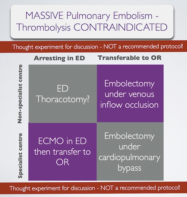
In summary, some patients with massive pulmonary embolism may benefit from surgery (contraindication to ‘lysis or failed ‘lysis). Getting them to surgery alive, or operating on them during cardiac arrest, is a challenge. Ideally they would undergo embolectomy under cardopulmonary bypass in the operating room, or could be placed on ECMO in the ED prior to going to the OR. If they present to a centre without these facilities, then the venous inflow occlusion technique could be used in the OR without bypass. Just rarely a patient may present in extremis with PE to an ED without these options. If that patient has major contraindications to thrombolysis, would an ED thoracotomy be something you would entertain?

I have done several thoracotomies for penetrating trauma but never for PE. I do not pretend to know how, and cannot find a case report of ED thoracotomy for pulmonary embolism in the literature. I’m therefore NOT recommending it. However, I would love to know people’s views on its feasibility. A possible approach could be summarised as:
Massive pulmonary embolism fascinates me, because it’s seen in the ‘talk and die’ patient. It is a single, treatable pathology that if diagnosed and treated appropriately truly makes the difference between life and death. When medicine presents us with an opportunity ‘on a plate’ like that to save a life, we need to be prepared. I have had great saves with this diagnosis and sadly have seen disastrous failures to act. When the time comes, we need to ask: ‘have we explored all options?’.

1. Surgical treatment of acute pulmonary embolism–a 12-year retrospective analysis.
Scand Cardiovasc J. 2012 Jun;46(3):172-6. Epub 2012 Mar 27.

OBJECTIVES: Surgical embolectomy for acute pulmonary embolism (PE) is considered to be a high risk procedure and therefore a last treatment option. We wanted to evaluate the procedures role in modern treatment of acute PE.
DESIGN: All data on patients treated with surgical embolectomy for acute PE were retrieved from our clinical database. The mortality was extracted from the Danish mortality register.
RESULTS: From October 1998 to July 2010, 33 patients underwent surgical embolectomy. All procedures were done through a median sternotomy and extracorporeal circulation. Twenty-six patients were diagnosed with a high risk PE and 7 with an intermediate risk PE and intracardial pathology. Six patients had been insufficiently treated with thrombolysis. Thirteen patients had contraindication for thrombolysis. Six patients were brought to the operating theatre in cardiogenic shock, 8 needed ventilator support, and 1 was in cardiac arrest. The postoperative 30-day mortality was 6% and during the 12-year follow-up the cumulative survival was 80% with 4 late deaths.
CONCLUSION: Surgical pulmonary embolectomy can be performed with low mortality although the treated patients belong to the most compromised part of the PE population. The results support surgical embolectomy as a vital part of the treatment algorithm for acute PE.

2. Pulmonary embolectomy with venous inflow-occlusion.
The Lancet 1972;1(7754):767–769

Massive pulmonary emboli have been removed surgically from 26 patients. The technique of normothermic circulatory arrest by venous inflow-occlusion was used in 25 patients. 13 patients survived. There were 10 operative deaths and 3 hospital deaths. Diagnosis was based upon clinical findings supplemented by electrocardiography and a plain radiograph of the chest. Surgery was offered to patients having a pulmonary embolus sufficiently massive to produce sustained hypotension. All patients whose hearts stopped beating before the embolectomy died. 6 successful operations were performed in hospitals without facilities for cardiac surgery. The method is recommended for its simplicity.

3. Left Anterior Thoracotomy for Pulmonary Embolectomy With 29-Year Follow-up
The Annals of Thoracic Surgery 1998, 66(4):1420-1421

Pulmonary embolectomy is usually performed in cardiopulmonary bypass. In acute situations too much time can be lost in setting up and connecting the pump oxygenator; this delay can cause cerebral damage in a patient with circulatory arrest. In such a situation left anterior thoracotomy can provide an ideal approach. An emergency thoracotomy can be performed in a few seconds. The lung automatically retracts. The phrenic nerve, pulmonary artery, and pericardium are clearly seen, and they outline the area for embolectomy. A case in which such an approach was successfully used is described.

4.ECMO treatment saved life of a young woman with acute pulmonary embolism
Lakartidningen 2004, 101(44):3420-3421

A 42-year old obese female using contraceptive medication was admitted to the emergency room because of sudden onset of dyspnoea and hypoxia. Computed tomography showed massive pulmonary emboli. Despite initial treatment with thrombolysis her condition deteriorated further and she was referred for acute surgery to our clinic. Before putting the patient to sleep extracorporeal circulation was instituted with access from the groin. After anaesthesia a median sternotomy was performed. With the heart beating, the main pulmonary artery was incised and a 9 cm long thrombus was removed. Immediate weaning from the heart-lung machine was not possible, mainly because of bleeding to the airways. The right atrium and the aorta was therefore cannulated and an extracorporeal circulation membrane oxygenator (ECMO) was used for three days. The patient required several re-entries for bleeding and a tracheotomy during the postoperative course. She was fully recovered three months after the operation.

5. Extracorporeal membrane oxygenator for pulmonary embolism.
The Annals of Thoracic Surgery 1997, 64(3):883-884 Free full text

6. Peripheral Extracorporeal Membrane Oxygenation: Comprehensive Therapy for High-Risk Massive Pulmonary Embolism
Ann Thorac Surg 2012;94:104–8

Background: Although commonly reserved as a last line of defense, experienced centers have reported excellent results with pulmonary embolectomy for massive and submassive pulmonary embolism (PE). We present a contemporary surgical series for PE that demonstrates the utility of peripheral extracorporeal membrane oxygenation (pECMO) for high-risk surgical candidates.
Methods: Between June 2005 and April 2011, 29 patients were treated for massive or submassive pulmonary embolism, with surgical embolectomy performed in 26. Four high-risk patients were placed on pECMO, established by percutaneously cannulating the right atrium through a femoral vein and perfusing by a Dacron graft anastomosed to the axillary artery. A small, extracorporeal, rotary assist device was used, interposing a compact oxygenator in the circuit, and maintaining anticoagulation with heparin.
Results: Extracorporeal membrane oxygenation was weaned in 3 of 4 patients after 5.3 days (5, 5, and 6), with normalization of right ventricular dysfunction and pulmonary artery pressure (44.0 ± 2.0 to 24.5 ± 5.5 mm Hg) by ECHO. Follow-up computed tomographies showed several peripheral, nearly resorbed emboli in 1 case and complete resolution in 2 others. The fourth patient, not improving after 10 days, underwent surgery where an embolic liposarcoma was extracted. For all 29 cases, hospital and 30-day mortality was 0% and all patients were discharged, with average postoperative length of stay of 15 days for embolectomy and 17 days for pECMO.
Conclusions: Heparin therapy with pECMO support is a rapid, effective option for patients who might benefit from pulmonary embolectomy but are at high risk for surgery.

Facilitated pericardial drainage

Drs Wyatt and Haugh describe a modified resuscitative thoracotomy technique which provided surgically facilitated pericardial drainage. A patient with a ruptured pseudoaneurysm of the right ventricular outflow tract presented in shock and arrested in the ED. She had had a prior history of idiopathic ventricular tachycardia and had undergone cardiac ablation of the posteroseptal wall of the right ventricular outflow tract. Sonographically identified tamponade was treated with pericardiocentesis which failed due to clotted blood, so a left lateral thoracotomy was performed by the emergency physician. Rather than fully expose the heart for repair in the ED, they elected to just make a 2cm incision in the pericardium which allowed drainage of blood and restoration of circulation. This was combined with blood product transfusion to buy time for the arrival of a cardiothoracic surgeon and transfer to the operating room.
Useful learning points from this paper are:

  • Ablation procedures are becoming more common
  • Serious complications such as atrioesophageal fistula, pseudoaneurysm, pericardial effusion, and cardiac tamponade occur approximately 3% of the time
  • When tamponade is suspected or confirmed ask patient about recent cardiac procedures such as catheterisations, surgery, and ablation procedures
  • Radiofrequency ablation procedures are often performed on the right side of the heart in areas that may be relatively inaccessible from a left-sided lateral thoracotomy approach.

Modified Emergency Department Thoracotomy for Postablation Cardiac Tamponade
Ann Emerg Med. 2012 Apr;59(4):265-7
[EXPAND Click for abstract]


Cardiac dysrhythmias are a common problem in the United States. Radiofrequency ablation is being used more frequently as a treatment for these diagnoses. Although rare, serious complications such as cardiac tamponade have been reported as a result of ablation procedures. Traditionally, emergency department (ED) thoracotomy has been reserved for cases of traumatic arrest only. We report a case of a successful modified ED thoracotomy in a patient with postablation cardiac tamponade and subsequent obstructive shock who failed intravenous fluid resuscitation, pressor administration, and multiple attempts at pericardiocentesis. In this case, a modified approach was used to incise the pericardium. Although this was associated with large blood loss, we believed that using the traditional method of completely removing the pericardium would have resulted in uncontrolled hemorrhage. Instead, our method led to successful resuscitation of the patient until definitive care was available. A smaller pericardial incision than is traditionally used during ED thoracotomy deserves further consideration and research to determine whether and when it may be most useful as a temporizing treatment of cardiac tamponade when other methods have failed.

[/EXPAND]

Superglue for CVCs

In resuscitation situations, the securing of vascular catheters is an important but sometimes cumbersome process, particular when sutures are required for central lines or arterial lines.

Medical grade ‘superglue’ (cyanoacrylate) can be used and this has been described in the anaesthetic literature before(1). Now, further in vitro work shows the glue does not weaken the intravenous catheter and is not associated with bacterial colonisation(2).

I think this is perfect for resuscitation lines. Just last night I used this technique to secure a femoral arterial line during a cardiac arrest resuscitation. It was great not to have to faff around with sharp suture needles during CPR and the line felt very secure after just a few seconds.

1. Tissue adhesive as an alternative to sutures for securing central venous catheters
Anaesthesia. 2007 Sep;62(9):969-70

2. Cyanoacrylate tissue adhesives – effective securement technique for intravascular catheters: in vitro testing of safety and feasibility
Anaesth Intensive Care. 2012 May;40(3):460-6

Size matters when you're sick

A nice example of a difference between elective anaesthesia and critical care practice when it comes to airway management is the selection of appropriate tracheal tube size when intubating, which is highlighted in a recent Anaesthesia article.
In recent years progressively smaller tubes have been used in anaesthesia in pursuit of decreased tracheal injury, sore throat, and hoarseness and increased ease of placement.
Patients likely to remain intubated for some time due to critical illness, however, may benefit from larger diameter tubes for the following reasons:

  • Accumulation of biofilm debris, which increases with duration of intubation – this can significantly decrease the luminal internal diameter, but is less likely to be significant with larger tubes.
  • Work of breathing during weaning: spontaneous breathing trials prior to extubation require patients to breathe through tracheal tubes. Volunteer studies have demonstrated that work of breathing increases as tube diameter decreases.
  • Bronchoscopes and suction catheters: the standard adult ICU fibreoptic bronchoscope has a diameter of 5.7 mm with a 2-mm suction channel to enable adequate suction, which limits the tracheal tube to those larger than 7.5–8.0 mm, and even with an 8.0-mm tube, the bronchoscope occupies more than 50% of the tube diameter, which can lead to ventilation issues during bronchoscopy.

The authors conclude by recommending:


‘If admission to ICU is contemplated then the time-honoured ‘8.0 for females, 9.0 for males’ is a reasonable rule of thumb, unless circumstances dictate otherwise, e.g. in difficult airways or particularly small patients.’

Size matters: choosing the right tracheal tube
Anaesthesia. 2012 Aug;67(8):815-9

Life, limb and sight-saving procedures

The challenge of competence in the face of rarity

by Dr Cliff Reid FCEM, and Dr Mike Clancy FCEM
This article is to be published in Emergency Medicine Journal (EMJ), and is reproduced here with permission of the BMJ Group.
Emergency physicians require competence in procedures which are required to preserve life, limb viability, or sight, and whose urgency cannot await referral to another specialist.
Some procedures that fit this description, such as tracheal intubation after neuromuscular blockade in a hypoxaemic patient with trismus, or placement of an intercostal catheter in a patient with a tension pneumothorax, are required sufficiently frequently in elective clinical practice that competence can be acquired simply by training in emergency department, intensive care, or operating room environments.
Other procedures, such as resuscitative thoracotomy, may be required so infrequently that the first time a clinician encounters a patient requiring such an intervention may be after the completion of specialist training, or in the absence of colleagues with prior experience in the technique.
Some techniques that might be considered limb or life saving may be too technically complex to acquire outside specialist surgical training programs. Examples are damage control laparotomy and limb fasciotomy. One could however argue that these are rarely too urgent to await arrival of the appropriate specialist.
The procedures which might fit the description of a time­‐critical life, limb, or sight saving procedure in which it is technically feasible to acquire competence within or alongside an emergency medicine residency, and that cannot await another specialist, include:

  • limb amputation for the entrapped casualty with life-­threatening injuries;
  • escharotomy for a burns patient with compromised ventilation or limb perfusion;

 
Defining competence for emergency physicians
A major challenge is the acquisition of competence in the face of such clinical rarity. One medical definition of competence is ‘the knowledge, skill, attitude or combination of these, that enables one to effectively perform the activities of a particular occupation or role to the standards expected’[1]; in essence the ability to perform to a standard, but where are these standards defined?
If we look to the curricula which are used to assess specialist emergency physicians in several English-­speaking nations, all the procedures in the short list above are included, although no one single nation’s curriculum includes the entire list (Table 1).

 
So an emergency physician is expected to be able to conduct these procedures, and a competent emergency physician effectively performs them to the ‘standards’ expected. It appears then that the question is not whether emergency physicians should perform them, but to what standard should they be trained? Only then can the optimal approach to training be decided.
There are convincing arguments that even after minimal training the performance of these procedures by emergency physicians is justifiable:

  • All the abovementioned interventions could be considered to carry 100% morbidity or mortality if not performed, with some chance of benefit whose magnitude depends on the timeliness of intervention. In some cases that risk is quantifiable: cardiac arrest due to penetrating thoracic trauma has 100% mortality if untreated, but an 18% survival to discharge rate, with a high rate of neurologically intact survivors, if performed by prehospital emergency medicine doctors in the field according to defined indications[2] and using a simple operative procedure[3]. In this extreme clinical example, no further harm to the patient can result from the procedure but a chance of supreme benefit exists. Thus, the ethical requirements of beneficence and non-­maleficence are both met even in the circumstance of very limited training for the procedure. It is hard to conceive of many other circumstances in medicine where the benefit:harm ratio approaches infinity.
  • The procedures in question are technically straightforward and can be executed without specialist equipment in non-­operating room environments. These factors appear to be underappreciated by non-­emergency specialist opponents of emergency physician-­provided thoracotomy whose practice and experience is likely to be predominantly operating room-­based[4].
  • Some of the procedures are recommended or mandated by official guidelines[5], raising the possibility of medicolegal consequences of failure to perform them.
  • The procedures are time-­critical and cannot await the arrival of an alternative specialist not already present. Simple pragmatism dictates that emergency physicians be trained to provide the necessary interventions.

 
The challenge of training
So how does one best train for these procedures? High volume trauma experience provided by a registrar term with the London Helicopter Emergency Medical Service or at a South African trauma centre will be an option for a very limited subset of trainees. Alternative training can be provided using simulation, animal labs, and cadaver labs, without risk to patients or requiring dedicated surgical specialty attachments.
Simulation manikins are not yet available for all the procedures mentioned, and lack realistic operable tissue. Human cadaver labs and live animal training bring administrative, legal, ethical and financial challenges that may be prohibitive to time and cash‐limited training schemes, or be less available to the ‘already trained’ providers in existing consultant posts. Even excellent focused cadaver-­based courses such as the Royal College of Surgeons’ Definitive Surgical Trauma Skills course[6] may not be appropriate for the emergency medicine environment: on such a course one of the authors (CR) was publicly castigated by a cardiothoracic surgeon instructor for inexpert suture technique during the resuscitative thoracotomy workshop, despite the former having successfully performed the procedure on several occasions ‘in the field’ without need of elaborate needlework.
An additional training challenge is that of metacompetence: the decision and ability to apply the competence at the right time. In the light of the relative technical simplicity of the practical procedures under discussion, this may indeed be the greatest challenge. Both authors can recount sad tales of colleagues failing to provide indicated life-­saving interventions despite being technically capable of intervening. Reasons for reticence include ‘I haven’t been properly trained’, and ‘I wouldn’t feel supported if it went wrong’.
 
Where do we go from here?
We have presented clinical, ethical, practical, and medicolegal arguments in favour of emergency physicians providing these procedures. Collectively, the emergency medicine curricula of English-­speaking nations mandate competence in them. The relative technical simplicity and overwhelming benefit:harm equation obviate the need to match the competence of a surgical subspecialist; these factors suggest training can be limited in time and cost as long as the metacompetences of ‘decision to act and knowing when to act’ are taught, simulated, and tested.
While we should capitalise on the technical expertise of surgical colleagues in the training situation, it is imperative that emergency physicians appreciative of the emergency department environment and equipment are directly involved in translating this training to emergency medicine practice. The rarity of the situations requiring these procedures requires that training should be revisited on a regular basis, preferably in the context of local departmental simulation in order to optimise equipment and teamwork preparation.
Finally, the College of Emergency Medicine needs to make it clear to its members and fellows that these procedures lie unquestionably within the domain of emergency medicine, and that emergency physicians are supported in performing them to the best of their abilities with limited training when circumstances dictate that this in the best interests of preserving a patient’s life, limb, or sight.
 
 
References
1. British Medical Association. Competency-­based assessment discussion paper for consultants, May 2008. http://www.bma.org.uk/employmentandcontracts/doctors_performance/1_app raisal/CompetencyBasedAssessment.jsp Accessed 22nd March 2012
2. Davies GE, Lockey DJ. Thirteen Survivors of Prehospital Thoracotomy for Penetrating Trauma: A Prehospital Physician‐Performed Resuscitation Procedure That Can Yield Good Results. J Trauma. 2011;70(5):E75-­8
3. Wise D, Davies G, Coats T, et al. Emergency thoracotomy: “how to do it”. Emerg Med J. 2005; 22(1):22–24 Free full text
4. Civil I. Emergency room thoracotomy: has availability triumphed over advisability in the care of trauma patients in Australasia? Emerg Med Australas. 2010;22(4):257­‐9
5. Soar J, Perkins GD, Abbas G, et al. European Resuscitation Council Guidelines for Resuscitation 2010 Section 8. Cardiac arrest in special circumstances: Electrolyte abnormalities, poisoning, drowning, accidental hypothermia, hyperthermia, asthma, anaphylaxis, cardiac surgery, trauma, pregnancy, electrocution. Resuscitation. 2010;81(10):1400-­33 Full text
6. Definitive Surgical Trauma Skills course. http://www.rcseng.ac.uk/courses/course-search/dsts.html Accessed 22nd March 2012
7. http://www.collemergencymed.ac.uk/Training-Exams/Curriculum/Curriculum%20from%20August%202010/ Accessed 22nd March 2012
8. http://www.eusem.org/cms/assets/1/pdf/european_curriculum_for_em-aug09-djw.pdf accessed 17 May 2012
9. The Model of the Clinical Practice of Emergency Medicine http://www.abem.org/PUBLIC/portal/alias__Rainbow/lang__en-%C2%AD%20US/tabID__4223/DesktopDefault.aspx Accessed 22nd March 2012
10. http://rcpsc.medical.org/residency/certification/objectives/emergmed_e.pdf Accessed 22nd March 2012
11. http://www.acem.org.au/media/publications/15_Fellowship_Curriculum.pdf accessed 17 May 2012
12. http://www.collegemedsa.ac.za/Documents/doc_173.pdf accessed 17 May 2012
Life, limb and sight-saving procedures-the challenge of competence in the face of rarity
Emerg Med J. 2012 Jul 16. [Epub ahead of print]

Unknown unknowns and pleural effusions

There are plenty of unknowns when it comes to management of pleural effusions on the ICU, which led to a paper with an eye-catching title1.
Mechanically ventilated patients frequently have pleural effusions detected by radiological investigations. Whether to drain them is a common conundrum for intensivists. A systematic review of the literature showed that drainage often improves oxygenation and has a low complication rate2.
While it may have the added advantage of assisting diagnosis and guiding therapy, there is a paucity of literature demonstrating improved patient-orientated outcomes with the routine drainage of pleural effusions in ventilated patients.
 
1. A pseudo-Rumsfeldian approach to pleural effusions in mechanically ventilated patients.
Crit Care. 2011 Mar 11;15(2):132 Free Full Text
2. Utility and safety of draining pleural effusions in mechanically ventilated patients: a systematic review and meta-analysis.
Crit Care. 2011;15(1):R46 Free Full Text
[EXPAND Click to read abstract]

INTRODUCTION: Pleural effusions are frequently drained in mechanically ventilated patients but the benefits and risks of this procedure are not well established.

METHODS: We performed a literature search of multiple databases (MEDLINE, EMBASE, HEALTHSTAR, CINAHL) up to April 2010 to identify studies reporting clinical or physiological outcomes of mechanically ventilated critically ill patients who underwent drainage of pleural effusions. Studies were adjudicated for inclusion independently and in duplicate. Data on duration of ventilation and other clinical outcomes, oxygenation and lung mechanics, and adverse events were abstracted in duplicate independently.

RESULTS: Nineteen observational studies (N = 1,124) met selection criteria. The mean PaO2:FiO2 ratio improved by 18% (95% confidence interval (CI) 5% to 33%, I2 = 53.7%, five studies including 118 patients) after effusion drainage. Reported complication rates were low for pneumothorax (20 events in 14 studies including 965 patients; pooled mean 3.4%, 95% CI 1.7 to 6.5%, I2 = 52.5%) and hemothorax (4 events in 10 studies including 721 patients; pooled mean 1.6%, 95% CI 0.8 to 3.3%, I2 = 0%). The use of ultrasound guidance (either real-time or for site marking) was not associated with a statistically significant reduction in the risk of pneumothorax (OR = 0.32; 95% CI 0.08 to 1.19). Studies did not report duration of ventilation, length of stay in the intensive care unit or hospital, or mortality.

CONCLUSIONS: Drainage of pleural effusions in mechanically ventilated patients appears to improve oxygenation and is safe. We found no data to either support or refute claims of beneficial effects on clinically important outcomes such as duration of ventilation or length of stay.

[/EXPAND]

International recommendations for lung ultrasound

A multidisciplinary panel of 28 experts from eight countries reviewed the literature and came up with consensus guidelines in point-of-care lung ultrasound. There were some big names involved – all the big players in emergency/critical care ultrasound from around the World. Conspicuously absent were Matt and Mike from the Emergency Ultrasound Podcast, but maybe there was a maximum awesomeness limit or something.

Here are some snippets, taken out of context and without the grade of recommendation attached. Try to get hold of the original if you can, which might not be easy. I never understand it when ‘international recommendations’ are published as subscription-only articles. Either they want people to follow them or not. Oh well – here are some of their recommendations:
Pneumothorax

  • The sonographic signs of pneumothorax include the following: Presence of lung point(s); Absence of lung sliding; Absence of B-lines; Absence of lung pulse
  • The lung pulse refers to the subtle rhythmic movement of the visceral upon the parietal pleura with cardiac oscillations and is a rule-out sign for pneumothorax
  • In the supine patient, the sonographic technique consists of exploration of the least gravitationally dependent areas progressing more laterally.
  • Bedside lung ultrasound is a useful tool to differentiate between small and large pneumothorax, using detection of the lung point.

Interstitial syndrome

  • B-lines are defined as discrete laser-like vertical hyperechoic reverberation artifacts that arise from the pleural line (previously described as ‘‘comet tails’’), extend to the bottom of the screen without fading, and move synchronously with lung sliding.
  • The presence of multiple diffuse bilateral B-lines indicates interstitial syndrome. Causes of interstitial syndrome include the following conditions: Pulmonary edema of various causes; Interstitial pneumonia or pneumonitis; Diffuse parenchymal lung disease (pulmonary fibrosis)

Lung consolidation

  • The sonographic sign of lung consolidation is a subpleural echo-poor region or one with tissue-like echotexture.
  • Lung ultrasound is a clinically useful tool to rule in pneumonia; however, lung ultrasound does not rule out consolidations that do not reach the pleura.
  • In mechanically ventilated patients lung ultrasound should be considered as it is more accurate than portable chest radiography in the detection of consolidation.

Pleural effusion

  • Both of the following signs are present in almost all free effusions: A space (usually anechoic) between the parietal and visceral pleura; Respiratory movement of the lung within the effusion (‘‘sinusoid sign’’)
  • In opacities identified by chest radiography, lung ultrasound should be used because it is more accurate than chest radiography in distinguishing between effusion and consolidation.
  • Visualization of internal echoes, either of mobile particles or septa, is highly suggestive of exudate or hemothorax


BACKGROUND: The purpose of this study is to provide evidence-based and expert consensus recommendations for lung ultrasound with focus on emergency and critical care settings.

METHODS: A multidisciplinary panel of 28 experts from eight countries was involved. Literature was reviewed from January 1966 to June 2011. Consensus members searched multiple databases including Pubmed, Medline, OVID, Embase, and others. The process used to develop these evidence-based recommendations involved two phases: determining the level of quality of evidence and developing the recommendation. The quality of evidence is assessed by the grading of recommendation, assessment, development, and evaluation (GRADE) method. However, the GRADE system does not enforce a specific method on how the panel should reach decisions during the consensus process. Our methodology committee decided to utilize the RAND appropriateness method for panel judgment and decisions/consensus.

RESULTS: Seventy-three proposed statements were examined and discussed in three conferences held in Bologna, Pisa, and Rome. Each conference included two rounds of face-to-face modified Delphi technique. Anonymous panel voting followed each round. The panel did not reach an agreement and therefore did not adopt any recommendations for six statements. Weak/conditional recommendations were made for 2 statements, and strong recommendations were made for the remaining 65 statements. The statements were then recategorized and grouped to their current format. Internal and external peer-review processes took place before submission of the recommendations. Updates will occur at least every 4 years or whenever significant major changes in evidence appear.

CONCLUSIONS: This document reflects the overall results of the first consensus conference on “point-of-care” lung ultrasound. Statements were discussed and elaborated by experts who published the vast majority of papers on clinical use of lung ultrasound in the last 20 years. Recommendations were produced to guide implementation, development, and standardization of lung ultrasound in all relevant settings.

International evidence-based recommendations for point-of-care lung ultrasound
Intensive Care Med. 2012 Apr;38(4):577-91

Simple emergency haemorrhage control

I had the honour of attending trauma rounds with leading South African trauma surgeons today at Groote Schuur Hospital in Cape Town. This was the first day of an intense week-long trauma education tour that I have organised for myself and three of my Sydney HEMS colleagues.
A technique for haemorrhage control in penetrating trauma is to place a Foley catheter (FC) in the wound and inflate the balloon to try to achieve compression of bleeding vascular structures. This has been life-saving in many cases and buys time to get the patient to a trauma or vascular surgeon or in some cases an interventional radiologist.

Catheter is knotted (black arrow) to occlude lumen. The wound is sutured around the catheter (white arrow).

First described by Gilroy and colleagues from Baragwanath Hospital in Johannesburg1, another, larger case series was subsequently reported by Cape Town’s Navsaria2, the Professor who conducted today’s trauma round I attended. In his paper he describes:


An 18- or 20-G FC was introduced into the bleeding neck wound. An attempt was made to follow the wound tract. The balloon was inflated with 5 ml of water or until resistance was felt. The FC was either clamped or knotted on itself to prevent bleeding through the lumen. The neck wound was sutured in two layers around the catheter. Continued bleeding around the catheter was an indication to proceed to surgery.

There were no deaths attributable to the use of FC balloon tamponade.
Prof. Navsaria describes the following algorithm for the subsequent investigation and management of these patients:

 I’ve been teaching this technique as an option in penetrating trauma for a few years but have never actually done it for real. Nice to finally see examples of its successful implementation by people who do this all the time. I’ve seen four patients with Foleys sticking out of their necks in the first 24 hours of being here.
1. Control of life-threatening haemorrhage from the neck: a new indication for balloon tamponade.
Injury. 1992;23(8):557-9
[EXPAND Click to read abstract]


We report the use of a Foley catheter, placed through the wound, to provide balloon tamponade of major bleeding from the neck and supraclavicular fossae. In 10 consecutive explorations for exsanguinating injury in these regions balloon tamponade was used eight times, and was judged to be fully effective in four patients, partly effective in one, and ineffective in three patients.

[/EXPAND]
2. Foley catheter balloon tamponade for life-threatening hemorrhage in penetrating neck trauma
World J Surg. 2006 Jul;30(7):1265-8
[EXPAND Click to read abstract]


BACKGROUND: Foley catheter (FC) balloon tamponade is a well-recognized technique employed to arrest hemorrhage from penetrating wounds. The aim of this study was to review our experience with this technique in penetrating neck wounds and to propose a management algorithm for patients with successful FC tamponade.

METHODS: A retrospective chart review (July 2004-June 2005 inclusive) was performed of patients identified from a prospectively collected penetrating neck injury computer database in whom FC balloon tamponade was used. The units’ policy for penetrating neck injuries is one of selective nonoperative management. All patients with successful FC tamponade underwent angiography. A venous injury was diagnosed if angiography was normal. Ancillary tests were performed as indicated. Removal of the FC was performed in the OR.

RESULTS: During the study period, 220 patients with penetrating neck injuries were admitted to our unit. Foley catheter balloon tamponade was used in 18 patients and was successful in 17 patients. Angiography was positive in 3 patients, all of whom underwent surgery. The FC was successfully removed in 13 patients at a mean of 72 (range 48-96) hours. One patient bled after removal of the catheter, mandating emergency surgery.

CONCLUSION: Foley catheter balloon tamponade remains a useful adjunct in the management of selective patients with penetrating, bleeding neck wounds.

[/EXPAND]