Category Archives: Resus

Life-saving medicine

Reassurance: difficult laryngoscopy in children remains rare

I was taught a useful principle by a paediatric anaesthetist 10 years ago which has proven true in my experience and has contributed to keeping me calm when intubating sick kids. Unlike adults, in whom difficulty in intubation can often be unexpected, the vast majority of normal looking children are easy to intubate, and the ones who are difficult usually have obvious indicators such as dysmorphism.
This appears to be supported by recent evidence: in a large retrospective series of 11.219 anaesthesia patients, the overall incidence of difficult laryngoscopy [Cormack and Lehane (CML) grade III and IV] was only 1.35%, although was much higher in infants less than one year compared with older children. This low percentage is in the same ball park as two other paediatric studies. Besides younger age, their database suggested underweight, ASA III and IV physical status and, if obtainable, Mallampati III and IV findings as predictors for difficult laryngoscopy. The authors point out:

…the oromaxillofacial surgery department with a high proportion of cleft palate interventions and pediatric cardiac surgery contributed substantially to the total number of difficult laryngoscopies. In patients undergoing pediatric cardiac surgery, a possible explanation for the higher incidence of CML III/IV findings might be that some congenital heart defects are associated with chromosomal anomalies like microdeletion 22q11.2 syndrome. This syndrome is also associated with extracardiac anomalies like cranio-facial dysmorphism

Take home message: As a very rough rule of thumb to illustrate the difference between the ease/difficulty of laryngoscopy between adults and kids, I think it’s fair to say grade III or IV views occur in about 10% of adults but only about 1% of children.

Incidence and predictors of difficult laryngoscopy in 11.219 pediatric anesthesia procedures
Paediatr Anaesth. 2012 Aug;22(8):729-36
[EXPAND Click for abstract]


OBJECTIVE: Difficult laryngoscopy in pediatric patients undergoing anesthesia.

AIM: This retrospective analysis was conducted to investigate incidence and predictors of difficult laryngoscopy in a large cohort of pediatric patients receiving general anesthesia with endotracheal intubation.

BACKGROUND: Young age and craniofacial dysmorphy are predictors for the difficult pediatric airway and difficult laryngoscopy. For difficult laryngoscopy, other general predictors are not yet described.

METHODS: Retrospectively, from a 5-year period, data from 11.219 general anesthesia procedures in pediatric patients with endotracheal intubation using age-adapted Macintosh blades in a single center (university hospital) were analyzed statistically.

RESULTS: The overall incidence of difficult laryngoscopy [Cormack and Lehane (CML) grade III and IV] was 1.35%. In patients younger than 1 year, the incidence of CML III or IV was significantly higher than in the older patients (4.7% vs 0.7%). ASA Physical Status III and IV, a higher Mallampati Score (III and IV) and a low BMI were all associated (P < 0.05) with difficult laryngoscopy. Patients undergoing oromaxillofacial surgery and cardiac surgery showed a significantly higher rate of CML III/IV findings.

CONCLUSION: The general incidence of difficult laryngoscopy in pediatric anesthesia is lower than in adults. Our results show that the risk of difficult laryngoscopy is much higher in patients below 1 year of age, in underweight patients and in ASA III and IV patients. The underlying disease might also contribute to the risk. If the Mallampati score could be obtained, prediction of difficult laryngoscopy seems to be reliable. Our data support the existing recommendations for a specialized anesthesiological team to provide safe anesthesia for infants and neonates.

[/EXPAND]

Transtracheal airways in kids. Well, pigs' kids anyway

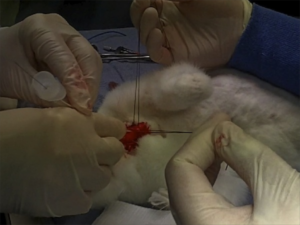
Ever had to do a surgical airway in a child? Thought not. They’re pretty rare. Bill Heegaard MD from Henepin County Medical Center taught me a few approaches (with the help of an anaesthetised rabbit) which really got me thinking. It’s something I’d often trained for in my internal simulator, and I even keep the equipment for it in my house (listen out for an upcoming podcast on that). Research and experience has demonstrated that open surgical airway techniques are more reliable than transtracheal needle techniques in adults, but what about kids, in whom traditional teaching cautions against open techniques?

Australian investigators who were experienced airway proceduralists evaluated transtracheal needle techniques using a rabbit model (an excellent model for the infant airway). Their success rate was only 60% and they perforated the posterior tracheal wall in 42% of attempts. Of 13 attempts to insert a dedicated paediatric tracheotomy device, the Quicktrach Child, none were successful(1) (they did not use the Quicktrach Infant model as it is not available in Australia).

Danish investigators used fresh piglet cadavers weighing around 8 kg to assess two transtracheal cannulas, in which they achieved success rates of 65.6% and 68.8%(2). There was also a very high rate of posterior tracheal wall perforation. Using an open surgical tracheostomy technique, they were successful in 97% of attempts. These were also experienced operators, with a median anaesthetic experience of 12.5 years.
Their tracheotomy technique was nice and simple, and used just a scalpel, scissors, and surgical towel clips. Here’s their technique:

Simple tracheotomy procedure described by Holm-Knudsen et al

  1. Identify larynx and proximal trachea by palpation
  2. Vertical incision through the skin and subcutaneous tissue from the upper part of larynx to the sternal notch
  3. Grasp strap muscles with two towel forceps and separate in the midline
  4. Palpate and identify the trachea (palpate rather than look for tracheal rings, as in a live patient one would expect bleeding to obscure the view)
  5. Stabilise the trachea by grasping it with a towel forceps
  6. Insert sharp tip of the scissors between two tracheal rings and lift the trachea anteriorly to avoid damage to the posterior wall
  7. Cut vertically in the midline of the trachea with the scissors – they chose to use the scissors to cut the tracheal rings to facilitate tube insertion
  8. Insert the tracheal tube

Using ultrasound and CT to evaluate comparative airway dimensions, the authors concluded that the pig model is most useful for training emergency airway management in older children aged 5–10 years.
Why were they doing a tracheotomy rather than a cricothyroidotomy? Reasons given by the authors include:

  • The infant cricothyroid membrane is very small
  • Palpation of the thyroid notch may be hindered by the overlying hyoid bone
  • The mandible may obstruct needle access to the cricothyroid membrane given the cephalad position in the neck of the infant larynx.

From an emergency medicine point of view, there are a couple of other reasons why we need to be able to access the trachea lower than the cricothyroid membrane. One is fractured larynx or other blunt or penetrating airway injury where there may be anatomical disruption at the cricothyroid level. The other situation is foreign body airway obstruction, when objects may lodge at the level of the cricoid ring which is functionally the narrowest part of the pediatric upper airway. Of course, alternative methods might be considered to remove the foreign body prior to tracheotomy, such as employing basic choking algorithms, and other techniques depending on whether you do or don’t have equipment.

Take home messages

  • Transtracheal airways in kids are so rare, we can’t avoid extrapolating animal data
  • Whichever infant or paediatric model is used, transtracheal needle techniques have a high rate of failure even by ‘experienced’ operators
  • The small size and easy compressibility of the airway probably contributes to this failure rate, including the high rate of posterior wall puncture
  • In keeping with adult audit data, open surgical techniques may have a higher success rate
  • Tracheotomy may be necessary rather than cricothyroidotomy in infants and children depending on clinical scenario and accessibility of anatomy
  • The stress and blood that is not simulated in cadaveric animal models will make open tracheotomy harder in a live patient, and so these success rates may not translate. However these factors do mean that whatever technique is used must be kept simple and should employ readily available and familiar equipment
  • Something to maintain control and anterior position of the anterior trachea wall should be used during incision and intubation of the trachea. The study reported here used towel clips; sutures around the tracheal rings may also be used (see image below)

I recommend you add ‘paediatric tracheotomy’ to the list of procedures you might need to do (if it’s not already there). Identify what equipment you would use and run the simulation in your head and in your work environment.
Have fun.

1. The ‘Can’t Intubate Can’t Oxygenate’ scenario in Pediatric Anesthesia: a comparison of different devices for needle cricothyroidotomy
Paediatr Anaesth. 2012 Dec;22(12):1155-8

BACKGROUND: Little evidence exists to guide the management of the ‘Can’t Intubate, Can’t Oxygenate’ (CICO) scenario in pediatric anesthesia.

OBJECTIVES: To compare two intravenous cannulae for ease of use, success rate and complication rate in needle tracheotomy in a postmortem animal model of the infant airway, and trial a commercially available device using the same model.

METHODS: Two experienced proceduralists repeatedly attempted cannula tracheotomy in five postmortem rabbits, alternately using 18-gauge (18G) and 14-gauge (14G) BD Insyte(™) cannulae (BD, Franklin Lakes, NJ, USA). Attempts began at the first tracheal cartilage, with subsequent attempts progressively more caudad. Success was defined as intratracheal cannula placement. In each rabbit, an attempt was then made by each proceduralist to perform a cannula tracheotomy using the Quicktrach Child(™) device (VBM Medizintechnik GmbH, Sulz am Neckar, Germany).

RESULTS: The rabbit tracheas were of similar dimensions to a human infant. 60 attempts were made at cannula tracheotomy, yielding a 60% success rate. There was no significant difference in success rate, ease of use, or complication rate between cannulae of different gauge. Successful aspiration was highly predictive (positive predictive value 97%) and both sensitive (89%) and specific (96%) for tracheal cannulation. The posterior tracheal wall was perforated in 42% of tracheal punctures. None of 13 attempts using the Quicktrach Child(™) were successful.

CONCLUSION: Cannula tracheotomy in a model comparable to the infant airway is difficult and not without complication. Cannulae of 14- and 18-gauge appear to offer similar performance. Successful aspiration is the key predictor of appropriate cannula placement. The Quicktrach Child was not used successfully in this model. Further work is required to compare possible management strategies for the CICO scenario

2. Emergency airway access in children – transtracheal cannulas and tracheotomy assessed in a porcine model
Paediatr Anaesth. 2012 Dec;22(12):1159-65

OBJECTIVES: In the rare scenario when it is impossible to oxygenate or intubate a child, no evidence exists on what strategy to follow.

AIM: The aim of this study was to compare the time and success rate when using two different transtracheal needle techniques and also to measure the success rate and time when performing an emergency tracheotomy in a piglet cadaver model.

METHODS: In this randomized cross-over study, we included 32 anesthesiologists who each inserted two transtracheal cannulas (TTC) using a jet ventilation catheter and an intravenous catheter in a piglet model. Second, they performed an emergency tracheotomy. A maximum of 2 and 4 min were allowed for the procedures, respectively. The TTC procedures were recorded using a video scope.

RESULTS: Placement of a transtracheal cannula was successful in 65.6% and 68.8% of the attempts (P = 0.76), and the median duration of the attempts was 69 and 42 s (P = 0.32), using the jet ventilation catheter and the intravenous catheter, respectively. Complications were frequent in both groups, especially perforation of the posterior tracheal wall. Performing an emergency tracheotomy was successful in 97%, in a median of 88 s.

CONCLUSIONS: In a piglet model, we found no significant difference in success rates or time to insert a jet ventilation cannula or an intravenous catheter transtracheally, but the incidence of complications was high. In the same model, we found a 97% success rate for performing an emergency tracheotomy within 4 min with a low rate of complications.

Externally rotate leg for femoral vein access

Want to access the femoral vein? Externally rotate the leg at the hip and things might be a bit easier. This study was done in adult patients, with the knee straight and no abduction applied. External rotation is also helpful in kids, with abduction up to sixty degrees.


Objective: To determine if external rotation of the leg increases the size and accessibility of the femoral vein compared with a neutral position.

Methods: One hundred patients presenting to a tertiary teaching hospital were prospectively recruited. The right common femoral vein of each subject was scanned with a linear probe (5–10 MHz) inferior to the inguinal ligament, with the leg in a neutral position and then in the externally rotated position. The transverse diameter of the femoral vein, the accessible diameter of the vein (lying medial to the femoral artery) and the depth of the vein were measured.

Results: The mean diameter of the femoral vein in the externally rotated leg was greater than with the leg in the neutral position (15.4 mm vs 13.8 mm); the mean difference was 1.6 mm (95% CI 1.3–1.9). The mean accessible diameter of the femoral vein was larger with the leg externally rotated (13.8 mm vs 11.7 mm, mean difference 2.1 mm, 95% CI 1.8–2.5). The depth from the skin to the femoral vein was less with the leg in external rotation (20.9 mm vs 22.6 mm, mean difference 1.7 mm, 95% CI 1.2–2.2). The mean diameter and depth were greater in patients with overweight or obese body mass index (BMI) measurements in both leg positions. The increase in femoral vein diameter and accessibility with external rotation was observed in all BMI groups.

Conclusion: The total and accessible femoral vein diameter is increased and the surface depth of the vein is decreased by placing the leg in external rotation compared with the neutral position.

Simple external rotation of the leg increases the size and accessibility of the femoral vein
Emerg Med Australas. 2012 Aug;24(4):408-13

Hyperglycaemia & mortality in sepsis – lactate dependent?

I like this paper for introducing a new concept to me. For years the critical care community has recognised the link between hyperglycaemia and mortality, leading to early recommendations of intensive insulin regimens subsequently shown not to be of benefit. Now it appears that the association between hyperglycaemia and mortality may be less relevant in patients with a normal lactate.
In a study of adult nondiabetic critically ill patients, hyperglycaemia had a significant association with increased mortality risk using simple univariate analysis. When they adjusted for concurrent hyperlactataemia however, hyperglycaemia was not significantly associated with increased mortality risk.
The authors discuss several known or postulated aspects of interplay between lactate and glucose in sepsis:

  • Hyperlactataemia appears to inhibit glucose uptake by muscle cells and decrease activity of the GLUT-4 transporters
  • Hyperlactataemia has also been shown to increase insulin resistance directly
  • Glucose and lactate levels tend to be elevated simultaneously in severe sepsis at baseline.
  • Experimentally it has been estimated that 45% of infused (radiolabelled) lactate is either converted into glucose via gluconeogenesis or is transformed into glycogen via the Cori cycle, representing a higher proportion of glucose formation from lactate than in nonseptic controls.
  • It is possible that elevated glucose and lactate levels in sepsis both may be measures of the same phenomenon: glucose accumulates due to the sympathomimetic response to a systemic infection with increased catecholamine levels leading to increased activity of the Na+K+-ATPase, resulting in accumulation of adenosine diphosphate (ADP). Increased levels of ADP in turn augment glycogenolysis.
  • Mitochondrial metabolism cannot meet the increased cellular energy needs of sepsis, resulting in accumulation of ADP and leading to cytosolic glycolysis and lactate production, even in an aerobic environment.

The augmented glycolysis of sepsis (and during adrenergic therapy such as epinephrine/adrenaline or albuterol/salbutamol) is one of the causes of a raised lactate to consider when applying the LACTATES mnemonic I like to use.
Hyperlactatemia affects the association of hyperglycemia with mortality in nondiabetic adults with sepsis
Acad Emerg Med. 2012 Nov;19(11):1268-75
[EXPAND Click for abstract]


BACKGROUND: Admission hyperglycemia has been reported as a mortality risk factor for septic nondiabetic patients; however, hyperglycemia’s known association with hyperlactatemia was not addressed in these analyses.

OBJECTIVES: The objective was to determine whether the association of hyperglycemia with mortality remains significant when adjusted for concurrent hyperlactatemia.

METHODS: This was a post hoc, nested analysis of a retrospective cohort study performed at a single center. Providers had identified study subjects during their emergency department (ED) encounters; all data were collected from the electronic medical record (EMR). Nondiabetic adult ED patients hospitalized for suspected infection, two or more systemic inflammatory response syndrome (SIRS) criteria, and simultaneous lactate and glucose testing in the ED were enrolled. The setting was the ED of an urban teaching hospital from 2007 to 2009. To evaluate the association of hyperglycemia (glucose > 200 mg/dL) with hyperlactatemia (lactate ≥ 4.0 mmol/L), a logistic regression model was created. The outcome was a diagnosis of hyperlactatemia, and the primary variable of interest was hyperglycemia. A second model was created to determine if coexisting hyperlactatemia affects hyperglycemia’s association with mortality; the main outcome was 28-day mortality, and the primary risk variable was hyperglycemia with an interaction term for simultaneous hyperlactatemia. Both models were adjusted for demographics; comorbidities; presenting infectious source; and objective evidence of renal, respiratory, hematologic, or cardiovascular dysfunction.

RESULTS: A total of 1,236 ED patients were included, and the median age was 77 years (interquartile range [IQR] = 60 to 87 years). A total of 115 (9.3%) subjects were hyperglycemic, 162 (13%) were hyperlactatemic, and 214 (17%) died within 28 days of their initial ED visits. After adjustment, hyperglycemia was significantly associated with simultaneous hyperlactatemia (odds ratio [OR] = 4.14, 95% confidence interval [CI] = 2.65 to 6.45). Hyperglycemia and concurrent hyperlactatemia were associated with increased mortality risk (OR = 3.96, 95% CI = 2.01 to 7.79), but hyperglycemia in the absence of simultaneous hyperlactatemia was not (OR = 0.78, 95% CI = 0.39 to 1.57).

CONCLUSIONS: In this cohort of septic adult nondiabetic patients, mortality risk did not increase with hyperglycemia unless associated with simultaneous hyperlactatemia. The previously reported association of hyperglycemia with mortality in nondiabetic sepsis may be due to the association of hyperglycemia with hyperlactatemia.

[/EXPAND]

Echo for cardiac arrest outcome prediction

A meta-analysis of studies evaluation transthoracic echo as a means of predicting return of spontaneous circulation in cardiac arrest (ROSC) provides some likelihood ratios to what we already know: absence of sonographic cardiac activity means a very low chance of ROSC.

The authors report a pooled negative LR of 0.18 (95% CI = 0.10 to 0.31), and a positive likelihood ratio of 4.26 (95% CI = 2.63 to 6.92).

They conclude that focused transthoracic echo is a fairly effective (although not definitive) test for predicting death if no cardiac activity is noted during resuscitation, and recommend interpreting the echo in the light of the test characteristics and the clinical pre-test probability, as one should do for all imaging investigations:


“An elderly patient with an unwitnessed cardiac arrest already has very poor odds for survival. Confirmation of asystole on echo lowers those pretest odds by a factor of 5.6 and therefore might lead to termination of resuscitation. However, in the case of a 50-year-old rescued from drowning, detection of cardiac contractility on echo would increase his already fair odds of survival by a factor of 4.3, prompting continued aggressive resuscitation.”

Only five relatively small studies contributed to the findings. A more definitive answer to this question should be provided in the future by the multi-centre REASON 1 trial.

Objectives:  The objective was to determine if focused transthoracic echocardiography (echo) can be used during resuscitation to predict the outcome of cardiac arrest.

Methods:  A literature search of diagnostic accuracy studies was conducted using MEDLINE via PubMed, EMBASE, CINAHL, and Cochrane Library databases. A hand search of references was performed and experts in the field were contacted. Studies were included for further appraisal and analysis only if the selection criteria and reference standards were met. The eligible studies were appraised and scored by two independent reviewers using a modified quality assessment tool for diagnostic accuracy studies (QUADAS) to select the papers included in the meta-analysis.

Results:  The initial search returned 2,538 unique papers, 11 of which were determined to be relevant after screening criteria were applied by two independent researchers. One additional study was identified after the initial search, totaling 12 studies to be included in our final analysis. The total number of patients in these studies was 568, all of whom had echo during resuscitation efforts to determine the presence or absence of kinetic cardiac activity and were followed up to determine return of spontaneous circulation (ROSC). Meta-analysis of the data showed that as a predictor of ROSC during cardiac arrest, echo had a pooled sensitivity of 91.6% (95% confidence interval [CI] = 84.6% to 96.1%), and specificity was 80.0% (95% CI = 76.1% to 83.6%). The positive likelihood ratio for ROSC was 4.26 (95% CI = 2.63 to 6.92), and negative likelihood ratio was 0.18 (95% CI = 0.10 to 0.31). Heterogeneity of the results (sensitivity) was nonsignificant (Cochran’s Q: χ(2) = 10.63, p = 0.16, and I(2) = 34.1%).

Conclusions:  Echocardiography performed during cardiac arrest that demonstrates an absence of cardiac activity harbors a significantly lower (but not zero) likelihood that a patient will experience ROSC. In selected patients with a higher likelihood of survival from cardiac arrest at presentation, based on established predictors of survival, echo should not be the sole basis for the decision to cease resuscitative efforts. Echo should continue to be used only as an adjunct to clinical assessment in predicting the outcome of resuscitation for cardiac arrest.

Bedside Focused Echocardiography as Predictor of Survival in Cardiac Arrest Patients: A Systematic Review
Acad Emerg Med. 2012 Oct;19(10):1119-1126

Aeromedical retrieval: invasive vs noninvasive blood pressure

The chaps from the Emergency Medical Retrieval Service in the UK compared invasive (IABP) and non-invasive blood pressure (NIBP) measurements on the ground and in the air. They concluded that NIBP was unreliable, although it was no worse in the aeromedical environment than in the hospital. Not surprisingly there was a better correlation between the mean IABP and NIBP than systolic or diastolic pressures (oscillometric NIBP devices measure the mean BP and derive systolic and diastolic using an algorithm specific to the device).
In their summary, they recommend:

  • IABP monitoring should be used in any unwell patient in whom accurate blood pressure measurement is desirable.
  • The aeromedical transport environment does not lead to less precise NIBP results than the non-transport environment.
  • Where NIBP measurement is the only option, the mean blood pressure should be used in preference to systolic measurements


Blood pressure measurement is an essential physiological measurement for all critically ill patients. Previous work has shown that non-invasive blood pressure is not an accurate reflection of invasive blood pressure measurement. In a transport environment, the effects of motion and vibration may make non-invasive blood pressure less accurate.

Consecutive critically ill patients transported by a dedicated aeromedical retrieval and critical care transfer service with simultaneous invasive and non-invasive blood pressure measurements were analysed. Two sets of measurements were recorded, first in a hospital environment before departure (pre-flight) and a second during aeromedical transport (in-flight).

A total of 56 complete sets of data were analysed. Bland-Altman plots showed limits of agreement (precision) for pre-flight systolic blood pressure were -37.3 mmHg to 30.0 mmHg, and for pre-flight mean arterial pressure -20.5 mmHg to 25.0 mmHg. The limits of agreement for in-flight systolic blood pressure were -40.6 mmHg to 33.1 mmHg, while those for in-flight mean blood pressure in-flight were -23.6 mmHg to 24.6 mmHg. The bias for the four conditions ranged from 0.5 to -3.8 mmHg. There were no significant differences in values between pre-flight and in-flight blood pressure measurements for all categories of blood pressure measurement.

Thus, our data show that non-invasive blood pressure is not a precise reflection of invasive intra-arterial blood pressure. Mean blood pressure measured non-invasively may be a better marker of invasive blood pressure than systolic blood pressure. Our data show no evidence of non-invasive blood pressures being less accurate in an aeromedical transport environment.

Comparison of non-invasive and invasive blood pressure in aeromedical care
Anaesthesia. 2012 Dec;67(12):1343-7

Etomidate & sepsis

A meta-analysis attempts to quantify etomidate’s effect on mortality and adrenal suppression. Of course, we all know a meta-analysis can only be as reliable as the original data it’s analysing. I think editorialists Lauzier and Turgeon have a point with their statement:

“Given the widespread use of etomidate in the emergency room, we believe that a RCT designed to evaluate the safety of etomidate as a hypnotic agent for endotracheal intubation of patients with sepsis is not only ethical but also urgently warranted”

For a critique of the paper and subsequent discussion, check out the Academic Life in EM blog post by Brian Hayes


OBJECTIVE: To evaluate the effects of single-dose etomidate on the adrenal axis and mortality in patients with severe sepsis and septic shock.

DESIGN: A systematic review of randomized controlled trials and observational studies with meta-analysis.

SETTING: Literature search of EMBASE, Medline, Cochrane Database, and Evidence-Based Medical Reviews.

SUBJECTS: Sepsis patients who received etomidate for rapid sequence intubation.

INTERVENTIONS: None.

MEASUREMENTS AND MAIN RESULTS: We conducted a systematic review of randomized controlled trials and observational studies with meta-analysis assessing the effects of etomidate on adrenal insufficiency and all-cause mortality published between January 1950 and February 2012. We only examined studies including septic patients. All-cause mortality served as our primary end point, whereas the prevalence of adrenal insufficiency was our secondary end point. Adrenal insufficiency was determined using a cosyntropin stimulation test in all studies. We used a random effects model for analysis; heterogeneity was assessed with the I statistic. Publication bias was evaluated with Begg’s test. Five studies were identified that assessed mortality in those who received etomidate. A total of 865 subjects were included. Subjects who received etomidate were more likely to die (pooled relative risk 1.20; 95% confidence interval 1.02-1.42; Q statistic, 4.20; I2 statistic, 4.9%). Seven studies addressed the development of adrenal suppression associated with the administration of etomidate; 1,303 subjects were included. Etomidate administration increased the likelihood of developing adrenal insufficiency (pooled relative risk 1.33; 95% confidence interval 1.22-1.46; Q statistic, 10.7; I2 statistic, 43.9%).

CONCLUSIONS: Administration of etomidate for rapid sequence intubation is associated with higher rates of adrenal insufficiency and mortality in patients with sepsis.

Etomidate is associated with mortality and adrenal insufficiency in sepsis: A meta-analysis Crit Care Med. 2012 Nov;40(11):2945-53

A whole bunch of trauma guidelines

The Eastern Association for the Surgery of Trauma has published a number of helpful evidence-based guidelines for trauma management, and many of them are included in this month’s Journal of Trauma and Acute Care Surgery
Here are brief snippets from some of them. All the guidelines can be viewed or downloaded in full for free here.
 
Nonoperative management of blunt hepatic injury

  • Nonoperative management of blunt hepatic injuries currently is the treatment modality of choice in hemodynamically stable patients, irrespective of the grade of injury or patient age.
  • Patients presenting with hemodynamic instability and peritonitis still warrant emergent operative intervention.
  • Intravenous contrast enhanced computed tomographic scan is the diagnostic modality of choice for evaluating blunt hepatic injuries.

 
Selective nonoperative management of blunt splenic injury

  • Nonoperative management of blunt splenic injuries is now the treatment modality of choice in hemodynamically stable patients, irrespective of the grade of injury, patient age, or the presence of associated injuries.
  • Patients presenting with hemodynamic instability and peritonitis still warrant emergent operative intervention.
  • Intravenous contrast enhanced computed tomographic scan is the diagnostic modality of choice for evaluating blunt splenic injuries.

 
Screening for blunt cardiac injury

  • Electrocardiogram (ECG) alone is not sufficient to rule out BCI.
  • BCI can be ruled out only if both ECG result and troponin I level are normal, a significant change from the previous guideline.
  • Patients with new ECG changes and/or elevated troponin I should be admitted for monitoring.
  • Echocardiogram is not beneficial as a screening tool for BCI and should be reserved for patients with hypotension and/or arrhythmias.
  • The presence of a sternal fracture alone does not predict BCI.
  • Cardiac computed tomography or magnetic resonance imaging can be used to differentiate acute myocardial infarction from BCI in trauma patients.

 
Evaluation and management of penetrating lower extremity arterial trauma

  • Expedited triage of patients is possible with physical examination and/or the measurement of ankle-brachial indices.
  • Computed tomographic angiography has become the diagnostic study of choice when imaging is required.
  • Tourniquets and intravascular shunts have emerged as adjuncts in the treatment of penetrating lower extremity arterial trauma.

 
Prophylactic antibiotic use in penetrating abdominal trauma

  • There is evidence to support a Level I recommendation that prophylactic antibiotics should only be administered for 24 hours in the presence of a hollow viscus injury.
  • There are no data to support continuing prophylactic antibiotics longer than 24 hours in damage control laparotomy.

 
Screening for thoracolumbar spinal injuries in blunt trauma

  • Multidetector computed tomographic scans have become the screening modality of choice and the criterion standard in screening for TLS injuries.
  • Patients without altered mentation or significant mechanism may be excluded by clinical examination without imaging.
  • Patients with gross neurologic deficits or concerning clinical examination findings with negative imaging should receive a magnetic resonance imaging expediently, and the spine service should be consulted

 
Emergency tracheal intubation immediately following traumatic injury

  • The decision to intubate a patient following traumatic injury is based on multiple factors, including the need for oxygenation and ventilation, the extent and mechanism of injury, predicted operative need, or progression of disease.
  • Rapid sequence intubation with direct laryngoscopy continues to be the recommended method for ETI, although the use of airway adjuncts such as blind insertion supraglottic devices and video laryngoscopy may be useful in facilitating successful ETI and may be preferred in certain patient populations.
  • There is no pharmacologic induction agent of choice for ETI; however, succinylcholine is the neuromuscular blockade agent recommended for rapid sequence intubation.

 
Presumptive antibiotic use in tube thoracostomy for traumatic hemopneumothorax

  • Routine presumptive antibiotic use to reduce the incidence of empyema and pneumonia in TT for traumatic hemopneumothorax is controversial; however, there is insufficient published evidence to support any recommendation either for or against this practice.

 
Evaluation and management of geriatric trauma

  • Effective evidence-based care of aging patients necessitates aggressive triage, correction of coagulopathy, and limitation of care when clinical evidence points toward an overwhelming likelihood of poor long-term prognosis

 
Management of pulmonary contusion and flail chest

  • Patients with PC-FC should not be excessively fluid restricted but should be resuscitated to maintain signs of adequate tissue perfusion.
  • Obligatory mechanical ventilation in the absence of respiratory failure should be avoided.
  • The use of optimal analgesia and aggressive chest physiotherapy should be applied to minimize the likelihood of respiratory failure.
  • Epidural catheter is the preferred mode of analgesia delivery in severe flail chest injury.
  • Paravertebral analgesia may be equivalent to epidural analgesia and may be appropriate in certain situations when epidural is contraindicated.
  • A trial of mask continuous positive airway pressure should be considered in alert patients with marginal respiratory status.
  • Patients requiring mechanical ventilation should be supported in a manner based on institutional and physician preference and separated from the ventilator at the earliest possible time.
  • Positive end-expiratory pressure or continuous positive airway pressure should be provided.
  • High-frequency oscillatory ventilation should be considered for patients failing conventional ventilatory modes. Independent lung ventilation may also be considered in severe unilateral pulmonary contusion when shunt cannot be otherwise corrected.
  • Surgical fixation of flail chest may be considered in cases of severe flail chest failing to wean from the ventilator or when thoracotomy is required for other reasons.
  • Self-activating multidisciplinary protocols for the treatment of chest wall injuries may improve outcome and should be considered where feasible.
  • Steroids should not be used in the therapy of pulmonary contusion.
  • Diuretics may be used in the setting of hydrostatic fluid overload in hemodynamically stable patients or in the setting of known concurrent congestive heart failure.

 
Evaluation and management of small-bowel obstruction

  • Level I evidence now exists to recommend the use of computed tomographic scan, especially multidetector computed tomography with multiplanar reconstructions, in the evaluation of patients with SBO because it can provide incremental clinically relevant information over plains films that may lead to changes in management.
  • Patients with evidence of generalized peritonitis, other evidence of clinical deterioration, such as fever, leukocytosis, tachycardia, metabolic acidosis, and continuous pain, or patients with evidence of ischemia on imaging should undergo timely exploration.
  • The remainder of patients can safely undergo initial nonoperative management for both partial and complete SBO.
  • Water-soluble contrast studies should be considered in patients who do not clinically resolve after 48 to 72 hours for both diagnostic and potential therapeutic purposes.
  • Laparoscopic treatment of SBO has been demonstrated to be a viable alternative to laparotomy in selected cases.

 
2012 Eastern Association for the Surgery of Trauma (EAST) Practice Management Guidelines Supplement
J Trauma Acute Care Surg. 2012 Nov;73(5 Suppl 4)

Needle decompression: it's still not going to work

A pet topic that keeps coming up here is management of tension pneumothorax. Plenty of studies demonstrate that traditionally taught needle thoracostomy may fail, and open, or ‘finger’ thoracostomy is recommended for the emergency management of tension pneumothorax in a patient who is being ventilated with positive pressure (including those patients in cardiac arrest).
A recent CT scan-based study of adult trauma patients makes the case that needle decompression with a standard iv cannula would be expected to fail in 42.5% of cases at the second intercostal space (ICS) compared with 16.7% at the fifth ICS at the anterior axillary line (AAL).
The authors add an important point: “As BMI increases, there is a stepwise increase in chest wall thickness, further compounding the difficulty of needle placement in all but the lowest BMI quartile for the second ICS.”
An accompanying editorial cautions that the proximity of the heart may confer a safety issue if a needle is inserted blindly into the left 5th ICS at the AAL.


Objective To compare the distance to be traversed during needle thoracostomy decompression performed at the second intercostal space (ICS) in the midclavicular line (MCL) with the fifth ICS in the anterior axillary line (AAL).

Design Patients were separated into body mass index (BMI) quartiles, with BMI calculated as weight in kilograms divided by height in meters squared. From each BMI quartile, 30 patients were randomly chosen for inclusion in the study on the basis of a priori power analysis (n = 120). Chest wall thickness on computed tomography at the second ICS in the MCL was compared with the fifth ICS in the AAL on both the right and left sides through all BMI quartiles.

Setting Level I trauma center.

Patients Injured patients aged 16 years or older evaluated from January 1, 2009, to January 1, 2010, undergoing computed tomography of the chest.

Results A total of 680 patients met the study inclusion criteria (81.5% were male and mean age was 41 years [range, 16-97 years]). Of the injuries sustained, 13.2% were penetrating, mean (SD) Injury Severity Score was 15.5 (10.3), and mean BMI was 27.9 (5.9) (range, 15.4-60.7). The mean difference in chest wall thickness between the second ICS at the MCL and the fifth ICS at the AAL was 12.9 mm (95% CI, 11.0-14.8; P < .001) on the right and 13.4 mm (95% CI, 11.4-15.3; P < .001) on the left. There was a stepwise increase in chest wall thickness across all BMI quartiles at each location of measurement. There was a significant difference in chest wall thickness between the second ICS at the MCL and the fifth ICS at the AAL in all quartiles on both the right and the left. The percentage of patients with chest wall thickness greater than the standard 5-cm decompression needle was 42.5% at the second ICS in the MCL and only 16.7% at the fifth ICS in the AAL.

Conclusions In this computed tomography–based analysis of chest wall thickness, needle thoracostomy decompression would be expected to fail in 42.5% of cases at the second ICS in the MCL compared with 16.7% at the fifth ICS in the AAL. The chest wall thickness at the fifth ICS AAL was 1.3 cm thinner on average and may be a preferred location for needle thoracostomy decompression.

Radiologic evaluation of alternative sites for needle decompression of tension pneumothorax
Arch Surg. 2012 Sep 1;147(9):813-8

Thoracoabdominal trauma outcomes

In what the authors describe as ‘the largest and most rigorous description of blunt thoracoabdominal injury to date’, we learn some interesting things and are reminded of some others:

Most solid organ injuries in the abdomen are managed non-operatively

Thoracotomy – especially non-resuscitative thoracotomy – is rare

In patients with thoracoabdominal trauma, the overwhelming majority of injuries requiring operative intervention were found in the abdomen, therefore..

…excluding those patients in extremis requiring a resuscitative thoracotomy, the initial incision, without directive radiological information, belongs in the abdomen

Concomitant thoracic injury did not preclude nonoperative management of abdominal solid organ injury

This is a really interesting paper providing important data on the outcomes and management of a patient group that frequently produces management dilemmas in trauma centres.
The double jeopardy of blunt thoracoabdominal trauma
Arch Surg. 2012 Jun;147(6):498-504
[EXPAND Click for abstract]


OBJECTIVES: To examine the specific injuries, need for operative intervention, and clinical outcomes of patients with blunt thoracoabdominal trauma.

DESIGN: Trauma registry and medical record review.

SETTING: Level I trauma center in Los Angeles, California.

PATIENTS: All patients with thoracoabdominal injuries from January 1996 to December 2010.

MAIN OUTCOME MEASURES: Injuries, incidence and type of operative intervention, clinical outcomes, and risk factors for mortality.

RESULTS: Blunt thoracoabdominal injury occurred in 1661 patients. Overall, 474 (28.5%) required laparotomy, 31 (1.9%) required thoracotomy (excluding resuscitative thoracotomy), and 1146 (69.0%) required no thoracic or abdominal operation. Overall incidence of intraabdominal solid organ injury was 59.7% and hollow viscus injury, 6.0%. Blunt cardiac trauma occurred in 6.3%; major thoracic vessel injury, in 4.6%; and diaphragmatic trauma, in 6.0%. The majority of solid organ injuries were managed nonoperatively (liver, 83.9%; spleen, 68.3%; and kidney, 91.2%). Excluding patients with severe head trauma, mortality ranged from 4.5% with nonoperative management to 18.1% and 66.7% in those requiring laparotomy and dual cavitary exploration, respectively. Age 55 years or older, Injury Severity Score of 25 or more, Glasgow Coma Scale score of 8 or less, initial hypotension, massive transfusion, and liver, cardiac, or abdominal vascular trauma were all independent risk factors for mortality.

CONCLUSIONS: Most patients with blunt thoracoabdominal trauma are managed nonoperatively. The need for non-resuscitative thoracotomy or combined thoracoabdominal operation is rare. The abdomen contains the overwhelming majority of injuries requiring operative intervention and should be the initial cavity of exploration in the patient requiring emergent surgery without directive radiologic data.

[/EXPAND]