Category Archives: Trauma

Care of severely injured patient

London Calling – part 2

Notes from Days 2 & 3 of the London Trauma Conference
Day 2 of the LTC was really good. There were some cracking speakers who clearly had the ‘gift’ when it comes to entertaining the audience. No death by PowerPoint here (although it seems Keynote is now the presentation software of choice!). The theme of the day was prehospital care and major incidents.
The golden nuggets to take away include: (too many to list all of course)

  • ‘Pull’ is the key to rapid extrication from cars if time critical from the Norweigan perspective. Dr Lars Wik of the Norweigen air ambulance presented their method of rapid extrication. Essentially they drag the car back on the road or away from what ever it has crashed into to control the environment and make space (360 style). They put a paramedic in the car whilst this is happening. They then make a cut in the A post near the roof, secure the rear of the car to a fire truck or fixed object with a chain and put another chain around the lower A post and steering wheel that is then winched tight. This has the effect of ‘reversing’ the crash and a few videos showed really fast access to the patient. The car seems to peel open. As they train specifically for it, there doesn’t seem to be any safety problems so far and its much quicker than their old method. I guess it doesnt matter really how you organise a rapid extrication method as long as it is trained for and everyone is on the same page.
  • Dr Bob Winter presented his thoughts on hangings – to date no survivor of a non-judicial hanging has had a C-spine injury, so why do we collar them? Also there seems no point in cooling them. All imaging and concern for these patients should be based on the significant soft tissue injury that can be caused around the neck.
  • Drownings – if the patient is totally submerged probably reasonable to search for 30mins in water that is >6 degrees or 90mins if <6 degrees. After that it becomes a body recovery (unless there is an air pocket or some exceptional circumstance). Patients that have drowned should have early ventilatory support if they show any signs of resp distress.
  • Drs Julian Thompson and Mark Byers reassured us on a variety of safety issues at major incidents. It seems the risk to rescuers from secondary bombs at scene is low. Very few terrorist attacks world wide, ever, have had secondary devices so rescuers should be reassured (a bit). Greatest risk to the rescuer, like always, are the silly simple things that are a risk every day, like tripping over your own feet! With reference to chemical incidents, simple PPE seems to be sufficient for the vast majority of incidents, even fairly significant chemical ones, all this mucking about in full air tight suits is probably pointless and means patients cant be treated (at all). This led to the debate of how much risk should we, as rescue staff, accept? Clearly there are no absolute answers but minimising all risk to the rescuer is often at conflict with your ability to rescue. Where the balance should lie is a matter for organisations and individuals I guess.
  • Sir Prof Keith Porter also gave us an update on the future of Prehospital emergency medicine as a recognised medical specialty. As those in the know, know, the specialty has been recognised by the GMC and the first draft of trainees are currently in post. More deaneries will be following suit soon to begin training but it is likely to take some time to build up large numbers of trained specialists. Importantly for those of us who already have completed our training there will be an option to sub specialise in PHEM but it will involve undertaking the FIMC exam. Great, more exams – see you there.

 
Day 3 – Major trauma
The focus of day 3 was that of damage control. Damage control surgery and damage control resucitation. We had indepth discussions about how to manage pelvic trauma and some of the finer points of trauma resuscitation.
Specific points raised were:

  • Pelvic binders are great and can replace an ex fix if the abdomen needs opening to fix a spleen for example.
  • You can catheterise patients with pelvic fractures (one gentle try).
  • Most pelvic bleeds are venous which is why surgeons who can pack a pelvis is better than a radiologist who can mainly only treat arterial bleeds.
  • Coagulopathy in trauma is not DIC and is probably caused by peripheral hypoperfusion.
  • All the standard clotting tests that we use (INR etc) are useless and take too long to do. ROTEM or TEG is much better but still not perfect.

Also, as I am sure will please many – pressure isn’t flow so dont use pressors in trauma!


Chris Hill is an emergency and prehospital care physician based in the United Kingdom

London Calling!!

Notes from Day 1 of the London Trauma Conference
I’ve always fancied trying my hand at journalism so when this opportunity to cover the London Trauma Conference (LTC) presented itself how could I resist? The LTC is well established now running into its sixth year. So what little gems does it have left to offer?
The Air Ambulance Symposium opened the conference with strong representation from Norway.
Dr Marius Rehn presented a thought provoking talk on pre-hospital trauma triage. Pragmatically there will always be a proportion of patients that are mistriaged. So is under triage worse than over triage? It depends on whose point of view you take. If you’re the trauma victim then under triage is your greatest fear. But as clinicians we display loyalty bias (preferential consideration for our current patient over those we have no involvement with) which leads to over triage. The consequences are usually unseen as they manifest in other areas of the health system – studies have demonstrated a detrimental effect in cardiac patients arriving in units where a trauma patient is treated concurrently. Commonly under triaged are older patients that have low mechanism falls and children involved in RTC’s are over triaged. Triage protocols aren’t perfect but those based on physiology and anatomy are the best; even better still an experienced clinician (physicians better than paramedics) and in the future we should think about using lactate clearance.
I have never needed any convincing that ultrasound has a role in pre-hospital care. However Dr Nils Petter Oveland presented some of his research (due for publication next year) which reinforces this belief. He studied chest ultrasound for the detection of pneumothoraces. Plain radiography interpreted by a consultant radiologist can detect a 500ml pneumothorax; ultrasonography can detect a mere 50ml. Using pig models he demonstrated a linear relationship between the volume of the pneumothorax and the sternal – lung point distance (lung point = where the lung edge remains in contact with the pleura). Practically how can we use this? A small pneumothorax may be detected by ultrasound but have no clinical consequence. Prior to aero medical transfer the lung point can be marked and if clinical deterioration occurs en route repeat US can accurately determine an increase in pneumothorax volume and guide treatment. Genius!
Prof Hans Morten Lossius provided a convincing argument for pre-hospital stroke thrombolysis. If you believe in this treatment, then it is more efficacious the sooner it is delivered (see photo). So why are we aiming for a thrombolysis time that is suboptimal? The thrombolysis times for a central Norwegian hospital were in the region of 3.5hrs, this reduced to 2.5hrs with rapid transportation. Approaching the problem from a different angle they trialled pre-hospital management with a mobile unit (CT scanner + neuroradiologist + neurologist) reducing time to thrombolysis to 72min (Lancet Neurology 2012, Walter). The next step is a multicentre RCT comparing standard treatment against a mobile CT + pre-hospital team with telemedical links to the Stroke centre……..
The Keynote address from Dr Gareth Davies took a look at the past and then a look to the future – the focus remained the same; providing the intervention patients need when they need it! Could this lead us into a future of Resuscitative Emergency Balloon Occlusion of the Aorta (REBOA) or Emergency Preservation Resuscitation (EPR) or emergency pre-hospital burr holes? Only time will tell.
Dr Steven Solid presented a double bill on patient safety. Admission to hospital is a high risk activity (as risky as bungee jumping!). Patient harm in aviation occurs 2 per 1000 flights. Only 25% were aviation related; mostly they are communication or equipment failures. He suggests medical line checks and team simulation training.

Dr Anne Weaver finished the first day with the story of her quest to get pre-hospital blood onto London HEMS to compliment the pre-hospital haemostatic resuscitation strategy they have for exsanguinating haemorrhage (tranexamic acid, prothrombin complex concentrate (for rapid warfarin reversal), POC INR machine, Buddy Lite™ blood warmers). Initial observations after the first six months are that ROSC is achieved more frequently in traumatic cardiac arrests although it’s too early to comment on mortality benefit. But this isn’t then end of the story – the next challenge is fresh frozen plasma.

Transtracheal airways in kids. Well, pigs' kids anyway

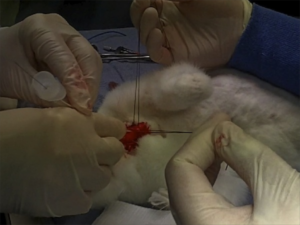
Ever had to do a surgical airway in a child? Thought not. They’re pretty rare. Bill Heegaard MD from Henepin County Medical Center taught me a few approaches (with the help of an anaesthetised rabbit) which really got me thinking. It’s something I’d often trained for in my internal simulator, and I even keep the equipment for it in my house (listen out for an upcoming podcast on that). Research and experience has demonstrated that open surgical airway techniques are more reliable than transtracheal needle techniques in adults, but what about kids, in whom traditional teaching cautions against open techniques?

Australian investigators who were experienced airway proceduralists evaluated transtracheal needle techniques using a rabbit model (an excellent model for the infant airway). Their success rate was only 60% and they perforated the posterior tracheal wall in 42% of attempts. Of 13 attempts to insert a dedicated paediatric tracheotomy device, the Quicktrach Child, none were successful(1) (they did not use the Quicktrach Infant model as it is not available in Australia).

Danish investigators used fresh piglet cadavers weighing around 8 kg to assess two transtracheal cannulas, in which they achieved success rates of 65.6% and 68.8%(2). There was also a very high rate of posterior tracheal wall perforation. Using an open surgical tracheostomy technique, they were successful in 97% of attempts. These were also experienced operators, with a median anaesthetic experience of 12.5 years.
Their tracheotomy technique was nice and simple, and used just a scalpel, scissors, and surgical towel clips. Here’s their technique:

Simple tracheotomy procedure described by Holm-Knudsen et al

  1. Identify larynx and proximal trachea by palpation
  2. Vertical incision through the skin and subcutaneous tissue from the upper part of larynx to the sternal notch
  3. Grasp strap muscles with two towel forceps and separate in the midline
  4. Palpate and identify the trachea (palpate rather than look for tracheal rings, as in a live patient one would expect bleeding to obscure the view)
  5. Stabilise the trachea by grasping it with a towel forceps
  6. Insert sharp tip of the scissors between two tracheal rings and lift the trachea anteriorly to avoid damage to the posterior wall
  7. Cut vertically in the midline of the trachea with the scissors – they chose to use the scissors to cut the tracheal rings to facilitate tube insertion
  8. Insert the tracheal tube

Using ultrasound and CT to evaluate comparative airway dimensions, the authors concluded that the pig model is most useful for training emergency airway management in older children aged 5–10 years.
Why were they doing a tracheotomy rather than a cricothyroidotomy? Reasons given by the authors include:

  • The infant cricothyroid membrane is very small
  • Palpation of the thyroid notch may be hindered by the overlying hyoid bone
  • The mandible may obstruct needle access to the cricothyroid membrane given the cephalad position in the neck of the infant larynx.

From an emergency medicine point of view, there are a couple of other reasons why we need to be able to access the trachea lower than the cricothyroid membrane. One is fractured larynx or other blunt or penetrating airway injury where there may be anatomical disruption at the cricothyroid level. The other situation is foreign body airway obstruction, when objects may lodge at the level of the cricoid ring which is functionally the narrowest part of the pediatric upper airway. Of course, alternative methods might be considered to remove the foreign body prior to tracheotomy, such as employing basic choking algorithms, and other techniques depending on whether you do or don’t have equipment.

Take home messages

  • Transtracheal airways in kids are so rare, we can’t avoid extrapolating animal data
  • Whichever infant or paediatric model is used, transtracheal needle techniques have a high rate of failure even by ‘experienced’ operators
  • The small size and easy compressibility of the airway probably contributes to this failure rate, including the high rate of posterior wall puncture
  • In keeping with adult audit data, open surgical techniques may have a higher success rate
  • Tracheotomy may be necessary rather than cricothyroidotomy in infants and children depending on clinical scenario and accessibility of anatomy
  • The stress and blood that is not simulated in cadaveric animal models will make open tracheotomy harder in a live patient, and so these success rates may not translate. However these factors do mean that whatever technique is used must be kept simple and should employ readily available and familiar equipment
  • Something to maintain control and anterior position of the anterior trachea wall should be used during incision and intubation of the trachea. The study reported here used towel clips; sutures around the tracheal rings may also be used (see image below)

I recommend you add ‘paediatric tracheotomy’ to the list of procedures you might need to do (if it’s not already there). Identify what equipment you would use and run the simulation in your head and in your work environment.
Have fun.

1. The ‘Can’t Intubate Can’t Oxygenate’ scenario in Pediatric Anesthesia: a comparison of different devices for needle cricothyroidotomy
Paediatr Anaesth. 2012 Dec;22(12):1155-8

BACKGROUND: Little evidence exists to guide the management of the ‘Can’t Intubate, Can’t Oxygenate’ (CICO) scenario in pediatric anesthesia.

OBJECTIVES: To compare two intravenous cannulae for ease of use, success rate and complication rate in needle tracheotomy in a postmortem animal model of the infant airway, and trial a commercially available device using the same model.

METHODS: Two experienced proceduralists repeatedly attempted cannula tracheotomy in five postmortem rabbits, alternately using 18-gauge (18G) and 14-gauge (14G) BD Insyte(™) cannulae (BD, Franklin Lakes, NJ, USA). Attempts began at the first tracheal cartilage, with subsequent attempts progressively more caudad. Success was defined as intratracheal cannula placement. In each rabbit, an attempt was then made by each proceduralist to perform a cannula tracheotomy using the Quicktrach Child(™) device (VBM Medizintechnik GmbH, Sulz am Neckar, Germany).

RESULTS: The rabbit tracheas were of similar dimensions to a human infant. 60 attempts were made at cannula tracheotomy, yielding a 60% success rate. There was no significant difference in success rate, ease of use, or complication rate between cannulae of different gauge. Successful aspiration was highly predictive (positive predictive value 97%) and both sensitive (89%) and specific (96%) for tracheal cannulation. The posterior tracheal wall was perforated in 42% of tracheal punctures. None of 13 attempts using the Quicktrach Child(™) were successful.

CONCLUSION: Cannula tracheotomy in a model comparable to the infant airway is difficult and not without complication. Cannulae of 14- and 18-gauge appear to offer similar performance. Successful aspiration is the key predictor of appropriate cannula placement. The Quicktrach Child was not used successfully in this model. Further work is required to compare possible management strategies for the CICO scenario

2. Emergency airway access in children – transtracheal cannulas and tracheotomy assessed in a porcine model
Paediatr Anaesth. 2012 Dec;22(12):1159-65

OBJECTIVES: In the rare scenario when it is impossible to oxygenate or intubate a child, no evidence exists on what strategy to follow.

AIM: The aim of this study was to compare the time and success rate when using two different transtracheal needle techniques and also to measure the success rate and time when performing an emergency tracheotomy in a piglet cadaver model.

METHODS: In this randomized cross-over study, we included 32 anesthesiologists who each inserted two transtracheal cannulas (TTC) using a jet ventilation catheter and an intravenous catheter in a piglet model. Second, they performed an emergency tracheotomy. A maximum of 2 and 4 min were allowed for the procedures, respectively. The TTC procedures were recorded using a video scope.

RESULTS: Placement of a transtracheal cannula was successful in 65.6% and 68.8% of the attempts (P = 0.76), and the median duration of the attempts was 69 and 42 s (P = 0.32), using the jet ventilation catheter and the intravenous catheter, respectively. Complications were frequent in both groups, especially perforation of the posterior tracheal wall. Performing an emergency tracheotomy was successful in 97%, in a median of 88 s.

CONCLUSIONS: In a piglet model, we found no significant difference in success rates or time to insert a jet ventilation cannula or an intravenous catheter transtracheally, but the incidence of complications was high. In the same model, we found a 97% success rate for performing an emergency tracheotomy within 4 min with a low rate of complications.

A whole bunch of trauma guidelines

The Eastern Association for the Surgery of Trauma has published a number of helpful evidence-based guidelines for trauma management, and many of them are included in this month’s Journal of Trauma and Acute Care Surgery
Here are brief snippets from some of them. All the guidelines can be viewed or downloaded in full for free here.
 
Nonoperative management of blunt hepatic injury

  • Nonoperative management of blunt hepatic injuries currently is the treatment modality of choice in hemodynamically stable patients, irrespective of the grade of injury or patient age.
  • Patients presenting with hemodynamic instability and peritonitis still warrant emergent operative intervention.
  • Intravenous contrast enhanced computed tomographic scan is the diagnostic modality of choice for evaluating blunt hepatic injuries.

 
Selective nonoperative management of blunt splenic injury

  • Nonoperative management of blunt splenic injuries is now the treatment modality of choice in hemodynamically stable patients, irrespective of the grade of injury, patient age, or the presence of associated injuries.
  • Patients presenting with hemodynamic instability and peritonitis still warrant emergent operative intervention.
  • Intravenous contrast enhanced computed tomographic scan is the diagnostic modality of choice for evaluating blunt splenic injuries.

 
Screening for blunt cardiac injury

  • Electrocardiogram (ECG) alone is not sufficient to rule out BCI.
  • BCI can be ruled out only if both ECG result and troponin I level are normal, a significant change from the previous guideline.
  • Patients with new ECG changes and/or elevated troponin I should be admitted for monitoring.
  • Echocardiogram is not beneficial as a screening tool for BCI and should be reserved for patients with hypotension and/or arrhythmias.
  • The presence of a sternal fracture alone does not predict BCI.
  • Cardiac computed tomography or magnetic resonance imaging can be used to differentiate acute myocardial infarction from BCI in trauma patients.

 
Evaluation and management of penetrating lower extremity arterial trauma

  • Expedited triage of patients is possible with physical examination and/or the measurement of ankle-brachial indices.
  • Computed tomographic angiography has become the diagnostic study of choice when imaging is required.
  • Tourniquets and intravascular shunts have emerged as adjuncts in the treatment of penetrating lower extremity arterial trauma.

 
Prophylactic antibiotic use in penetrating abdominal trauma

  • There is evidence to support a Level I recommendation that prophylactic antibiotics should only be administered for 24 hours in the presence of a hollow viscus injury.
  • There are no data to support continuing prophylactic antibiotics longer than 24 hours in damage control laparotomy.

 
Screening for thoracolumbar spinal injuries in blunt trauma

  • Multidetector computed tomographic scans have become the screening modality of choice and the criterion standard in screening for TLS injuries.
  • Patients without altered mentation or significant mechanism may be excluded by clinical examination without imaging.
  • Patients with gross neurologic deficits or concerning clinical examination findings with negative imaging should receive a magnetic resonance imaging expediently, and the spine service should be consulted

 
Emergency tracheal intubation immediately following traumatic injury

  • The decision to intubate a patient following traumatic injury is based on multiple factors, including the need for oxygenation and ventilation, the extent and mechanism of injury, predicted operative need, or progression of disease.
  • Rapid sequence intubation with direct laryngoscopy continues to be the recommended method for ETI, although the use of airway adjuncts such as blind insertion supraglottic devices and video laryngoscopy may be useful in facilitating successful ETI and may be preferred in certain patient populations.
  • There is no pharmacologic induction agent of choice for ETI; however, succinylcholine is the neuromuscular blockade agent recommended for rapid sequence intubation.

 
Presumptive antibiotic use in tube thoracostomy for traumatic hemopneumothorax

  • Routine presumptive antibiotic use to reduce the incidence of empyema and pneumonia in TT for traumatic hemopneumothorax is controversial; however, there is insufficient published evidence to support any recommendation either for or against this practice.

 
Evaluation and management of geriatric trauma

  • Effective evidence-based care of aging patients necessitates aggressive triage, correction of coagulopathy, and limitation of care when clinical evidence points toward an overwhelming likelihood of poor long-term prognosis

 
Management of pulmonary contusion and flail chest

  • Patients with PC-FC should not be excessively fluid restricted but should be resuscitated to maintain signs of adequate tissue perfusion.
  • Obligatory mechanical ventilation in the absence of respiratory failure should be avoided.
  • The use of optimal analgesia and aggressive chest physiotherapy should be applied to minimize the likelihood of respiratory failure.
  • Epidural catheter is the preferred mode of analgesia delivery in severe flail chest injury.
  • Paravertebral analgesia may be equivalent to epidural analgesia and may be appropriate in certain situations when epidural is contraindicated.
  • A trial of mask continuous positive airway pressure should be considered in alert patients with marginal respiratory status.
  • Patients requiring mechanical ventilation should be supported in a manner based on institutional and physician preference and separated from the ventilator at the earliest possible time.
  • Positive end-expiratory pressure or continuous positive airway pressure should be provided.
  • High-frequency oscillatory ventilation should be considered for patients failing conventional ventilatory modes. Independent lung ventilation may also be considered in severe unilateral pulmonary contusion when shunt cannot be otherwise corrected.
  • Surgical fixation of flail chest may be considered in cases of severe flail chest failing to wean from the ventilator or when thoracotomy is required for other reasons.
  • Self-activating multidisciplinary protocols for the treatment of chest wall injuries may improve outcome and should be considered where feasible.
  • Steroids should not be used in the therapy of pulmonary contusion.
  • Diuretics may be used in the setting of hydrostatic fluid overload in hemodynamically stable patients or in the setting of known concurrent congestive heart failure.

 
Evaluation and management of small-bowel obstruction

  • Level I evidence now exists to recommend the use of computed tomographic scan, especially multidetector computed tomography with multiplanar reconstructions, in the evaluation of patients with SBO because it can provide incremental clinically relevant information over plains films that may lead to changes in management.
  • Patients with evidence of generalized peritonitis, other evidence of clinical deterioration, such as fever, leukocytosis, tachycardia, metabolic acidosis, and continuous pain, or patients with evidence of ischemia on imaging should undergo timely exploration.
  • The remainder of patients can safely undergo initial nonoperative management for both partial and complete SBO.
  • Water-soluble contrast studies should be considered in patients who do not clinically resolve after 48 to 72 hours for both diagnostic and potential therapeutic purposes.
  • Laparoscopic treatment of SBO has been demonstrated to be a viable alternative to laparotomy in selected cases.

 
2012 Eastern Association for the Surgery of Trauma (EAST) Practice Management Guidelines Supplement
J Trauma Acute Care Surg. 2012 Nov;73(5 Suppl 4)

Prehospital ketamine analgesia

A prospective open-label randomised controlled trial in prehospital patients receiving morphine for traumatic pain compared intravenous ketamine with additional morphine.
Analgesia was superior in the ketamine group, with some minor (expected) adverse effects.
This small study adds to the prehospital ketamine literature and provides some support to ambulance services considering introducing ketamine analgesia.


Study objective: We assess the efficacy of intravenous ketamine compared with intravenous morphine in reducing pain in adults with significant out-of-hospital traumatic pain.

Methods: This study was an out-of-hospital, prospective, randomized, controlled, open-label study. Patients with trauma and a verbal pain score of greater than 5 after 5 mg intravenous morphine were eligible for enrollment.
Patients allocated to the ketamine group received a bolus of 10 or 20 mg, followed by 10 mg every 3 minutes thereafter. Patients allocated to the morphine alone group received 5 mg intravenously every 5 minutes until pain free. Pain scores were measured at baseline and at hospital arrival.

Results: A total of 135 patients were enrolled between December 2007 and July 2010. There were no differences between the groups at baseline. After the initial 5-mg dose of intravenous morphine, patients allocated to ketamine received a mean of 40.6 mg (SD 25 mg) of ketamine. Patients allocated to morphine alone received a mean of 14.4 mg (SD 9.4 mg) of morphine. The mean pain score change was 5.6 (95% confidence interval [CI] 6.2 to 5.0) in the ketamine group compared with 3.2 (95% CI 3.7 to 2.7) in the morphine group. The difference in mean pain score change was 2.4 (95% CI 3.2 to 1.6) points. The intravenous morphine group had 9 of 65 (14%; 95% CI 6% to 25%) adverse effects reported (most commonly nausea [6/65; 9%]) compared with 27 of 70 (39%; 95% CI 27% to 51%) in the ketamine group (most commonly disorientation [8/70; 11%]).

Conclusion: Intravenous morphine plus ketamine for out-of-hospital adult trauma patients provides analgesia superior to that of intravenous morphine alone but was associated with an increase in the rate of minor adverse effects.

Morphine and Ketamine Is Superior to Morphine Alone for Out-of-Hospital Trauma Analgesia: A Randomized Controlled Trial
Ann Emerg Med. 2012 Jun;59(6):497-503

Needle decompression: it's still not going to work

A pet topic that keeps coming up here is management of tension pneumothorax. Plenty of studies demonstrate that traditionally taught needle thoracostomy may fail, and open, or ‘finger’ thoracostomy is recommended for the emergency management of tension pneumothorax in a patient who is being ventilated with positive pressure (including those patients in cardiac arrest).
A recent CT scan-based study of adult trauma patients makes the case that needle decompression with a standard iv cannula would be expected to fail in 42.5% of cases at the second intercostal space (ICS) compared with 16.7% at the fifth ICS at the anterior axillary line (AAL).
The authors add an important point: “As BMI increases, there is a stepwise increase in chest wall thickness, further compounding the difficulty of needle placement in all but the lowest BMI quartile for the second ICS.”
An accompanying editorial cautions that the proximity of the heart may confer a safety issue if a needle is inserted blindly into the left 5th ICS at the AAL.


Objective To compare the distance to be traversed during needle thoracostomy decompression performed at the second intercostal space (ICS) in the midclavicular line (MCL) with the fifth ICS in the anterior axillary line (AAL).

Design Patients were separated into body mass index (BMI) quartiles, with BMI calculated as weight in kilograms divided by height in meters squared. From each BMI quartile, 30 patients were randomly chosen for inclusion in the study on the basis of a priori power analysis (n = 120). Chest wall thickness on computed tomography at the second ICS in the MCL was compared with the fifth ICS in the AAL on both the right and left sides through all BMI quartiles.

Setting Level I trauma center.

Patients Injured patients aged 16 years or older evaluated from January 1, 2009, to January 1, 2010, undergoing computed tomography of the chest.

Results A total of 680 patients met the study inclusion criteria (81.5% were male and mean age was 41 years [range, 16-97 years]). Of the injuries sustained, 13.2% were penetrating, mean (SD) Injury Severity Score was 15.5 (10.3), and mean BMI was 27.9 (5.9) (range, 15.4-60.7). The mean difference in chest wall thickness between the second ICS at the MCL and the fifth ICS at the AAL was 12.9 mm (95% CI, 11.0-14.8; P < .001) on the right and 13.4 mm (95% CI, 11.4-15.3; P < .001) on the left. There was a stepwise increase in chest wall thickness across all BMI quartiles at each location of measurement. There was a significant difference in chest wall thickness between the second ICS at the MCL and the fifth ICS at the AAL in all quartiles on both the right and the left. The percentage of patients with chest wall thickness greater than the standard 5-cm decompression needle was 42.5% at the second ICS in the MCL and only 16.7% at the fifth ICS in the AAL.

Conclusions In this computed tomography–based analysis of chest wall thickness, needle thoracostomy decompression would be expected to fail in 42.5% of cases at the second ICS in the MCL compared with 16.7% at the fifth ICS in the AAL. The chest wall thickness at the fifth ICS AAL was 1.3 cm thinner on average and may be a preferred location for needle thoracostomy decompression.

Radiologic evaluation of alternative sites for needle decompression of tension pneumothorax
Arch Surg. 2012 Sep 1;147(9):813-8

Thoracoabdominal trauma outcomes

In what the authors describe as ‘the largest and most rigorous description of blunt thoracoabdominal injury to date’, we learn some interesting things and are reminded of some others:

Most solid organ injuries in the abdomen are managed non-operatively

Thoracotomy – especially non-resuscitative thoracotomy – is rare

In patients with thoracoabdominal trauma, the overwhelming majority of injuries requiring operative intervention were found in the abdomen, therefore..

…excluding those patients in extremis requiring a resuscitative thoracotomy, the initial incision, without directive radiological information, belongs in the abdomen

Concomitant thoracic injury did not preclude nonoperative management of abdominal solid organ injury

This is a really interesting paper providing important data on the outcomes and management of a patient group that frequently produces management dilemmas in trauma centres.
The double jeopardy of blunt thoracoabdominal trauma
Arch Surg. 2012 Jun;147(6):498-504
[EXPAND Click for abstract]


OBJECTIVES: To examine the specific injuries, need for operative intervention, and clinical outcomes of patients with blunt thoracoabdominal trauma.

DESIGN: Trauma registry and medical record review.

SETTING: Level I trauma center in Los Angeles, California.

PATIENTS: All patients with thoracoabdominal injuries from January 1996 to December 2010.

MAIN OUTCOME MEASURES: Injuries, incidence and type of operative intervention, clinical outcomes, and risk factors for mortality.

RESULTS: Blunt thoracoabdominal injury occurred in 1661 patients. Overall, 474 (28.5%) required laparotomy, 31 (1.9%) required thoracotomy (excluding resuscitative thoracotomy), and 1146 (69.0%) required no thoracic or abdominal operation. Overall incidence of intraabdominal solid organ injury was 59.7% and hollow viscus injury, 6.0%. Blunt cardiac trauma occurred in 6.3%; major thoracic vessel injury, in 4.6%; and diaphragmatic trauma, in 6.0%. The majority of solid organ injuries were managed nonoperatively (liver, 83.9%; spleen, 68.3%; and kidney, 91.2%). Excluding patients with severe head trauma, mortality ranged from 4.5% with nonoperative management to 18.1% and 66.7% in those requiring laparotomy and dual cavitary exploration, respectively. Age 55 years or older, Injury Severity Score of 25 or more, Glasgow Coma Scale score of 8 or less, initial hypotension, massive transfusion, and liver, cardiac, or abdominal vascular trauma were all independent risk factors for mortality.

CONCLUSIONS: Most patients with blunt thoracoabdominal trauma are managed nonoperatively. The need for non-resuscitative thoracotomy or combined thoracoabdominal operation is rare. The abdomen contains the overwhelming majority of injuries requiring operative intervention and should be the initial cavity of exploration in the patient requiring emergent surgery without directive radiologic data.

[/EXPAND]

Still no oil painting

During a Krav Maga self defence drill a middle aged martial arts enthusiast cleverly blocked a full contact punch with his nose. Following a suggestion to pause training to control the bleeding, he noticed some lateral nasal displacement which was easily manually reduced with an audible click. He was able to resume training with a piece of toilet paper stuffed up the bleeding nostril, and no ill effects were noticed on subsequent training nights that week.
The day after injury he demonstrated a characteristic bruising pattern:

As he was working an aeromedical retrieval shift, he was able to use the Retrieval Service Sonosite M-Turbo ultrasound machine to identify the cortical disruption from his nasal bone fracture.

Ultrasound compares favourably with both plain radiography(1) and computed tomography(2) in the diagnosis of nasal bone fractures.

Personal access to sonography and full board certification in emergency medicine help to decrease health care costs and emergency department load when individuals sustain fractures that do not require operative management(3).

1. Comparison of ultrasonography and conventional radiography in the diagnosis of nasal fractures.
Arch Otolaryngol Head Neck Surg. 2005 May;131(5):434-9
2. Comparison of high-resolution ultrasonography and computed tomography in the diagnosis of nasal fractures.
J Ultrasound Med. 2009 Jun;28(6):717-23
3. Only when I laugh
When the same bloke broke his rib

What is 'hypotension' in penetrating trauma?

I previously noted an article demonstrating that a ‘lowish’ – as opposed to a low – systolic blood pressure is a reason to be vigilant in blunt trauma patients, as a significant increase in mortality has been demonstrated with a systolic blood pressure (SBP) < 110 mmHg.
The same researchers have found similar results in patients with penetrating trauma.
Compared with the reference group with SBP 110-129mmHg, mortality was doubled at SBP 90-109mmHg, was four-fold higher at 70-89mmHg and 10-fold higher at <70mmHg. SBP values ≥150mmHg were associated with decreased mortality.
Systolic blood pressure below 110 mmHg is associated with increased mortality in penetrating major trauma patients: Multicentre cohort study
Resuscitation. 2012 Apr;83(4):476-81
[EXPAND Click for abstract]


INTRODUCTION: Non-invasive systolic blood pressure (SBP) measurement is a commonly used triaging tool for trauma patients. A SBP of <90mmHg has represented the threshold for hypotension for many years, but recent studies have suggested redefining hypotension at lower levels. We therefore examined the association between SBP and mortality in penetrating trauma patients.

METHODS: We conducted a prospective cohort study in adult (≥16 years) penetrating trauma patients. Patients were admitted to hospitals belonging to the Trauma Audit and Research Network (TARN) between 2000 and 2009. The main outcome measure was the association between SBP and mortality at 30 days. Multivariate logistic regression models adjusted for the influence of age, gender, Injury Severity Score (ISS) and Glasgow Coma Score (GCS) on mortality were used. RESULTS: 3444 patients with a median age of 30 years (IQR 22.5-41.4), SBP of 126mmHg (IQR 107-142), ISS of 9 (IQR 9-14) and GCS of 15 (IQR 15-15), were analysed. Multivariable logistic regression analysis adjusted for age, gender, severity of injury and level of consciousness showed a cut-off for SBP at <110mmHg, after which increased mortality was observed. Compared with the reference group with SBP 110-129mmHg, mortality was doubled at SBP 90-109mmHg, was four-fold higher at 70-89mmHg and 10-fold higher at <70mmHg. SBP values ≥150mmHg were associated with decreased mortality.

CONCLUSION: We recommend that penetrating trauma patients with a SBP<110mmHg are triaged to resuscitation areas within dedicated, appropriately specialised, high-level care trauma centres.

[/EXPAND]

Superglue for CVCs

In resuscitation situations, the securing of vascular catheters is an important but sometimes cumbersome process, particular when sutures are required for central lines or arterial lines.

Medical grade ‘superglue’ (cyanoacrylate) can be used and this has been described in the anaesthetic literature before(1). Now, further in vitro work shows the glue does not weaken the intravenous catheter and is not associated with bacterial colonisation(2).

I think this is perfect for resuscitation lines. Just last night I used this technique to secure a femoral arterial line during a cardiac arrest resuscitation. It was great not to have to faff around with sharp suture needles during CPR and the line felt very secure after just a few seconds.

1. Tissue adhesive as an alternative to sutures for securing central venous catheters
Anaesthesia. 2007 Sep;62(9):969-70

2. Cyanoacrylate tissue adhesives – effective securement technique for intravascular catheters: in vitro testing of safety and feasibility
Anaesth Intensive Care. 2012 May;40(3):460-6