Tag Archives: Guidelines

Severe Traumatic Brain Injury in Children

The Brain Trauma Foundation has released updated guidelines on traumatic brain injury in children.
Most of the recommendations are Grade C and therefore based on limited evidence.

Indications for ICP monitoring

Use of intracranial pressure (ICP) monitoring may be considered in infants and children with severe traumatic brain injury (TBI) (Grade C).
Four lines of evidence support the use of ICP monitoring in children with severe TBI:

  • a frequently reported high incidence of intracranial hypertension in children with severe TBI
  • a widely reported association of intracranial hypertension and poor neurologic outcome
  • the concordance of protocol-based intracranial hypertension therapy and best-reported clinical outcomes
  • and improved outcomes associated with successful ICP-lowering therapies.

Threshold for treatment of intracranial hypertension
Treatment of intracranial pressure (ICP) may be considered at a threshold of 20 mm Hg (Grade C).
Sustained elevations in ICP (>20 mm Hg) are associated with poor outcome in children after severe TBI.
Normal values of blood pressure and ICP are age-dependent (lower at younger ages), so it is anticipated that the optimal ICP treatment threshold may be age-dependent.
Cerebral perfusion pressure thresholds
A CPP threshold 40–50 mm Hg may be considered. There may be age-specific thresholds with infants at the lower end and adolescents at the upper end of this range (Grade C).
Survivors of severe pediatric TBI undergoing ICP monitoring consistently have higher CPP values vs. nonsurvivors, but no study demonstrates that active maintenance of CPP above any target threshold in pediatric TBI reduces mortality or morbidity.
CPP should be determined in a standard fashion with ICP zeroed to the tragus (as an indicator of the foramen of Monro and midventricular level) and MAP zeroed to the right atrium with the head of the bed elevated 30°.
Advanced neuromonitoring
If brain oxygenation monitoring is used, maintenance of partial pressure of brain tissue oxygen (PbtO2) >10 mm Hg may be considered.
Neuroimaging
In the absence of neurologic deterioration or increasing intracranial pressure (ICP), obtaining a routine repeat computed tomography (CT) scan >24 hrs after the admission and initial follow-up study may not be indicated for decisions about neurosurgical intervention (Grade C).
Hyperosmolar therapy
Hypertonic saline should be considered for the treatment of severe pediatric traumatic brain injury (TBI) associated with intracranial hypertension. Effective doses for acute use range between 6.5 and 10 mL/kg (of 3%) (Grade B).
Hypertonic saline should be considered for the treatment of severe pediatric TBI associated with intracranial hypertension. Effective doses as a continuous infusion of 3% saline range between 0.1 and 1.0 mL/kg of body weight per hour administered on a sliding scale. The minimum dose needed to maintain intracranial pressure (ICP)
Temperature control
Moderate hypothermia (32–33°C) beginning early after severe traumatic brain injury (TBI) for only 24 hrs’ duration should be avoided.
Moderate hypothermia (32–33°C) be- ginning within 8 hrs after severe TBI for up to 48 hrs’ duration should be considered to reduce intracranial hypertension.
If hypothermia is induced for any indication, rewarming at a rate of >0.5°C/hr should be avoided (Grade B).
Moderate hypothermia (32–33°C) be- ginning early after severe TBI for 48 hrs, duration may be considered (Grade C).
Note: after completion of these guidelines, the committee became aware that the Cool Kids trial of hypothermia in pediatric TBI was stopped because of futility. The implications of this development on the recommendations in this section may need to be considered by the treating physician when details of the study are published.
Cerebrospinal fluid drainage
Cerebrospinal fluid (CSF) drainage through an external ventricular drain may be considered in the management of increased intracranial pressure (ICP) in children with severe traumatic brain injury (TBI).
The addition of a lumbar drain may be considered in the case of refractory intracranial hypertension with a functioning external ventricular drain, open basal cis- terns, and no evidence of a mass lesion or shift on imaging studies (Grade C).
Barbiturates
High-dose barbiturate therapy may be considered in hemodynamically stable patients with refractory intracranial hypertension despite maximal medical and surgical management.
When high-dose barbiturate therapy is used to treat refractory intracranial hy- pertension, continuous arterial blood pressure monitoring and cardiovascular support to maintain adequate cerebral perfusion pressure are required (Grade C).
Decompressive craniectomy for the treatment of intracranial hypertension
Decompressive craniectomy (DC) with duraplasty, leaving the bone flap out, may be considered for pediatric patients with traumatic brain injury (TBI) who are showing early signs of neurologic deterioration or herniation or are developing intracranial hypertension refractory to medical management during the early stages of their treatment (Grade C).
Hyperventilation
Avoidance of prophylactic severe hyperventilation to a PaCO2 If hyperventilation is used in the management of refractory intracranial hypertension, advanced neuromonitoring for evaluation of cerebral ischemia may be considered (Grade C).
Corticosteroids
The use of corticosteroids is not recommended to improve outcome or reduce intracranial pressure (ICP) for children with severe traumatic brain injury (TBI) (Grade B).
Analgesics, sedatives, and neuromuscular blockade
Etomidate may be considered to control severe intracranial hypertension; however, the risks resulting from adrenal suppression must be considered.
Thiopental may be considered to control intracranial hypertension.
In the absence of outcome data, the specific indications, choice and dosing of analgesics, sedatives, and neuromuscular-blocking agents used in the management of infants and children with severe traumatic brain injury (TBI) should be left to the treating physician.
*As stated by the Food and Drug Administration, continuous infusion of propofol for either sedation or the management of refractory intracranial hypertension in infants and children with severe TBI is not recommended (Grade C).
The availability of other sedatives and analgesics that do not suppress adrenal function, small sample size and single-dose administration in the study discussed previously, and limited safety profile in pediatric TBI limit the ability to endorse the general use of etomidate as a sedative other than as an option for single-dose administration in the setting of raised ICP.
Glucose and nutrition
The evidence does not support the use of an immune-modulating diet for the treatment of severe traumatic brain injury (TBI) to improve outcome (Grade B).
In the absence of outcome data, the specific approach to glycemic control in the management of infants and children with severe TBI should be left to the treating physician (Grade C).
Antiseizure prophylaxis
Prophylactic treatment with phenytoin may be considered to reduce the incidence of early posttraumatic seizures (PTS) in pediatric patients with severe traumatic brain injury (TBI) (Grade C).
The incidence of early PTS in pediatric patients with TBI is approximately 10% given the limitations of the available data. Based on a single class III study (4), prophylactic anticonvulsant therapy with phenytoin may be considered to reduce the incidence of early posttraumatic seizures in pediatric patients with severe TBI. Concomitant monitoring of drug levels is appropriate given the potential alterations in drug metabolism described in the context of TBI. Stronger class II evidence is available supporting the use of prophylactic anticonvulsant treatment to reduce the risk of early PTS in adults. There are no compelling data in the pediatric TBI literature to show that such treatment reduces the long-term risk of PTS or improves long-term neurologic outcome.
Guidelines for the Acute Medical Management of Severe Traumatic Brain Injury in Infants, Children, and Adolescents-Second Edition
Pediatr Crit Care Med 2012 Vol. 13, No. 1 (Suppl.)
Read online
Download PDF (617k)
Other Brain Trauma Foundation Guidelines

Two new anaphylaxis guidelines

Many local and national guidelines for the management of anaphylaxis exist, but did you know there was a World Allergy Organization, and it has a very detailed guideline on this important life threatening condition?

Some interesting snippets from the guideline are included here

Anaphylaxis and cardiac disease

  • Anaphylaxis can precipitate acute myocardial infarction in susceptible individuals: in patients with ischemic heart disease, the number and density of cardiac mast cells is increased, including in the atherosclerotic plaques. Mediators released during anaphylaxis contribute to vasoconstriction and coronary artery spasm.
  • Epinephrine is not contraindicated in the treatment of anaphylaxis in patients with known or suspected cardiovascular disease, or in middle-aged or elderly patients without any history of coronary artery disease who are at increased risk of ACS only because of their age. Through its beta-1 adrenergic effects, epinephrine actually increases coronary artery blood flow because of an increase in myocardial contractility and in the duration of diastole relative to systole.
  • Glucagon has noncatecholamine-dependent inotropic and chronotropic cardiac effects, and is sometimes needed in patients taking a beta-adrenergic blocker who have hypotension and bradycardia and who do not respond optimally to epinephrine.
  • Anticholinergic agents are sometimes needed in beta-blocked patients, for example, atropine in those with persistent bradycardia or ipratropium in those with epinephrine-resistant bronchospasm.

How quickly can untreated anaphylaxis kill you?

Studies show median times to cardiorespiratory arrest after exposure to the offending stimulus were 5 minutes after administration of contrast media or drugs, 15 minutes after an insect sting, and 30 minutes after food ingestion.

What about confirming the diagnosis with serum tryptase measurements?

  • Blood samples for measurement of tryptase levels are optimally obtained 15 minutes to 3 hours after symptom onset.
  • Blood samples for measurement of histamine levels are optimally obtained 15–60 minutes after symptom onset. These tests are not specific for anaphylaxis.
  • Increased serum tryptase levels are often found in patients with anaphylaxis from insect stings or injected medications, and in those who are hypotensive
  • However, levels are often within normal limits in patients with anaphylaxis triggered by food and in those who are normotensive
  • Serial measurement of tryptase levels during an anaphylactic episode, and measurement of a baseline level after recovery are reported to be more useful than measurement at only one point in time.
  • Normal levels of either tryptase or histamine do not rule out the clinical diagnosis of anaphylaxis


How does epinephrine help?

  • Epinephrine is life-saving because of its alpha-1 adrenergic vasoconstrictor effects in most body organ systems (skeletal muscle is an important exception) and its ability to prevent and relieve airway obstruction caused by mucosal edema, and to prevent and relieve hypotension and shock.
  • Other relevant properties in anaphylaxis include its beta-1 adrenergic agonist inotropic and chronotropic properties leading to an increase in the force and rate of cardiac contractions, and its beta-2 adrenergic agonist properties such as decreased mediator release, bronchodilation and relief of urticaria
  • Epinephrine in a dose of 0.01 mg/kg of a 1:1,000 (1 mg/mL) solution injected promptly by the intramuscular route is effective and safe in the initial treatment of anaphylaxis. In other anaphylaxis scenarios, this low first-aid dose is unlikely to be effective. For example, if shock is imminent or has already developed, epinephrine needs to be given by slow intravenous infusion, ideally with the dose titrated according to noninvasive continuous cardiac monitoring.

What is the empty ventricle syndrome?

  • Patients with anaphylaxis should not suddenly sit, stand, or be placed in the upright position.
  • Instead, they should be placed on the back with their lower extremities elevated or, if they are experiencing respiratory distress or vomiting, they should be placed in a position of comfort with their lower extremities elevated.
  • This accomplishes 2 therapeutic goals: 1) preservation of fluid in the circulation (the central vascular compartment), an important step in managing distributive shock; and 2) prevention of the empty vena cava/empty ventricle syndrome, which can occur within seconds when patients with anaphylaxis suddenly assume or are placed in an upright position.
  • Patients with this syndrome are at high risk for sudden death. They are unlikely to respond to epinephrine regardless of route of administration, because it does not reach the heart and therefore cannot be circulated throughout the body

Should we give antihistamines, beta 2 agonists, and steroids?

The evidence base for use of these second line medications in the initial management of anaphylaxis, is extrapolated mainly from their use in treatment of other diseases such as urticaria (antihistamines) or acute asthma (beta-2 adrenergic agonists and glucocorticoids). Concerns have been raised that administering one or more second-line medications potentially delays prompt injection of epinephrine, the first-line treatment


Is ‘biphasic anaphylaxis’ a real phenomenon we should be concerned about?

  • Biphasic anaphylaxis occurs when symptoms recur within 1–72 hours (usually within 8–10 hours) after the initial symptoms have resolved, despite no further exposure to the trigger.
  • It occurs in up to 23% of adults and up to 11% of children.
  • After apparent resolution of symptoms, duration of monitoring in a medically supervised setting should be individualized. For example, patients with moderate respiratory or cardiovascular compromise should be monitored for at least 4 hours, and if indicated, for 8–10 hours or longer.
  • Protracted uniphasic anaphylaxis is uncommon, but can last for days.

World Allergy Organization Guidelines for the Assessment and Management of Anaphylaxis
World Allergy Organization Journal 2011;4(2):13-37 Full Text
[EXPAND click for abstract]

The illustrated World Allergy Organization (WAO) Anaphylaxis Guidelines were created in response to absence of global guidelines for anaphylaxis. Uniquely, before they were developed, lack of worldwide availability of essentials for the diagnosis and treatment of anaphylaxis was documented. They incorporate contributions from more than 100 allergy/immunology specialists on 6 continents. Recommendations are based on the best evidence available, supported by references published to the end of December 2010. The Guidelines review patient risk factors for severe or fatal anaphylaxis, co-factors that amplify anaphylaxis, and anaphylaxis in vulnerable patients, including pregnant women, infants, the elderly, and those with cardiovascular disease. They focus on the supreme importance of making a prompt clinical diagnosis and on the basic initial treatment that is urgently needed and should be possible even in a low resource environment. This involves having a written emergency protocol and rehearsing it regularly; then, as soon as anaphylaxis is diagnosed, promptly and simultaneously calling for help, injecting epinephrine (adrenaline) intramuscularly, and placing the patient on the back or in a position of comfort with the lower extremities elevated. When indicated, additional critically important steps include administering supplemental oxygen and maintaining the airway, establishing intravenous access and giving fluid resuscitation, and initiating cardiopulmonary resuscitation with continuous chest compressions. Vital signs and cardiorespiratory status should be monitored frequently and regularly (preferably, continuously). The Guidelines briefly review management of anaphylaxis refractory to basic initial treatment. They also emphasize preparation of the patient for self-treatment of anaphylaxis recurrences in the community, confirmation of anaphylaxis triggers, and prevention of recurrences through trigger avoidance and immunomodulation. Novel strategies for dissemination and implementation are summarized. A global agenda for anaphylaxis research is proposed.

[/EXPAND]
This month has also seen the publication guidelines from the UK’s National Institute for Health & Clinical Excellence, entitled ‘Anaphylaxis: assessment to confirm an anaphylactic episode and the decision to refer after emergency treatment for a suspected anaphylactic episode’
Their guideline summary is as follows:

After a suspected anaphylactic reaction in adults or young people aged 16 years or older, take timed blood samples for mast cell tryptase testing as follows:

  • a sample as soon as possible after emergency treatment has started
  • a second sample ideally within 1–2 hours (but no later than 4 hours) from the onset of symptoms.

After a suspected anaphylactic reaction in children younger than 16 years, consider taking blood samples for mast cell tryptase testing as follows if the cause is thought to be venom-related, drug-related or idiopathic:

  • a sample as soon as possible after emergency treatment has started
  • a second sample ideally within 1–2 hours (but no later than 4 hours) from the onset of symptoms.

Patients who have had emergency treatment for suspected anaphylaxis should be observed for 6–12 hours from the onset of symptoms, depending on their response to emergency treatment
After emergency treatment for suspected anaphylaxis, offer people a referral to a specialist allergy service (age-appropriate where possible) consisting of healthcare professionals with the skills and competencies necessary to accurately investigate, diagnose, monitor and provide ongoing management of, and patient education about, suspected anaphylaxis.
After emergency treatment for suspected anaphylaxis, offer people (or, as appropriate, their parent and/or carer) an appropriate adrenaline injector as an interim measure before the specialist allergy service appointment.
Before discharge a healthcare professional with the appropriate skills and competencies should offer people (or, as appropriate, their parent and/or carer) the following:

  • information about anaphylaxis, including the signs and symptoms of an anaphylactic reaction
  • information about the risk of a biphasic reaction
  • information on what to do if an anaphylactic reaction occurs (use the adrenaline injector and call emergency services)

Anaphylaxis: assessment to confirm an anaphylactic episode and the decision to refer after emergency treatment for a suspected anaphylactic episode
CG134 Anaphylaxis: NICE guideline

2011 Asthma Guidelines

The British Thoracic Society / SIGN Guidelines on asthma have been updated for 2011. There don’t seem to be any modificiations to the sections on acute severe asthma which were updated in 2009 and blogged here, although the treatment algorithms seem to be presented in a slightly different format and therefore are reproduced here:

Management of acute severe asthma in adults in hospital

Management of acute asthma in children in hospital

Offensive medicine: CT before LP

I’m getting worn down by clinicians – often other specialists – who insist that CT imaging of the brain is mandatory prior to lumbar puncture in all patients. There is surely a subgroup of patients (especially young ones) in whom the benefit:harm balance of CT comes out in favour of NOT doing the imaging. In these cases, getting the scan is not ‘defensive medicine’ but ‘offensive medicine’ – offending the principle of primum non nocere. During ED shifts I have recently had to perform online searches in order to furnish colleagues and patients’ medically qualified relatives with printouts of the literature on this. This page is here to save me having to repeat those searches. Regarding the practice of performing a routine head CT prior to lumbar puncture to rule out risk of herniation:

  • Mass effect on CT does not predict herniation
  • Lack of mass effect on CT does not rule out raised ICP or herniation
  • Herniation has occurred in patients who did not undergoing lumbar puncture because of CT findings
  • Clinical predictors of raised ICP are more reliable than CT findings
  • CT may delay diagnosis and treatment of meningitis
  • Even in patients in whom LP may be considered contraindicated (cerebral abscess, mass effect on CT), complications from LP were rare in several studies

Best practice, it would seem, is the following

  • If you think CT will show a cause for the headache, do a CT
  • If a CT is indicated for other reasons (depressed conscious level, focal neurology), do a CT
  • If a GCS 15 patient is to undergo LP for suspected (or to rule out) meningitis, and they have a normal neurological exam (including fundi), and are not elderly or immunosuppressed, there is no need to do a CT first.
  • If you’re seriously worried about meningitis and are intent on getting a CT prior to LP, don’t let the imaging delay antimicrobial therapy.

Here are some useful references:

1. The CT doesn’t help

CT head before lumbar puncture in suspected meningitis BestBET evidence summary: In cases of suspected meningitis it is very unlikely that patients without clinical risk factors (immunocompromise/ history of CNS disease/seizures) or positive neurological findings will have a contraindication to lumbar puncture on their CT scan If CT scan is deemed to be necessary, administration of antibiotics should not be delayed. BestBETS website

Computed Tomography of the Head before Lumbar Puncture in Adults with Suspected Meningitis Much cited NEJM paper from 2001 which concludes: “In adults with suspected meningitis, clinical features can be used to identify those who are unlikely to have abnormal findings on CT of the headN Engl J Med. 2001 Dec 13;345(24):1727-33 Full Text

Cranial CT before Lumbar Puncture in Suspected Meningitis Correspondence in 2002 NEJM including study of 75 patients with pneumococcal meningitis: CT cannot rule out risk of herniation Cranial CT before Lumbar Puncture in Suspected Meningitis N Engl J Med. 2002 Apr 18;346(16):1248-51 Full Text

2. The CT may harm

Cancer risk from CT Paucis verbis card, from the wonderful Academic Life in EM

3. Guidelines say CT is not always needed

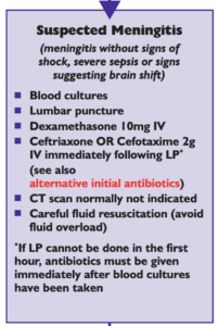
National (UK) guidelines on meningitis (community acquired meningitis in the immunocompetent host) available from meningitis.org. , including this box:

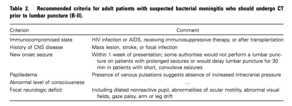
Practice Guidelines for the Management of Bacterial Meningitis These 2004 guidelines from the Infectious Diseases Society of America provide the following table listing the recommended criteria for adult patients with suspected bacterial meningitis who should undergo CT prior to lumbar puncture:

Clin Infect Dis. (2004) 39 (9): 1267-1284 Full text

4. This is potentially even more of an issue with paediatric patients

Fatal Lumbar Puncture: Fact Versus Fiction—An Approach to a Clinical Dilemma

An excellent summary of the above mentioned issues presented in a paediatric context, including the following:

On initial consideration a cranial CT would seem to be an appropriate and potentially useful diagnostic study for confirming the diagnosis of cerebral herniataion. The fallacy in this assessment has been emphasized by the finding that no clinically significant CT abnormalities are found that are not suspected on clinical assessments. Further, as previously noted, a normal CT examination may be found at about the time of a fatal herniation. Thus, the practical usefulness of a cranial CT in the majority of pediatric patients is limited to those rare patients whose increased ICP is secondary to mass lesions, not in the initial approach to acute meningitis.

Pediatrics. 2003 Sep;112(3 Pt 1):e174-6 Full Text

The last words should go to Dr Brad Spellberg, who in response to the IDSA’s guidelines wrote an excellent letter summarising much of the evidence at the time, confessed:

Why do we persist in using the CT scan for this purpose, despite the lack of supportive data? I am as guilty of this practice as anyone else, and the reason is simple: I am a chicken.

Clin Infect Dis. (2005) 40 (7): 1061 Full Text

Prehospital Spine Immobilisation for Penetrating Trauma

The Executive Committee of Prehospital Trauma Life Support, comprised of surgeons, emergency physicians, and paramedics, has reviewed the literature and produced the following recommendations on Prehospital Spine Immobilisation for Penetrating Trauma:


PHTLS Recommendations

  • There are no data to support routine spine immobilization in patients with penetrating trauma to the neck or torso.
  • There are no data to support routine spine immobilization in patients with isolated penetrating trauma to the cranium.
  • Spine immobilization should never be done at the expense of accurate physical examination or identification and correction of life-threatening conditions in patients with penetrating trauma.
  • Spinal immobilization may be performed after penetrating injury when a focal neurologic deficit is noted on physical examination although there is little evidence of benefit even in these cases.

Prehospital Spine Immobilization for Penetrating Trauma—Review and Recommendations From the Prehospital Trauma Life Support Executive Committee
Journal of Trauma-Injury Infection & Critical Care September 2011;71(3):763-770

Pre-hospital NIV

"The driver's dyspneic - pass the Boussignac valve!"

Pre-hospital non-invasive ventilation for patients with acute dyspnoea features in two journals this month. Researchers at the Ambulance Service New South Wales published an evidence-based review of pre-hospital NIV for acute cardiogenic pulmonary oedema, concluding that there are probable benefits.

Background Non-invasive ventilation (NIV) is increasingly being implemented by many ambulance jurisdictions as a standard of care in the out-of-hospital management of acute cardiogenic pulmonary oedema (ACPO). This implementation appears to be based on the body of evidence from the emergency department (ED) setting, with the assumption that earlier administration by paramedics would give benefits with regard to inhospital mortality and the rate of endotracheal intubation beyond those seen when initiated in the ED. This paper sought to identify and review the current level of evidence supporting NIV in the prehospital setting.

Methods Electronic searches of Medline, EMBASE, CINAHL, Cochrane Database of Systematic Reviews and Cochrane Database of Controlled Trials were conducted and reference lists of relevant articles were hand searched.

Results The search identified 12 primary studies documenting the use of NIV, either continuous positive airway pressure or bi-level non-invasive ventilation, for ACPO in the out-of-hospital setting. Only three studies were randomised controlled trials, with none addressing inhospital mortality as a primary outcome measure. The majority of articles were non-comparative descriptive studies.

Conclusion Early prehospital NIV appears to be a safe and feasible therapy that results in faster improvement in physiological status and may decrease the need for intubation when compared with delayed administration in the ED. There is weak evidence that is may decrease mortality. The cost versus benefit equation of system-wide prehospital implementation of NIV is unclear and, based on the current evidence, should be considered with caution.

Prehospital non-invasive ventilation for acute cardiogenic pulmonary oedema: an evidence-based review.
Emerg Med J. 2011 Jul;28(7):609-12
At the same time, the National Association of EMS Physicians has published a position statement on noninvasive positive pressure ventilation, for the general indication of acute dyspnoea. It states:

The general indication for NIPPV is dyspnea accompanied by early respiratory failure in patients with intact protective airway reflexes and mental status. The majority of NIPPV studies have focused on patients with acute pulmonary edema.

However, NIPPV may prove useful with other reversible disease processes such as chronic obstructive pulmonary disease or asthma exacerbations. While utilized in in-hospital practice, the role of NIPPV for pneumonia-associated respiratory failure is less clear. While some clinicians advocate the use of NIPPV to augment oxygenation prior to ETI in the in- hospital setting, there are no studies evaluating this strategy in the prehospital setting.

 
Noninvasive positive pressure ventilation: resource document for the National Association of EMS Physicians position statement.
Prehosp Emerg Care. 2011 Jul-Sep;15(3):432-8
Full Text of Position Statement

UK Radiology guidelines for trauma


The Royal College of Radiologists in the UK has published a guideline document to set standards related to diagnostic and interventional radiology for use by major trauma centres (MTCs) and trauma units (TUs). The standards are:

  1. The trauma team leader is in overall charge in acute care
  2. Protocol-driven imaging and intervention must be available and delivered by experienced staff. Acute care for SIPs must be consultant delivered
  3. MDCT should be adjacent to, or in, the emergency room
  4. Digital radiography must be available in the emergency room
  5. If there is an early decision to request MDCT, FAST and DR should not cause any delay
  6. MRI must be available with safe access for the SIP
  7. A CT request in the trauma setting should comply with the Ionising Radiation (Medical Exposure) Regulations 2000 (IR(ME)R) justification regulations like any other request for imaging involving ionising radiation
  8. There should be clear written protocols for MDCT preparation and transfer to the scan room
  9. Whole-body contrast-enhanced MDCT is the default imaging procedure of choice in the SIP. Imaging protocols should be clearly defined and uniform across a regional trauma network
  10. Future planning and design of emergency rooms should concentrate on increasing the numbers of SIPs stable enough for MDCT and intervention
  11. The primary survey report should be issued immediately to the trauma team leader
  12. On-call consultant radiologists should provide the final report on the SIP within one hour of MDCT image acquisition
  13. On-call consultant radiologists must have teleradiology facilities at home that allow accurate reports to be issued within one hour of MDCT image acquisition
  14. IR facilities should be co-located to the emergency department
  15. Angiographic facilities and endovascular theatres in MTCs should be safe environments for SIPs and should be of theatre standard
  16. Agreed written transfer protocols between the emergency department and imaging/interventional facilities internally or externally must be available
  17. IR trauma teams should be in place within 60 minutes of the patient’s admission or 30 minutes of referral
  18. Any deficiency in consumable equipment should be reported at the debriefing and be the subject of an incident report

Some interesting snippets include:
IV access
Right antecubital access is preferred for contrast administration (left-sided injections compromise interpretation of mediastinal vasculature). However, if arm vein access is not possible and a central line is in situ, it should be of a type that can accept 4 ml contrast/ second via a power injector. This might require local negotiation with emergency department doctors beforehand

Pelvic fracture
If a pelvic fracture is suspected, a temporary pelvic stabilisation (wrap, binder and so on) should be applied before MDCT.
Limb fractures
Rapid immobilisation such as air splints. Only immediately limb conserving manipulations/splinting should be performed prior to CT.
Urinary catheter
All significantly injured patients without obvious contraindications should be catheterised unless this would delay transfer to CT. The catheter should be clamped prior to MDCT.
Standards of practice and guidance for trauma radiology in severely injured patients
Royal College of Radiologists – Full Text Link

Targeted temperature management guidelines


Okay – rather than ‘therapeutic hypothermia’, the recommended phrase now is ‘targeted temperature management’. Several critical care authorities got together to produce clinical recommendations on this topic. Here are a few interesting points from the document:
On coagulation:
Hypothermia affects platelet function and prolongs the prothrombin time and partial thromboplastin time. These effects are masked when laboratory analysis is performed at 37°C, suggesting that any risk will be mitigated by rewarming.
Although not mentioned in the abstract, the authors examined the role of TTM in raised intracranial pressure (ICP):
Sufficient evidence exists to conclude that TTM does decrease ICP compared to unstructured temperature management. However, there is no sufficient evidence to make a recommendation regarding the use of targeted hypothermia to control elevated ICP to improve patent-important outcomes in TBI. The jury makes NO RECOMMENDATION regarding the use of TTM as an ICP control strategy to improve outcomes in brain injuries regardless of cause (trauma, hemorrhage, or ischemic stroke).
Regarding acute liver failure with severe cerebral edema:
there are currently no RCTs. There is a case series suggesting a strongly favorable effect. This is a powerful argument for support of an RCT evaluating TTM alone or in combination with hepatic dialysis strategies

OBJECTIVE: Representatives of five international critical care societies convened topic specialists and a nonexpert jury to review, assess, and report on studies of targeted temperature management and to provide clinical recommendations.
DATA SOURCES: Questions were allocated to experts who reviewed their areas, made formal presentations, and responded to questions. Jurors also performed independent searches. Sources used for consensus derived exclusively from peer-reviewed reports of human and animal studies.
STUDY SELECTION: Question-specific studies were selected from literature searches; jurors independently determined the relevance of each study included in the synthesis.
CONCLUSIONS AND RECOMMENDATIONS:

  1. The jury opines that the term “targeted temperature management” replace “therapeutic hypothermia.”
  2. The jury opines that descriptors (e.g., “mild”) be replaced with explicit targeted temperature management profiles.
  3. The jury opines that each report of a targeted temperature management trial enumerate the physiologic effects anticipated by the investigators and actually observed and/or measured in subjects in each arm of the trial as a strategy for increasing knowledge of the dose/duration/response characteristics of temperature management. This enumeration should be kept separate from the body of the report, be organized by body systems, and be made without assertions about the impact of any specific effect on the clinical outcome.
  4. The jury STRONGLY RECOMMENDS targeted temperature management to a target of 32°C-34°C as the preferred treatment (vs. unstructured temperature management) of out-of-hospital adult cardiac arrest victims with a first registered electrocardiography rhythm of ventricular fibrillation or pulseless ventricular tachycardia and still unconscious after restoration of spontaneous circulation (strong recommendation, moderate quality of evidence).
  5. The jury WEAKLY RECOMMENDS the use of targeted temperature management to 33°C-35.5°C (vs. less structured management) in the treatment of term newborns who sustained asphyxia and exhibit acidosis and/or encephalopathy (weak recommendation, moderate quality of evidence).

Targeted temperature management in critical care: A report and recommendations from five professional societies
Crit Care Med. 2011 May;39(5):1113-1125

ACEP policy on PE

The American College of Emergency Physicians has revised its 2003 clinical policy on pulmonary embolism.


Among the areas considered is the the role of thrombolytic medication. The policy provides the following recommendations to this question:

What are the indications for thrombolytic therapy in patients with PE?
Level B recommendations
Administer thrombolytic therapy in hemodynamically unstable patients with confirmed PE for whom the benefits of treatment outweigh the risks of life-threatening bleeding complications.*
*In centers with the capability for surgical or mechanical thrombectomy, procedural intervention may be used as an alternative therapy.
Level C recommendations
(1) Consider thrombolytic therapy in hemodynamically unstable patients with a high clinical suspicion for PE for whom the diagnosis of PE cannot be confirmed in a timely manner.
(2) At this time, there is insufficient evidence to make any recommendations regarding use of thrombolytics in any subgroup of hemodynamically stable patients. Thrombolytics have been demonstrated to result in faster improvements in right ventricular function and pulmonary perfusion, but these benefits have not translated to improvements in mortality.

The document contains a detailed appraisal of the literature to date on benefits and harms from thrombolysis. Of interest is the Pulmonary Embolism Severity Index (PESI) – a scoring system that appears to reliably predict mortality and thus has the potential to assist physicians in making risk-benefit decisions when considering administration of thrombolytics. The full text of the policy, which covers far more than just thrombolysis, can be found by following the link below.
Critical Issues in the Evaluation and Management of Adult Patients Presenting to the Emergency Department With Suspected Pulmonary Embolism
Annals of Emergency Medicine 2011 June 57(6):628-652 – Free Full Text