Category Archives: ICU

Stuff relevant to patients on ICU

Dabigatran Reversal Agent – Idarucizumab

Thanks to Rob MacSweeney‘s fantastic Critical Care Reviews I learned of Idarucizumab, a monoclonal antibody fragment that binds the (pesky) anticoagulant dabigatran. Two industry-supported studies this week show rapid, complete reversal of anticoagulation in healthy volunteers(1) and patients who were either bleeding or undergoing procedures(2). The dose given to patients was 5g intravenously.
An accompanying editorial(3) highlights that the clinical study did not have a control group, and these patients had a high mortality. Further controlled studies examining patient-orientated outcomes will be helpful.
Of interest, another editorialist(4) lists other potential antidotes for Non-vitamin-K antagonist oral anticoagulants (NOACs) that have been or are being tested: an antidote against all oral direct factor Xa inhibitors called andexanet alpha (a recombinant activated factor X that binds direct factor Xa inhibitors), and a modified thrombin has been shown to be effective in vitro and in animals for reversal of dabigatran and potentially also other direct thrombin inhibitors.
1. Safety, tolerability, and efficacy of idarucizumab for the reversal of the anticoagulant effect of dabigatran in healthy male volunteers: a randomised, placebo-controlled, double-blind phase 1 trial
The Lancet Volume 386, No. 9994, p680–690, 15 August 2015
[EXPAND Abstract]


BACKGROUND: Idarucizumab is a monoclonal antibody fragment that binds dabigatran with high affinity in a 1:1 molar ratio. We investigated the safety, tolerability, and efficacy of increasing doses of idarucizumab for the reversal of anticoagulant effects of dabigatran in a two-part phase 1 study (rising-dose assessment and dose-finding, proof-of-concept investigation). Here we present the results of the proof-of-concept part of the study.

METHODS: In this randomised, placebo-controlled, double-blind, proof-of-concept phase 1 study, we enrolled healthy volunteers (aged 18-45 years) with a body-mass index of 18·5-29·9 kg/m2 into one of four dose groups at SGS Life Sciences Clinical Research Services, Belgium. Participants were randomly assigned within groups in a 3:1 ratio to idarucizumab or placebo using a pseudorandom number generator and a supplied seed number. Participants and care providers were masked to treatment assignment. All participants received oral dabigatran etexilate 220 mg twice daily for 3 days and a final dose on day 4. Idarucizumab (1 g, 2 g, or 4 g 5-min infusion, or 5 g plus 2·5 g in two 5-min infusions given 1 h apart) was administered about 2 h after the final dabigatran etexilate dose. The primary endpoint was incidence of drug-related adverse events, analysed in all randomly assigned participants who received at least one dose of dabigatran etexilate. Reversal of diluted thrombin time (dTT), ecarin clotting time (ECT), activated partial thromboplastin time (aPTT), and thrombin time (TT) were secondary endpoints assessed by measuring the area under the effect curve from 2 h to 12 h (AUEC2-12) after dabigatran etexilate ingestion on days 3 and 4. This trial is registered with ClinicalTrials.gov, number NCT01688830.

FINDINGS: Between Feb 23, and Nov 29, 2013, 47 men completed this part of the study. 12 were enrolled into each of the 1 g, 2 g, or 5 g plus 2·5 g idarucizumab groups (nine to idarucizumab and three to placebo in each group), and 11 were enrolled into the 4 g idarucizumab group (eight to idarucizumab and three to placebo). Drug-related adverse events were all of mild intensity and reported in seven participants: one in the 1 g idarucizumab group (infusion site erythema and hot flushes), one in the 5 g plus 2·5 g idarucizumab group (epistaxis); one receiving placebo (infusion site haematoma), and four during dabigatran etexilate pretreatment (three haematuria and one epistaxis). Idarucizumab immediately and completely reversed dabigatran-induced anticoagulation in a dose-dependent manner; the mean ratio of day 4 AUEC2-12 to day 3 AUEC2-12 for dTT was 1·01 with placebo, 0·26 with 1 g idarucizumab (74% reduction), 0·06 with 2 g idarucizumab (94% reduction), 0·02 with 4 g idarucizumab (98% reduction), and 0·01 with 5 g plus 2·5 g idarucizumab (99% reduction). No serious or severe adverse events were reported, no adverse event led to discontinuation of treatment, and no clinically relevant difference in incidence of adverse events was noted between treatment groups.

INTERPRETATION: These phase 1 results show that idarucizumab was associated with immediate, complete, and sustained reversal of dabigatran-induced anticoagulation in healthy men, and was well tolerated with no unexpected or clinically relevant safety concerns, supporting further testing. Further clinical studies are in progress.

[/EXPAND]
2. Idarucizumab for Dabigatran Reversal
N Engl J Med. 2015 Aug 6;373(6):511-20
[EXPAND Abstract]


BACKGROUND: Specific reversal agents for non-vitamin K antagonist oral anticoagulants are lacking. Idarucizumab, an antibody fragment, was developed to reverse the anticoagulant effects of dabigatran.

METHODS: We undertook this prospective cohort study to determine the safety of 5 g of intravenous idarucizumab and its capacity to reverse the anticoagulant effects of dabigatran in patients who had serious bleeding (group A) or required an urgent procedure (group B). The primary end point was the maximum percentage reversal of the anticoagulant effect of dabigatran within 4 hours after the administration of idarucizumab, on the basis of the determination at a central laboratory of the dilute thrombin time or ecarin clotting time. A key secondary end point was the restoration of hemostasis.

RESULTS: This interim analysis included 90 patients who received idarucizumab (51 patients in group A and 39 in group B). Among 68 patients with an elevated dilute thrombin time and 81 with an elevated ecarin clotting time at baseline, the median maximum percentage reversal was 100% (95% confidence interval, 100 to 100). Idarucizumab normalized the test results in 88 to 98% of the patients, an effect that was evident within minutes. Concentrations of unbound dabigatran remained below 20 ng per milliliter at 24 hours in 79% of the patients. Among 35 patients in group A who could be assessed, hemostasis, as determined by local investigators, was restored at a median of 11.4 hours. Among 36 patients in group B who underwent a procedure, normal intraoperative hemostasis was reported in 33, and mildly or moderately abnormal hemostasis was reported in 2 patients and 1 patient, respectively. One thrombotic event occurred within 72 hours after idarucizumab administration in a patient in whom anticoagulants had not been reinitiated.

CONCLUSIONS: Idarucizumab completely reversed the anticoagulant effect of dabigatran within minutes. (Funded by Boehringer Ingelheim; RE-VERSE AD ClinicalTrials.gov number, NCT02104947.).

[/EXPAND]
3. Targeted Anti-Anticoagulants
N Engl J Med. 2015 Aug 6;373(6):569-71
4. Antidotes for anticoagulants: a long way to go
The Lancet Volume 386, No. 9994, p634–636, 15 August 2015

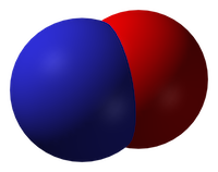
Inhaled nitric oxide: a tool for all resuscitationists?

The use of inhaled nitric oxide is established in certain groups of patients: it improves oxygenation (but not survival) in patients with acute respiratory distress syndrome(1), and it is used in neonatology for management of persistent pulmonary hypertension of the newborn(2). But it can be applied in other resuscitation settings: in arrested or peri-arrest patients with pulmonary hypertension.

Read this (modified) description of a case managed by one of my resuscitationist friends from an overseas location:

A young lady suffered a placental abruption requiring emergency hysterectomy. She arrested twice in the operating room after suspected amniotic fluid embolism. She had fixed dilated pupils.
She developed extreme pulmonary hypertension with suprasystemic pulmonary artery pressures, and she went down the pulmonary HT spiral as I stood there. On ultrasound her distended RV was making her LV totally collapse. She arrested. Futile CPR was started.
I have never had an extreme pulmonary HT survive an arrest. I grabbed a bag and rapidly set up a manual inhaled Nitric Oxide system and bagged and begged…
She achieved ROSC after some minutes. A repeat ultrasound showed a well functioning LV and less dilated RV.
Today, after 12 hours she is opening her eyes and obeying commands. Still a long way to go, but alive.

It sounds impressive. I don’t have more case details, and don’t know how confident they could be about the diagnosis of amniotic fluid embolism but the presentation certainly fits with acute pulmonary hypertension with RV failure. The use of inhaled nitric oxide has certainly been described for similar scenarios before(3). But it raises bigger questions: is this something we should all be capable of? Are there cardiac arrests involving or caused by pulmonary hypertension that will not respond to resuscitation without nitric oxide?

Nitric oxide
Inhaled nitric oxide is a pulmonary vasodilator. It decreases right-ventricular afterload and improves cardiac index by selectively decreasing pulmonary vascular resistance without causing systemic hypotension(4).

RV failure and pulmonary hypertension
Patients may become shocked or suffer cardiac arrest due to acute right ventricular dysfunction. This may be due to a primary cardiac cause such as right ventricular infarction (always consider this in a hypotensive patient with inferior STEMI, and confirm with a right ventricular ECG and/or echo). Alternatively it could be due to a pulmonary or systemic cause resulting in severe pulmonary hypertension, causing secondary right ventricular dysfunction. The commonest causes of acute pulmonary hypertension are massive PE, sepsis, and ARDS(5).

The haemodynamic consequences of RV failure are reduced pulmonary blood flow and inadequate left ventricular filling, leading to decreased cardiac output, shock, and arrest. In severe acute pulmonary hypertension the RV distends, resulting in a shift of the interventricular septum which compresses the LV and further inhibits LV filling (the concept of ventricular interdependence).

What’s wrong with standard ACLS?
In some patients with PHT who arrest, CPR may be ineffective due to a failure to achieve adequate pulmonary blood flow and ventricular filling. In one study of patients with known chronic PHT who arrested in the ICU, survival rates even for ventricular fibrillation were extremely poor and when measured end tidal carbon dioxide levels were very low. In the same study it was noted that some of the survivors had received an intravenous bolus administration of iloprost, a prostacyclin analogue (and pulmonary vasodilator) during CPR(6).

CPR may therefore be ineffective. Intubation and positive pressure ventilation may also be associated with haemodynamic deterioration in PHT patients(7), and intravenous epinephrine (adrenaline) has variable effects on the pulmonary circulation which could be deleterious(8).

If inhaled nitric oxide (iNO) can improve pulmonary blood flow and reduce right ventricular afterload, it could theoretically be of value in cases of shock or arrest with RV failure, especially in cases of pulmonary hypertension; these are patients who otherwise have poor outcomes and may not benefit from CPR.

Is the use of iNO described in shock or arrest?
Numerous case reports and series demonstrate recovery from shock or arrest following nitric oxide use in various situations of decompensated right ventricular failure from pulmonary hypertension secondary to pulmonary fibrotic disease(9), pneumonectomy surgery(10), and pulmonary embolism(11) including post-embolectomy(12).

Acute hemodynamic improvement was demonstrated following iNO therapy in a series of right ventricular myocardial infarction patients with cardiogenic shock(13).

A recent systematic review of inhaled nitric oxide in acute pulmonary embolism documented improvements in oxygenation and hemodynamic variables, “often within minutes of administration of iNO”. The authors state that these case reports underscore the need for randomised controlled trials to establish the safety and efficacy of iNO in the treatment of massive acute PE(14).

Why aren’t they telling us to use it?
If iNO may be helpful in certain cardiac arrest patients, why isn’t ILCOR recommending it? Actually it is mentioned – in the context of paediatric life support. The European Resuscitation Council states:

ERC Guideline: (Paediatric) Pulmonary hypertension
There is an increased risk of cardiac arrest in children with pulmonary hypertension.
Follow routine resuscitation protocols in these patients with emphasis on high FiO2 and alkalosis/hyperventilation because this may be as effective as inhaled nitric oxide in reducing pulmonary vascular resistance.
Resuscitation is most likely to be successful in patients with a reversible cause who are treated with intravenous epoprostenol or inhaled nitric oxide.
If routine medications that reduce pulmonary artery pressure have been stopped, they should be restarted and the use of aerosolised epoprostenol or inhaled nitric oxide considered.
Right ventricular support devices may improve survival

Should we use it?
So if acute (or acute on chronic) pulmonary hypertension can be suspected or demonstrated based on history, examination, and echo findings, and the patient is in extremis, it might be anticipated that standard ACLS approaches are likely to be futile (as they often are if the underlying cause is not addressed). One might consider attempts to induce pulmonary vasodilation to improve pulmonary blood flow and LV filling, improving oxygenation, and reducing RV afterload as means of reversing acute cor pulmonale.

Are there other pulmonary vasodilators we can use?
iNO is not the only means of inducing pulmonary vasodilation. Oxygen, hypocarbia (through hyperventilation)(15), and alkalosis are all known pulmonary vasodilators, the latter providing an argument for intravenous bicarbonate therapy from some quarters(16). Prostacyclin is a cheaper alternative to iNO(17) and can be given by inhalation or intravenously, although is more likely to cause systemic hypotension than iNO. Some inotropic agents such as milrinone and levosimendan can lower pulmonary vascular resistance(18).

What’s the take home message?
The take home message for me is that acute pulmonary hypertension provides yet another example of a condition that requires the resuscitationist to think beyond basic ACLS algorithms and aggressively pursue and manage the underlying cause(s) of shock or arrest. Inhaled pulmonary vasodilators may or may not be available but, as always, whatever resources and drugs are used, they need to be planned for well in advance. What’s your plan?

References
1. Adhikari NKJ, Dellinger RP, Lundin S, Payen D, Vallet B, Gerlach H, et al.
Inhaled Nitric Oxide Does Not Reduce Mortality in Patients With Acute Respiratory Distress Syndrome Regardless of Severity.
Critical Care Medicine. 2014 Feb;42(2):404–12
2. Steinhorn RH.
Neonatal pulmonary hypertension.
Pediatric Critical Care Medicine. 2010 Mar;11:S79–S84 Full text
3. McDonnell NJ, Chan BO, Frengley RW.
Rapid reversal of critical haemodynamic compromise with nitric oxide in a parturient with amniotic fluid embolism.
International Journal of Obstetric Anesthesia. 2007 Jul;16(3):269–73
4. Creagh-Brown BC, Griffiths MJ, Evans TW.
Bench-to-bedside review: Inhaled nitric oxide therapy in adults.
Critical Care. 2009;13(3):221 Full text
5. Tsapenko MV, Tsapenko AV, Comfere TB, Mour GK, Mankad SV, Gajic O.
Arterial pulmonary hypertension in noncardiac intensive care unit.
Vasc Health Risk Manag. 2008;4(5):1043–60 Full text
6. Hoeper MM, Galié N, Murali S, Olschewski H, Rubenfire M, Robbins IM, et al.
Outcome after cardiopulmonary resuscitation in patients with pulmonary arterial hypertension.
American Journal of Respiratory and Critical Care Medicine. 2002 Feb 1;165(3):341–4.
Full text
7. Höhn L, Schweizer A, Morel DR, Spiliopoulos A, Licker M.
Circulatory failure after anesthesia induction in a patient with severe primary pulmonary hypertension.
Anesthesiology. 1999 Dec;91(6):1943–5 Full text
8. Witham AC, Fleming JW.
The effect of epinephrine on the pulmonary circulation in man.
J Clin Invest. 1951 Jul;30(7):707–17 Full text
9. King R, Esmail M, Mahon S, Dingley J, Dwyer S.
Use of nitric oxide for decompensated right ventricular failure and circulatory shock after cardiac arrest.
Br J Anaesth. 2000 Oct;85(4):628–31. Full text
10. Fernández-Pérez ER, Keegan MT, Harrison BA.
Inhaled nitric oxide for acute right-ventricular dysfunction after extrapleural pneumonectomy.
Respir Care. 2006 Oct;51(10):1172–6 Full text
11. Summerfield DT, Desai H, Levitov A, Grooms DA, Marik PE.
Inhaled Nitric Oxide as Salvage Therapy in Massive Pulmonary Embolism: A Case Series.
Respir Care. 2012 Mar 1;57(3):444–8 Full text
12. Schenk P, Pernerstorfer T, Mittermayer C, Kranz A, Frömmel M, Birsan T, et al.
Inhalation of nitric oxide as a life-saving therapy in a patient after pulmonary embolectomy.
Br J Anaesth. 1999 Mar;82(3):444–7 Full text
13. Inglessis I, Shin JT, Lepore JJ, Palacios IF, Zapol WM, Bloch KD, et al.
Hemodynamic effects of inhaled nitric oxide in right ventricular myocardial infarction and cardiogenic shock.
Journal of the American College of Cardiology. 2004 Aug;44(4):793–8 Full text
14. Bhat T, Neuman A, Tantary M, Bhat H, Glass D, Mannino W, Akhtar M, Bhat A, Teli S, Lafferty J.
Inhaled nitric oxide in acute pulmonary embolism: a systematic review.
Rev Cardiovasc Med 2015;16(1):1–8.
15. Mahdi M, Joseph NJ, Hernandez DP, Crystal GJ, Baraka A, Salem MR.
Induced hypocapnia is effective in treating pulmonary hypertension following mitral valve replacement.
Middle East J Anaesthesiol. 2011 Jun;21(2):259-67
16. Evans S, Brown B, Mathieson M, Tay S.
Survival after an amniotic fluid embolism following the use of sodium bicarbonate.
BMJ Case Rep. 2014;2014
17. Fuller BM, Mohr NM, Skrupky L, Fowler S, Kollef MH, Carpenter CR.
The Use of Inhaled Prostaglandins in Patients With ARDS: A Systematic Review and Meta-analysis.
Chest. 2015 Jun;147(6):1510–22 Full text
18. LITFL: Right Ventricular Failure
Further reading
Life In The Fast Lane iNO info
LITFL on Pulmonary Hypertension

Be Like That Guy – Dr John Hinds

JHbannersm


The critical care and #FOAMed community lost our friend Dr John Hinds a few days ago.
We’re in the business of sudden death. As prehospital, emergency, acute medicine and intensive care clinicians, facing the reality of the tragic loss of a living person, loved by their friends and family, is our day job. This makes me think we shouldn’t really have any reason to be ‘shocked’ or ‘surprised’. But we have every right to be sad.
The news came in the same week as the tragic Flight for Life Helicopter Crash in Colorado, bringing us another unwelcome reminder of the dangers of prehospital work. My HEMS colleagues and I are always mindful of the possibility that every time we get in the helicopter it could be our last, and I’ve no doubt John appreciated this reality when responding on his motorcycle.
I admired John as he was the quintessential resuscitationist. He was not bound by specialty or location in his passion for excellence in life-saving medicine. He was a master (and innovator) of advanced prehospital emergency medicine in a region where it still barely exists. He was supportive of emergency physicians providing emergency anaesthesia. He performed the first thoracotomy for more than a decade in one hospital, prompting a review of systems, equipment and training and bringing specialties together to embrace multidisciplinary trauma management. He inspired our friends across the world with his approach to intensive care patients.
Two weeks ago John and I gave two of the opening talks at the SMACC conference in Chicago. My talk went first – entitled ‘Advice to a Young Resuscitationist’. I attempted to list a number of tips that could help a resuscitationist become more effective at saving lives while surviving and thriving in our often traumatic milieu. The talk will be uploaded soon, and I’ve listed the pieces of advice below. What strikes me now like a slap across the face with a large wet fish is the realisation that John exemplified every one of these characteristics and habits:
1. Carve your own path that takes you on a richer path than that worn by trainees in a single specialty
John was an anaesthetist, an intensivist, and prehospital doctor.
2. Never waste an opportunity to learn from other clinicians – leave your ego at the door. See any feedback as an opportunity to learn and to improve, no matter how painful it is to receive.
Despite being among the best in his field, John would on occasion discuss challenging cases and ask if we could think of anything else that should have been done (our answer being, without exception, “no”).
3. Have the confidence and self-belief to perform actions you are competent to perform when needed, to avoid the tragedy of acts of omission.
John’s amazing talk on “crack the chest – get crucified” (when no-one else would) shows how he embraced this mindset: do what needs to be done – with honourable intentions – and manage the consequences later.
4. You can’t save every one, but you can make each case count. When a case goes wrong you need to change something – yourself, your colleagues or the system.
John was a super-agent of change wherever he operated. One beautiful example is how in one hospital the thoracotomy tray ended up being named after him!

john-thoracotamysm

5. Caring is so critical to what we do, and is one of the most important things to patients and their families.
As Greg Henry taught me (quoting Theodore Roosevelt) : ‘Nobody cares how much you know until they know how much you care’

John was gentle, kind, and humble. So many of his tributes remark on his compassion and dedication to patients.
6. Choose your colleagues & your environment well. Greater team cohesiveness is protective against burnout and compassion fatigue.
John was proud of the teamwork he enjoyed with his ICU colleagues, and worked with forward thinking colleagues who contribute significantly to #FOAMed.
7. Strive for balance in your life and your work. Consider part time working or mixing your critical care with a non-clinical or non-critical care interest.
John was revered and loved within the world of motorcycle racing, a passion he managed to combine with his flair for critical care.
8. Train your brain to be aware of and to utilise strategies that protect it against cognitive traps and avoidable performance limitations under stress – learn the hacks for your MINDWARE.
Many of us now introduce stressors into our simulation training to help us learn to deal with the adrenal load of a difficult resuscitation. But I doubt many of us can hope to achieve the intense focus and concentration under pressure that is required of motorcycle racers. John sent me a link to this video of racer Michael Dunlop a few weeks ago with the comment ‘How about this for a scare!’

9. Maintain perspective. It’s not all about you or your resus room.The most effective resuscitationists save lives when they’re not there. They work on the systems – the processes, the training, the governance, the audit, the research.
John was a brilliant educator and systems thinker. The care given at the roadside, in the ED, the ICU and the operating room at many sites is better because of the teaching he gave and the approaches he developed.
10. Understand that everything you say and do in a resuscitation casts memorable impressions in trainees’ minds like the tossing of pebbles into a pond, whose ripples reach out and out to affect so many future lives and deaths in other resuscitation rooms.
You can imagine the obstacles and personalities John faced when trying to improve care in the environments in which he worked. But through it all he remained a gentleman. Always constructive, always collaborative, always supportive. I’ve never heard him say a bad word about any named individual or criticise another specialty. He truly embodied the non-tribal spirit of SMACC, which sets an example for us all to aspire to, and will influence future resuscitation room behaviour in far-reaching locations.
11. Behave as you would want to be remembered, and be mindful of the extent of the ripples in the pond. But don’t let that put you off throwing the pebbles – embrace the challenge of the highs and lows and above all enjoy the ride, for it is awesome.
In just 35 years of life John saved the lives of many and changed the lives of many more. He knew how to throw pebbles and wasn’t afraid to point out the lack of emperor’s clothes around many traditional aspects of medical practice. And that smile seen in all the pictures of him shows there’s no doubt John enjoyed the ride, and it was awesome. Thanks to his wit, intelligence, teaching, charm, and resuscitation brilliance, he helped us enjoy it all the more too.
I spent a lot of time preparing my talk ‘Advice to a Young Resuscitationist’. It’s clear to me now that I needn’t have bothered. Sharing the stage with John, I could have saved everyone’s time by simply saying: ‘Try to be like THIS guy’.
I am extremely privileged to know him, to have learned from him, and to have shared some moments from his days at smaccUS.
We will mourn, we will remember, and we will honour him by being the best resuscitationists we can.
You can also honour him by signing the Northern Ireland Air Ambulance petition

Pre-SMACC mini RAGE

Currently the RAGE Podcast site is recovering from a cold, so here are the show notes for the pre-SMACC mini RAGE episode released June 2015.
Here is the podcast
And here are the references:
SMACC Conference
It’s a knockout

GoodSAM
GoodSAM App
Oxygen therapy
AVOIDthis2
AVOID: Air Versus Oxygen in ST-Segment Elevation Myocardial Infarction.
HOT or NOT trial: HyperOxic Therapy OR NormOxic Therapy after out-of-hospital cardiac arrest (HOT OR NOT): a randomised controlled feasibility trial.
Helicopter Emergency Medical Services
Survival benefit of a physician-staffed HEMS assistance for severely injured patients
Willingness to pay for lives saved by HEMS
Intraosseous access
Intraosseous infusion rates under high pressure: a cadaveric comparison of anatomic sites.
Intraosseous hypertonic saline: myonecrosis in swine
Intraosseous hypertonic saline: safe in swine
Discussion post about intraosseous hypertonic saline at Sydney HEMS
Handstands
Handstands: a treatment for supraventricular tachycardia?
Impact of a modified Valsalva manoeuvre in the termination of paroxysmal supraventricular tachycardia
Handstands keep you awake
 
The next RAGE Podcast will air late August / early September

Post-arrest hypothermia in children did not improve outcome

Many clinicians extrapolate adult research findings to paediatric patients because there’s no alternative, and until now we’ve had to do that with post-cardiac arrest therapeutic hypothermia after paediatric cardiac arrest.
However the THAPCA trial in the New England Journal of Medicine now provides child-specific data.
It was a multicentre trial in the US which included children between 2 days and 18 years of age, who had had an out-of-hospital cardiac arrest and remained comatose after return of circulation. They were randomised to therapeutic hypothermia (target temperature, 33.0°C) or therapeutic normothermia (target temperature, 36.8°C) within 6 hours after the return of circulation.
Therapeutic hypothermia, as compared with therapeutic normothermia, did not confer a significant benefit with respect to survival with good functional outcome at 1 year, and survival at 12 months did not differ significantly between the treatment groups.
These findings are similar to the adult TTM trial, although there are some interesting differences. In the paediatric study, the duration of temperature control was longer (120 hrs vs 36 hrs in the adult study), respiratory conditions were the predominant cause of paediatric cardiac arrest (72%), and there were only 8% shockable rhythms in the paediatric patients, compared with 80% in the adult study.
The full text is available here.
Therapeutic Hypothermia after Out-of-Hospital Cardiac Arrest in Children
N Engl J Med. 2015 Apr 25
[EXPAND Abstract]


Background: Therapeutic hypothermia is recommended for comatose adults after witnessed out-of-hospital cardiac arrest, but data about this intervention in children are limited.

Methods: We conducted this trial of two targeted temperature interventions at 38 children’s hospitals involving children who remained unconscious after out-of-hospital cardiac arrest. Within 6 hours after the return of circulation, comatose patients who were older than 2 days and younger than 18 years of age were randomly assigned to therapeutic hypothermia (target temperature, 33.0°C) or therapeutic normothermia (target temperature, 36.8°C). The primary efficacy outcome, survival at 12 months after cardiac arrest with a Vineland Adaptive Behavior Scales, second edition (VABS-II), score of 70 or higher (on a scale from 20 to 160, with higher scores indicating better function), was evaluated among patients with a VABS-II score of at least 70 before cardiac arrest.

Results: A total of 295 patients underwent randomization. Among the 260 patients with data that could be evaluated and who had a VABS-II score of at least 70 before cardiac arrest, there was no significant difference in the primary outcome between the hypothermia group and the normothermia group (20% vs. 12%; relative likelihood, 1.54; 95% confidence interval [CI], 0.86 to 2.76; P=0.14). Among all the patients with data that could be evaluated, the change in the VABS-II score from baseline to 12 months was not significantly different (P=0.13) and 1-year survival was similar (38% in the hypothermia group vs. 29% in the normothermia group; relative likelihood, 1.29; 95% CI, 0.93 to 1.79; P=0.13). The groups had similar incidences of infection and serious arrhythmias, as well as similar use of blood products and 28-day mortality.

Conclusions: In comatose children who survived out-of-hospital cardiac arrest, therapeutic hypothermia, as compared with therapeutic normothermia, did not confer a significant benefit in survival with a good functional outcome at 1 year.

[/EXPAND]

Don't just pre-oxygenate: have an Oxygenation Strategy

A key component in the planning of intubation is pre-oxygenation. Recently apnoeic oxygenation during laryngoscopy has been adopted too. These are just two components of an overall oxygenation strategy to consider when intubating the critically ill. Some patients will require proactive preparation of the components of successful post-intubation oxygenation, especially those with severe lung pathology like ARDS.
Here’s a handy list of things to consider when planning a peri-intubation oxygenation strategy. Some people like their airway stuff to begin with ‘P’, so I’ve obliged:

oxygenation-strategy

CPR in Pectus Excavatum

nussSome pectus excavatum patients have a metal ‘Nuss bar’ inserted below the sternum which can make chest compressions more difficult. In those without one, standard compression depths compress the left ventricle more than in non-pectus subjects, and might lead to myocardial injury.
This has led to a recommendation in the journal Resuscitation:
Until further studies are available, we recommend strong chest compressions, according to the current guidelines, in PE patients with a sternal Nuss bar and, to minimize the risk of myocardial injury, we suggest a reduced chest compression depth (approximately 3–4 cm) at the level of lower half of the sternum in PE patients who have not had corrective surgery.
 
Cardiopulmonary resuscitation in pectus excavatum patients: Is it time to say more?
Resuscitation. 2014 Dec 10.[Epub ahead of print]

Esmolol for refractory VF

Already well publicised on social media, the team at Hennepin County published a retrospective comparison between patients with refractory VF who received esmolol with those who did not(1). The results are impressive and I look forward to further studies on this.
I work in an ED in a hospital with no cath lab and no access to extracorporeal life support, limiting our options for patients who remain in shockable rhythms despite ACLS interventions. We now have esmolol available in our resus room. You might want to keep it in your list of options for ACLS-refractory VF, which might also include double sequential external defibrillation(2) and even stellate ganglion block.
The dose of esmolol used was: loading dose 500 mcg/kg, followed by infusions of 0, 50, or 100 mcg/kg/min
An important point to note in the esmolol study is that almost all patients received high-quality mechanical CPR with the combined use of an impedence threshold device to augment venous return and cardiac output. The authors “speculate that this additional hemodynamic support may be essential given the hypotensive effects of esmolol.”
1. Use of esmolol after failure of standard cardiopulmonary resuscitation to treat patients with refractory ventricular fibrillation
Resuscitation. 2014 Oct;85(10):1337-41
[EXPAND Abstract]


INTRODUCTION: We compare the outcomes for patients who received esmolol to those who did not receive esmolol during refractory ventricular fibrillation (RVF) in the emergency department (ED).

METHODS: A retrospective investigation in an urban academic ED of patients between January 2011 and January 2014 of patients with out-of-hospital or ED cardiac arrest (CA) with an initial rhythm of ventricular fibrillation (VF) or ventricular tachycardia (VT) who received at least three defibrillation attempts, 300mg of amiodarone, and 3mg of adrenaline, and who remained in CA upon ED arrival. Patients who received esmolol during CA were compared to those who did not.

RESULTS: 90 patients had CA with an initial rhythm of VF or VT; 65 patients were excluded, leaving 25 for analysis. Six patients received esmolol during cardiac arrest, and nineteen did not. All patients had ventricular dysrhythmias refractory to many defibrillation attempts, including defibrillation after administration of standard ACLS medications. Most received high doses of adrenaline, amiodarone, and sodium bicarbonate. Comparing the patients that received esmolol to those that did not: 67% and 42% had temporary return of spontaneous circulation (ROSC); 67% and 32% had sustained ROSC; 66% and 32% survived to intensive care unit admission; 50% and 16% survived to hospital discharge; and 50% and 11% survived to discharge with a favorable neurologic outcome, respectively.

CONCLUSION: Beta-blockade should be considered in patients with RVF in the ED prior to cessation of resuscitative efforts.

[/EXPAND]
2. Double Sequential External Defibrillation in Out-of-Hospital Refractory Ventricular Fibrillation: A Report of Ten Cases.
Prehosp Emerg Care. 2015 January-March;19(1):126-130
[EXPAND Abstract]


Background. Ventricular fibrillation (VF) is considered the out-of-hospital cardiac arrest (OOHCA) rhythm with the highest likelihood of neurologically intact survival. Unfortunately, there are occasions when VF does not respond to standard defibrillatory shocks. Current American Heart Association (AHA) guidelines acknowledge that the data are insufficient in determining the optimal pad placement, waveform, or energy level that produce the best conversion rates from OOHCA with VF.

Objective. To describe a technique of double sequential external defibrillation (DSED) for cases of refractory VF (RVF) during OOHCA resuscitation.

Methods. A retrospective case series was performed in an urban/suburban emergency medical services (EMS) system with advanced life support care and a population of 900,000. Included were all adult OOHCAs having RVF during resuscitation efforts by EMS providers. RVF was defined as persistent VF following at least 5 unsuccessful single shocks, epinephrine administration, and a dose of antiarrhythmic medication. Once the patient was in RVF, EMS personnel applied a second set of pads and utilized a second defibrillator for single defibrillation with the new monitor/pad placement. If VF continued, EMS personnel then utilized the original and second monitor/defibrillator charged to maximum energy, and shocks were delivered from both machines simultaneously. Data were collected from electronic dispatch and patient care reports for descriptive analysis.

Results. From 01/07/2008 to 12/31/2010, a total of 10 patients were treated with DSED. The median age was 76.5 (IQR: 65-82), with median resuscitation time of 51minutes (IQR: 45-62). The median number of single shocks was 6.5 (IQR: 6-11), with a median of 2 (IQR: 1-3) DSED shocks delivered. VF broke after DSED in 7 cases (70%). Only 3 patients (30%) had ROSC in the field, and none survived to discharge.

Conclusion. This case series demonstrates that DSED may be a feasible technique as part of an aggressive treatment plan for RVF in the out-of-hospital setting. In this series, RVF was terminated 70% of the time, but no patient survived to discharge. Further research is needed to better understand the characteristics of and treatment strategies for RVF.

[/EXPAND]

Difficult airways can't be reliably predicted

This paper1 proves what Rich Levitan has been saying (and writing) for years – that there is no method of prediction of difficult intubation that is both highly sensitive (the test wouldn’t miss many difficult airways) and highly specific (meaning those predicted to be difficult would indeed turn out to be difficult). Most importantly, this means one should always have a plan for failure to intubate and failure to mask-ventilate regardless of how ‘easy’ the airway may appear.
This study of a large prospectively collected database captured anaesthetists’ clinical assessment of likelihood of difficult intubation and difficult mask-ventilation, and compared them with actual findings. These studies are always difficult, due in part to the lack of standard definitions of difficult airways, but the take home was clear – the large majority of difficulties were unanticipated and not suspected from pre-operative clinical assessment.
This issue was brilliantly summed up by Yentis in a 2002 Editorial2:
I dare to suggest that attempting to predict difficult intubation is unlikely to be useful – does that mean one shouldn’t do it at all? To this I say no, for there is another important benefit of this ritual: it forces the anaesthetist at least to think about the airway, and for this reason we should encourage our trainees (and ourselves) to continue doing it.”
1. Diagnostic accuracy of anaesthesiologists’ prediction of difficult airway management in daily clinical practice: a cohort study of 188 064 patients registered in the Danish Anaesthesia Database
Anaesthesia. 2014 Dec 16. doi: 10.1111/anae.12955. [Epub ahead of print]
[EXPAND Abstract]

Both the American Society of Anesthesiologists and the UK NAP4 project recommend that an unspecified pre-operative airway assessment be made. However, the choice of assessment is ultimately at the discretion of the individual anaesthesiologist. We retrieved a cohort of 188 064 cases from the Danish Anaesthesia Database, and investigated the diagnostic accuracy of the anaesthesiologists’ predictions of difficult tracheal intubation and difficult mask ventilation. Of 3391 difficult intubations, 3154 (93%) were unanticipated. When difficult intubation was anticipated, 229 of 929 (25%) had an actual difficult intubation. Likewise, difficult mask ventilation was unanticipated in 808 of 857 (94%) cases, and when anticipated (218 cases), difficult mask ventilation actually occurred in 49 (22%) cases. We present a previously unpublished estimate of the accuracy of anaesthesiologists’ prediction of airway management difficulties in daily routine practice. Prediction of airway difficulties remains a challenging task, and our results underline the importance of being constantly prepared for unexpected difficulties.

[/EXPAND]
2. Predicting difficult intubation–worthwhile exercise or pointless ritual?
Anaesthesia. 2002 Feb;57(2):105-9

High flow systems for apnoeic oxygenation

nascaniconApnoeic oxygenation during laryngoscopy via nasal prongs has really taken off in the last couple of years in emergency department RSI, and is associated with decreased desaturation rates in out-of-hospital RSI.
More effective oxygenation and a small amount of PEEP can be provided by high flow nasal cannulae with humidified oxygen (HFNC)
A logical step in the progression of this topic is to consider HFNC for apnoeic oxygenation, and Reuben Strayer wrote about this nearly three years ago.
In a Twitter conversation today, Dr Pete Sherren highlighted a new article describing its use in anaesthesia for patients with difficult airways. This is labelled Transnasal Humidified Rapid-Insufflation Ventilatory Exchange (THRIVE). A reply from Dr Neil Brain points out that when used in kids, the bulkiness of the apparatus may get in the way of bag-mask ventilation (if that becomes necessary).
But does HFNC apnoeic oxygenation confer any advantages over standard nasal cannulae?
In an apnoeic patient, 15l/min via standard cannulae should fill the pharyngeal space with 100% oxygen, and you can’t improve on 100%.
HFNC provide some continuous positive pressure, but this may be cancelled by the necessary mouth opening for laryngoscopy.
One issue with apnoea is of course a rise in carbon dioxide with consequent acidosis. The authors of the THRIVE paper (abstract below) point out that in previous apnoeic oxygenation studies, the rate of rise of carbon dioxide levels was between 0.35 and 0.45 kPa/min (2.7-3.4 mmHg/min), whereas with THRIVE the rise was 0.15 kPa/min (1.1 mmHg/min). They suggest that continuous insufflation with high flow oxygen facilitates oxygenation AND carbon dioxide clearance through gaseous mixing and flushing of the deadspace.
So should we switch from standard nasal cannula to high flow cannulae for apnoeic oxygenation? I think not routinely, but perhaps consider it in patients:
(1) with pressure-dependent oxygenation (eg. ARDS) although I’m not sure any CPAP effect would be sustained during laryngoscopy
and
(2) in patients with significant acidosis in whom a significant rise in carbon dioxide could be detrimental (eg. diabetic ketoacidosis).
I look forward to reading more studies on this, and to hearing from anyone with experience of this technique in the comments section.
Transnasal Humidified Rapid-Insufflation Ventilatory Exchange (THRIVE): a physiological method of increasing apnoea time in patients with difficult airways.
Anaesthesia. 2014 Nov 10. doi: 10.1111/anae.12923. [Epub ahead of print]
[EXPAND Abstract]

Emergency and difficult tracheal intubations are hazardous undertakings where successive laryngoscopy-hypoxaemia-re-oxygenation cycles can escalate to airway loss and the ‘can’t intubate, can’t ventilate’ scenario.
Between 2013 and 2014, we extended the apnoea times of 25 patients with difficult airways who were undergoing general anaesthesia for hypopharyngeal or laryngotracheal surgery. This was achieved through continuous delivery of transnasal high-flow humidified oxygen, initially to provide pre-oxygenation, and continuing as post-oxygenation during intravenous induction of anaesthesia and neuromuscular blockade until a definitive airway was secured. Apnoea time commenced at administration of neuromuscular blockade and ended with commencement of jet ventilation, positive-pressure ventilation or recommencement of spontaneous ventilation. During this time, upper airway patency was maintained with jaw-thrust.
Transnasal Humidified Rapid-Insufflation Ventilatory Exchange (THRIVE) was used in 15 males and 10 females. Mean (SD [range]) age at treatment was 49 (15 [25-81]) years. The median (IQR [range]) Mallampati grade was 3 (2-3 [2-4]) and direct laryngoscopy grade was 3 (3-3 [2-4]). There were 12 obese patients and nine patients were stridulous. The median (IQR [range]) apnoea time was 14 (9-19 [5-65]) min. No patient experienced arterial desaturation < 90%. Mean (SD [range]) post-apnoea end-tidal (and in four patients, arterial) carbon dioxide level was 7.8 (2.4 [4.9-15.3]) kPa. The rate of increase in end-tidal carbon dioxide was 0.15 kPa.min(-1) .
We conclude that THRIVE combines the benefits of ‘classical’ apnoeic oxygenation with continuous positive airway pressure and gaseous exchange through flow-dependent deadspace flushing. It has the potential to transform the practice of anaesthesia by changing the nature of securing a definitive airway in emergency and difficult intubations from a pressured stop-start process to a smooth and unhurried undertaking.

[/EXPAND]