Data on patients with moderate to severe traumatic brain injury from the San Diego Trauma Registry were analysed using modified TRISS methodology to determine predicted survival, from which an observed-predicted survival differential (OPSD) was calculated. The mean OPSD was calculated as the primary outcome for the following comparisons: intubated versus nonintubated, air versus ground transport, eucapnia (PCO2 30–50 mm Hg) versus noneucapnia, and hypoxemia (PO<90 mm Hg) versus nonhypoxemia. Of note in this region is that ground EMS staff intubate without drugs, whereas air medical services use rapid sequence intubation with suxamethonium plus either etomidate or midazolam.
The rationale behind this methodology was to eliminate the possible selection bias present in previous studies linking pre-hospital intubation with mortality (sicker patients are able to be intubated without drugs).
A total of 9,018 TBI patients had complete data to allow calculation of probability of survival using TRISS. A total of 16.7% of patients were intubated in the field; 49.6% of these were transported by air medical providers. Patients undergoing prehospital intubation, transported by ground, with arrival eucapnia, and without arrival hypoxemia had higher mean OPSD values.
Intubated patients were more likely to be “unexpected survivors” and live to hospital discharge despite low predicted survival values; patients transported by air medical personnel had higher OPSD values and had a higher proportion of unexpected survivors. No statistically significant differences were observed between air- and ground-transported patients with regard to arrival PCO2 values arrival PO2 values.
Prehospital Airway and Ventilation Management: A Trauma Score and Injury Severity Score-Based Analysis
J Trauma. 2010 Aug;69(2):294-301
Category Archives: Trauma
Care of severely injured patient
Decompressive craniectomy
Neuro-folks at LAC+USC Medical Centre describe outcomes for patients with traumatic brain injury without space-occupying haemorrhage who underwent decompressive craniectomy for intracranial hypertension refractory to maximal medical therapy. Of 43 included patients, 25.6% died (11 of 43), and 32.5% (14 of 43) remained in vegetative state or were severely disabled. Favourable outcome (Glasgow Outcome Scale 4 and 5) was observed in 41.9% (18 of 43). More evidence will result from two ongoing randomised multicentre trials: the European RescueICP study and the Australian DECRA trial.

Decompressive craniectomy: Surgical control of traumatic intracranial hypertension may improve outcome
Injury. 2010 Jul;41(7):934-8
Stab wounds to the neck
An algorithm for the management of patients with stab wounds to the neck has been proposed by authors of a review of the topic.
‘Hard’ signs of vascular injury include severe active bleeding, unresponsive shock, evolving stroke, and large/expanding haematoma. ‘Soft’ signs include a non-expanding moderate haematoma, a bruit/thrill, or a radial pulse deficit (although some consider the latter two to be hard signs). Mentioned in the text, but omitted from the algorithm, is the option of placing a Foley catheter into the wound and inflating the balloon to blindly control bleeding in a crashing haemodynamically unstable patient in order to buy time to get to the operating room.

Review article: Emergency department assessment and management of stab wounds to the neck.
Emerg Med Australas. 2010 Jun;22(3):201-10
Ultrasound of intracranial haematoma
Using a 2Mhz transducer insonating through the temporal acoustic bone window, Italian physicians detected the expansion of an extradural haematoma. In their discussion they highlight that transcranial sonography of brain parenchyma in adults has been proposed by several authors for the evaluation of the ventricular system, monitoring of midline shift, diagnosis and follow-up of intracranial mass lesions. In one study, of 151 patients, 133 (88%) had a sufficient acoustic bone window. Note that the skull contralateral to the acoustic bone window is visualised.

Bedside detection of acute epidural hematoma by transcranial sonography in a head-injured patient
Intensive Care Med. 2010 Jun;36(6):1091-2
Tactical Combat Casualty Care
The brave men and women of the military not only risk their lives for us – they also provide a wealth of trauma experience and publish interesting stuff.
This month’s Journal of Trauma contains a military trauma supplement. One of the articles describes the latest guidelines on Tactical Combat Casualty Care. These include:
- tourniquet use
- Quikclot Combat Gauze as the haemostatic agent which has replaced Quikclot powder and HemCon. This preference is based on field experience that powder and granular agents do not work well in wounds in which the bleeding vessel is at the bottom of a narrow wound tract or in windy environments. WoundStat was a backup agent but this has been removed because of concerns over possible embolic and thrombotic complications.
- longer catheters for decompression of tension pneumothorax (Harcke et al. found a mean chest wall thickness of 5.36 cm in 100 autopsy computed tomography studies of military fatalities. Several of the cases in their autopsy series were noted to have had unsuccessful attempts at needle thoracostomy because the needle/catheter units used for the procedure were too short to reach the pleural space*.)
- close open chest wounds immediately with an occlusive material, such as Vaseline gauze, plastic wrap, foil, or defibrillator pads
- a rigid eye shield and antibiotics for penetrating eye injury
Tactical Combat Casualty Care: Update 2009
The Journal of TRAUMA 2010;69(1):S10-13 (no abstract available)
Full text of guidelines in PDF at itstactical.com
*Harcke HT, Pearse LA, Levy AD, Getz JM, Robinson SR. Chest wall thickness in military personnel: implications for needle thoracentesis in tension pneumothorax. Mil Med. 2007;172:1260 –1263
Pre-hospital chest escharotomy
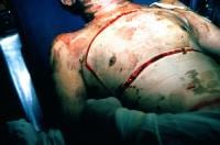
Two cases are described in Pre-hospital Emergency Care of severely burned patients who were impossible to adequately ventilate after tracheal intubation until they underwent escharotomy by a pre-hospital physician.
The review that follows reminds us of some intersting escharotomy facts:
- circumferential extremity burns can cause limb ischaemia
- abdominal burns can cause elevated intra-abdominal pressure and ischemic bowel
- neck burns can cause tracheal and jugular venous compression
- chest burns can cause respiratory compromise
- one previous study showed that chest and abdominal escharotomies significantly decreased intra-abdominal pressure, retention of carbon dioxide, and central venous and inferior vena caval pressures while significantly increasing serum oxygen concentration and systolic blood pressure.
- escharotomies may be performed on multiple body parts, including the extremities, the digits, the chest, the abdomen, the neck, and the penis
- neck escharotomy is a relatively simple procedure that involves an incision of the skin eschar longitudinally in the anterior midline from the chin to the sternal notch
- although different ways of doing chest escharotomies have been described, in the two reported cases in this article the procedure only involved longitudinal incisions, with good immediate effect.

Of note, neither of the physicians concerned had seen or done an escharotomy before. I’m adding this to my list of life-saving surgical interventions that are technically straightforward to perform, cannot always wait for another specialist to do, and happen too rarely to train for in the traditional way (ie being taught on a patient under supervision prior to the first time you do one).
Out-of-hospital chest escharotomy: a case series and procedure review
Prehosp Emerg Care. 2010 Jul-Sep;14(3):349-54
Two tier trauma team
Rather than activating a full trauma team based on traditional criteria, this team devised a two tier approach; if there were no positive anatomical or physiological criteria, a trauma team ‘consult’ approach was adopted, in which the patient was evaluated by emergency department and general surgery doctors only.

Of 1144 trauma activations, 468 (41%) were full trauma and 676 (59%) were consult trauma activations.. Sensitivity of the triage tool for the major trauma outcome (ISS>15, death, or needing critical care or urgent surgery) was 83%, specificity was 68%, undertriage was 3% and overtriage was 27%. There were no deaths in undertriaged patients.
This is an important study that has the potential to improve resource utilisation and even patient experience.
Prospective evaluation of a two-tiered trauma activation protocol in an Australian major trauma referral hospital
Injury. 2010 May;41(5):470-4
Military pre-hospital thoracotomy
Military doctors in Afghanistan reviewed their experience of thoracotomy done within 24 hours of admission to their hospital. The ballistic nature of thoracic penetrating trauma (mainly Afghan civilians without body armour) differs from the typical knife-wound related injury seen in survivors of thoracotomy reported in the pre-hospital literature.
Six of the patients presented in cardiac arrest – four PEA and two asystole. One of the PEA patients survived; this patient had sustained a thoracoabdominal GSW and had arrested 8 minutes from hospital. Following emergency thoracotomy, aortic control, and concomitant massive transfusion, return of spontaneous circulation (ROSC) was achieved and damage control surgery undertaken in both chest and abdomen.
The two patients in asystole had sustained substantial pulmonary and hilar injuries, and ROSC was never achieved. The patients in PEA all had arrested as a consequence of hypovolaemia from solid intra-abdominal visceral haemorrhage. All patients in PEA had ROSC achieved, albeit temporarily.
Following thoracotomy, patients required surgical manoeuvres such as pulmonary hilar clamping, packing and temporary aortic occlusion; hypovolaemia was the leading underlying cause of the cardiac arrest. These factors lead the authors to conclude that although isolated cardiac wounds do feature in war, they are unusual and the injury pattern of casualties in conflict zones are often complex and multifactorial.
Is pre-hospital thoracotomy necessary in the military environment?
Injury. 2010 Jul;41(7):1008-12
Pre-hospital RSI
Physicians from HEMS London document their experience of 400 pre-hospital rapid sequence induction / intubations. Their data are consistent with the experience of other similar services and with the emergency airway management literature in general:
- Failure to intubate is rare
- Removing cricoid pressure often improves the view
- A BURP manoeuvre can improve the view and facilitate intubation, but bimanual laryngoscopy / external laryngeal manipulation is better
- Having an SOP optimises first-pass success rate
Cricoid pressure and laryngeal manipulation in 402 pre-hospital emergency anaesthetics: Essential safety measure or a hindrance to rapid safe intubation?
Resuscitation 2010(81):810–816
Exsanguinating pelvis – occlude the aorta
Some patients with life-threatening arterial haemorrhage from a pelvic fracture may be peri-arrest prior to transfer to the angiography suite. French authors describe their use of a balloon catheter to occlude the infrarenal aorta to allow resuscitation to achieve sufficient stability for the transfer. As well as exsanguinating pelvic haemorrhage, intra-aortic balloon occlusion has already been described for the treatment of hemorrhagic shock in the case of ruptured abdominal aortic aneurysm, in abdominal trauma, in gastrointestinal bleeding, and in postpartum hemorrhage.
Features of note regarding the technique include:
- it can be done blind (without radiological guidance)
- it can be done prior to transfer to a centre with interventional radiology
- it can be done in cardiac arrest (and has resulted in ROSC and subsequent survival)
The authors are at pains to point out that the intra-aortic balloon occlusion method described in the study ‘should be reserved to patients in critically uncontrollable hemorrhagic shock (CUHS) and is not a first-line treatment of pelvic fractures in hemorrhagic shock.’
Intra-Aortic Balloon Occlusion to Salvage Patients With Life-Threatening Hemorrhagic Shocks From Pelvic Fractures
J Trauma. 2010 Apr;68(4):942-8.
