Tag Archives: outcome

Pharmaconutrition for Acute Lung Injury

A trial by the ARDS Clinical Trials Network of pharmaconutrition for acute lung injury1 was stopped early for futility – outcomes were worse in the intervention group that received the enteral supplementation of n-3 fatty acids, γ-linolenic acid, and antioxidants. It had been hypothesised that the immunomodulatory effects of these supplements would provide clinical benefit in acute lung injury.
An accompanying editorial2 reports benefits of pharmaconutrition in other areas of critical care:

  • arginine-supplemented diets are associated with reduced infections and lengths of hospital stay in patients undergoing elective operations
  • glutamine-supplemented parenteral nutrition is associated with reduced infection and mortality in critically ill patients
  • antioxidant supplementation is associated with reduced mortality among critically ill patients with systemic inflammation.


Context The omega-3 (n-3) fatty acids docosahexaenoic acid and eicosapentaenoic acid, along with γ-linolenic acid and antioxidants, may modulate systemic inflammatory response and improve oxygenation and outcomes in patients with acute lung injury.

Objective To determine if dietary supplementation of these substances to patients with acute lung injury would increase ventilator-free days to study day 28.

Design, Setting, and Participants The OMEGA study, a randomized, double-blind, placebo-controlled, multicenter trial conducted from January 2, 2008, through February 21, 2009. Participants were 272 adults within 48 hours of developing acute lung injury requiring mechanical ventilation whose physicians intended to start enteral nutrition at 44 hospitals in the National Heart, Lung, and Blood Institute ARDS Clinical Trials Network. All participants had complete follow-up.

Interventions Twice-daily enteral supplementation of n-3 fatty acids, γ-linolenic acid, and antioxidants compared with an isocaloric control. Enteral nutrition, directed by a protocol, was delivered separately from the study supplement.

Main Outcome Measure Ventilator-free days to study day 28.

Results The study was stopped early for futility after 143 and 129 patients were enrolled in the n-3 and control groups. Despite an 8-fold increase in plasma eicosapentaenoic acid levels, patients receiving the n-3 supplement had fewer ventilator-free days (14.0 vs 17.2; P = .02) (difference, −3.2 [95% CI, −5.8 to −0.7]) and intensive care unit–free days (14.0 vs 16.7; P = .04). Patients in the n-3 group also had fewer nonpulmonary organ failure–free days (12.3 vs 15.5; P = .02). Sixty-day hospital mortality was 26.6% in the n-3 group vs 16.3% in the control group (P = .054), and adjusted 60-day mortality was 25.1% and 17.6% in the n-3 and control groups, respectively (P = .11). Use of the n-3 supplement resulted in more days with diarrhea (29% vs 21%; P = .001).

Conclusions Twice-daily enteral supplementation of n-3 fatty acids, γ-linolenic acid, and antioxidants did not improve the primary end point of ventilator-free days or other clinical outcomes in patients with acute lung injury and may be harmful.

1. Enteral Omega-3 Fatty Acid, γ-Linolenic Acid, and Antioxidant Supplementation in Acute Lung Injury
JAMA. 2011; 306:1574-1581
2. Pharmaconutrition in Acute Lung Injury
JAMA. 2011;306(14):1599-1600

Pre-hospital hypertonic saline during ACLS

A newly published study examines pre-hospital hypertonic saline during CPR. A randomised trial compared 7.2% hypertonic saline / hydroxyethyl starch with hydroxyethyl starch alone in over 200 adult patients with non-traumatic out-of-hospital cardiac arrest. The volume infused was 2 ml /kg over 10 mins. All patients were resuscitated by the physicians of the Emergency Medical System (EMS) in Bonn, Germany.
There were no differences in survival to admission or discharge. There was a barely statistically significant increase in those survivors with higher cerebral performance categories (1 or 2) in the hypertonic saline group, inviting further study. The study was conducted from 2001 to 2004 (according to the 2000 CPR-Guidelines), so took an interestingly long time to see print.
Randomised study of hypertonic saline infusion during resuscitation from out-of-hospital cardiac arrest
Resuscitation. 2011 Sep 19. [Epub ahead of print]
[EXPAND Click to read abstract]


Aim of the study Animal models of hypertonic saline infusion during cardiopulmonary resuscitation (CPR) improve survival, as well as myocardial and cerebral perfusion during CPR. We studied the effect of hypertonic saline infusion during CPR (Guidelines 2000) on survival to hospital admission and hospital discharge, and neurological outcome on hospital discharge.

Methods The study was performed by the EMS of Bonn, Germany, with ethical committee approval. Study inclusion criteria were non-traumatic out-of-hospital cardiac arrest, aged 18–80 years, and given of adrenaline (epinephrine) during CPR. Patients were randomly infused 2 ml kg−1 HHS (7.2% NaCl with 6% hydroxyethyl starch 200,000/0.5 [HES]) or HES over 10 min.

Results 203 patients were randomised between May 2001 and June 2004. After HHS infusion, plasma sodium concentration increased significantly to 162 ± 36 mmol l−1 at 10 min after infusion and decreased to near normal (144 ± 6 mmol l−1) at hospital admission. Survival to hospital admission and hospital discharge was similar in both groups (50/100 HHS vs. 49/103 HES for hospital admission, 23/100 HHS vs. 22/103 HES for hospital discharge). There was a small improvement in neurological outcome in survivors on discharge (cerebral performance category 1 or 2) in the HHS group compared to the HES group (13/100 HHS vs. 5/100 HES, p < 0.05, odds-ratio 2.9, 95% confidence interval 1.004–8.5).
Conclusion Hypertonic saline infusion during CPR using Guidelines 2000 did not improve survival to hospital admission or hospital discharge. There was a small improvement with hypertonic saline in the secondary endpoint of neurological outcome on discharge in survivors. Further adequately powered studies using current guidelines are needed.

[/EXPAND]

Cardiac arrest caused by subarachnoid haemorrhage

We know that subarachnoid haemorrhage (SAH) can cause cardiac arrest. Some questions we may have about this are:

Questions

  • What proportion of out-of-hospital cardiac arrests (OOHCA) who achieve return of spontaneous circulation (ROSC) are caused by SAH?
  • What is the usual presenting arrest rhythm – VT/VF or non-shockable rhythms?
  • What is the outcome of these patients – do any survive?
  • Do they have other characteristic cardiac features, such as ECG or echo abnormalities?
  • Should we do a head CT on all survivors of out-of-hospital cardiac arrest of uncertain aetiology?

A recent Japanese article in Resuscitation1 is the third from that country to be published on the topic in three years, the other two2,3 coming from different centres and all demonstrating some consistent answers, as do papers published in recent years from Europe4 and North America5:

Answers

  • Rates of SAH in OOHCA patients who achieve ROSC and make it to CT range from 4-16% (even higher if other sources of intracranial haemorrhage are included).
  • Studies consistently demonstrate VT/VF to be very rare – PEA and asystole are by far the commonest presenting arrest rhythms.
  • Almost no patients with this presentation due to SAH survive to hospital discharge.
  • In the most recent study, all patients who survived long enough to get a 12 lead showed ST-T abnormalities and/or QT prolongation, although echocardiograms were mostly normal.
  • Rates of SAH in OOHCA patients who achieve ROSC seem to be sufficiently high to seriously consider head CT in these patients if there is no obvious alternate explanation for the arrest.

1. Clinical and cardiac features of patients with subarachnoid haemorrhage presenting with out-of-hospital cardiac arrest
Resuscitation. 2011 Oct;82(10):1294-7
[EXPAND Abstract]


Background Subarachnoid haemorrhage (SAH) is known as one of the aetiologies of out-of-hospital cardiac arrest (OHCA). However, the mechanisms of circulatory collapse in these patients have remained unclear.

Methods and results We examined 244 consecutive OHCA patients transferred to our emergency department. Head computed tomography was performed on all patients and revealed the existence of SAH in 14 patients (5.9%, 10 females). Among these, sudden collapse was witnessed in 7 patients (50%). On their initial cardiac rhythm, all 14 patients showed asystole or pulseless electrical activity, but no ventricular fibrillation (VF). Return of spontaneous circulation (ROSC) was obtained in 10 of the 14 patients (14.9% of all ROSC patients) although all resuscitated patients died later. The ROSC rate in patients with SAH (71%) was significantly higher than that of patients with either other types of intracranial haemorrhage (25%, n = 2/8) or presumed cardiovascular aetiologies (22%, n = 23/101) (p < 0.01). On electrocardiograms, ST-T abnormalities and/or QT prolongation were found in all 10 resuscitated patients. Despite their electrocardiographic abnormalities, only 3 patients showed echocardiographic abnormalities.

Conclusions The frequency of SAH in patients with all causes of OHCA was about 6%, and in resuscitated patients was about 15%. The initial cardiac rhythm revealed no VF even though half had a witnessed arrest. A high ROSC rate was observed in patients with SAH, although none survived to hospital discharge.

[/EXPAND]
2. Assessing outcome of out-of-hospital cardiac arrest due to subarachnoid hemorrhage using brain CT during or immediately after resuscitation
Signa Vitae 2010; 5(2): 21 – 24 Full Text
[EXPAND Abstract]


Objectives. The clinical course and outcome of out-of-hospital cardiopulmonary arrest (OHCPA) due to subarachnoid hemorrhage (SAH) is unclear. The objective of this study is to clarify them.

Study design. Single- center, observational study. Setting. We usually perform a brain computed tomography (CT) in OHCPA patients who present without a clear etiology (42% of all OHCPA), such as trauma, to determine the cause of OHCPA and to guide treatment.

Patients. The study included OHCPA patients without a clear etiology, who were transferred to our center and who underwent a brain CT during resuscitation.

Methods of measurement. Patients’ records were reviewed; initial cardiac rhythm, existence of a witness and bystander cardiopulmonary resuscitation efforts (CPR) were compared with patients’ outcomes.

Results. Sixty-six patients were enrolled. 72.7% achieved return of spontaneous circulation (ROSC), 71.2% were admitted, 30.3% survived more than 7 days, and 9.1. survived-to-discharge. In 41 witnessed OHCPA, 87.8% obtained ROSC, 85.4% were admitted, and 14.6% survived-to-discharge. All survivors were witnessed. In 25 non-witnessed OHCPA, 48% obtained ROSC and were admitted, and no patients were discharged. Initial cardiac rhythm was ventricular fibrillation (VF), pulseless electrical activity (PEA) and asystole in 3.0%, 39.4%, and 47.0%. In 2 VF patients 50.0% survived-to- discharge, and there was no survivor with PEA or asystole.

Conclusion. This study shows a high rate of ROSC and admission in OHCPA patients with a SAH, and also reveals their very poor neurological outcome. We conclude that the detection of a SAH in OHCPA patients is important to determine the accurate frequency of SAH in this patient group and to guide appropriate treatment of all OHCPA patients.

[/EXPAND]
3. Subarachnoid haemorrhage as a cause of out-of-hospital cardiac arrest: A prospective computed tomography study
Resuscitation. 2009 Sep;80(9):977-80
[EXPAND Abstract]


Aim Aneurysmal subarachnoid haemorrhage (SAH) is a relatively common cause of out-of-hospital cardiac arrest (OHCA). Early identification of SAH-induced OHCA with the use of brain computed tomography (CT) scan obtained immediately after resuscitation may help emergency physicians make therapeutic decision as quickly as they can.

Methods During the 4-year observation period, brain CT scan was obtained prospectively in 142 witnessed non-traumatic OHCA survivors who remained haemodynamically stable after resuscitation. Demographics and clinical characteristics of SAH-induced OHCA survivors were compared with those with “negative” CT finding.

Results Brain CT scan was feasible with an average door-to-CT time of 40.0min. SAH was found in 16.2% of the 142 OHCA survivors. Compared with 116 survivors who were negative for SAH, SAH-induced OHCA survivors were significantly more likely to be female, to have experienced a sudden headache, and trended to have achieved return of spontaneous circulation (ROSC) prior to arrival in the emergency department less frequently. Ventricular fibrillation (VF) was significantly less likely to be seen in SAH-induced than SAH-negative OHCA (OR, 0.06; 95% CI, 0.01–0.46). Similarly, Cardiac Trop-T assay was significantly less likely to be positive in SAH-induced OHCA (OR, 0.08; 95% CI, 0.01–0.61).

Conclusion Aneurysmal SAH causes OHCA more frequently than had been believed. Immediate brain CT scan may particularly be useful in excluding SAH-induced OHCA from thrombolytic trial enrollment, for whom the use of thrombolytics is contraindicated. The low VF incidence suggests that VF by itself may not be a common cause of SAH-induced OHCA.

[/EXPAND]
4. Spontaneous subarachnoid haemorrhage as a cause of out-of-hospital cardiac arrest
Resuscitation. 2001 Oct;51(1):27-32
[EXPAND Abstract]


Objective: Spontaneous subarachnoid haemorrhage as a cause of out-of-hospital cardiac arrest is poorly evaluated. We analyse disease-specific and emergency care data in order to improve the recognition of subarachnoid haemorrhage as a cause of cardiac arrest.

Design: We searched a registry of cardiac arrest patients admitted after primarily successful resuscitation to an emergency department retrospectively and analysed the records of subarachnoid haemorrhage patients for predictive features.

Results: Over 8.5 years, spontaneous subarachnoidal haemorrhage was identified as the immediate cause in 27 (4%) of 765 out-of-hospital cardiac arrests. Of these 27 patients, 24 (89%) presented with at least three or more of the following common features: female gender (63%), age under 40 years (44%), lack of co-morbidity (70%), headache prior to cardiac arrest (39%), asystole or pulseless electric activity as the initial cardiac rhythm (93%), and no recovery of brain stem reflexes (89%). In six patients (22%), an intraventricular drain was placed, one of them (4%) survived to hospital discharge with a favourable outcome.

Conclusions: Subarachnoid haemorrhage complicated by cardiac arrest is almost always fatal even when a spontaneous circulation can be restored initially. This is due to the severity of brain damage. Subarachnoid haemorrhage may present in young patients without any previous medical history with cardiac arrest masking the diagnosis initially.

[/EXPAND]
5. Cranial computed tomography in the resuscitated patient with cardiac arrest
Am J Emerg Med. 2009 Jan;27(1):63-7
[EXPAND Abstract]


Introduction The incidence of out-of-hospital and in-hospital cardiorespiratory arrest from all causes in the United States occurs not infrequently. Postresuscitation care should include the identification of the inciting arrest event as well as therapy tailored to support the patient and treat the primary cause of the decompensation. The application of one particular testing modality, cranial computed tomography (CT) of the head, has not yet been determined. We undertook an evaluation of the use of head CT in patients who were resuscitated from cardiac arrest.

Methods Prehospital (emergency medical services), ED, and hospital records were reviewed for patients of all ages with cardiorespiratory arrest over a 4-year period (July 1996-June 2000). Information regarding diagnosis, management, and outcome was recorded. The results of cranial CT, if performed, and any apparent resulting therapeutic changes were recorded. Patients with a known traumatic mechanism for the cardiorespiratory arrest were excluded.

Results A total of 454 patients (mean age 58.3 years with 60% male) with cardiorespiratory arrest were entered in the study with 98 (22%) individuals (mean age 58.5 years with 53% male) undergoing cranial CT. Arrest location was as follows: emergency medical services, 41 (42%); ED, 11 (11%); and hospital, 46 (47%). Seventy-eight (79%) patients demonstrated 111 CT abnormalities: edema, 35 (32%); atrophy, 24 (22%); extra-axial hemorrhage, 14 (13%); old infarct, 12 (11%); new infarct, 11 (10%); intraparenchymal hemorrhage, 6 (5%); skull fracture, 5 (4%); mass, 3 (2%); and foreign body, 1 (1%). Therapeutic and diagnostic alterations in care were made in 38 (39%) patients—35 abnormal and 3 normal CTs. The following alterations occurred: medication administration, 26; withdrawal of life support, 7; additional diagnostic study, 6; neurologic consultation, 6; and intracranial pressure monitoring. 4. No patient survived to discharge.

Conclusion In this subset of resuscitated patients with cardiac arrest, abnormalities on the head CT were not uncommon. Alterations in management did occur in those patients with abnormalities. The indications and impact of head CT in the population of resuscitated patients with cardiac arrest remain unknown, warranting further investigation.

[/EXPAND]

Offensive medicine: CT before LP

I’m getting worn down by clinicians – often other specialists – who insist that CT imaging of the brain is mandatory prior to lumbar puncture in all patients. There is surely a subgroup of patients (especially young ones) in whom the benefit:harm balance of CT comes out in favour of NOT doing the imaging. In these cases, getting the scan is not ‘defensive medicine’ but ‘offensive medicine’ – offending the principle of primum non nocere. During ED shifts I have recently had to perform online searches in order to furnish colleagues and patients’ medically qualified relatives with printouts of the literature on this. This page is here to save me having to repeat those searches. Regarding the practice of performing a routine head CT prior to lumbar puncture to rule out risk of herniation:

  • Mass effect on CT does not predict herniation
  • Lack of mass effect on CT does not rule out raised ICP or herniation
  • Herniation has occurred in patients who did not undergoing lumbar puncture because of CT findings
  • Clinical predictors of raised ICP are more reliable than CT findings
  • CT may delay diagnosis and treatment of meningitis
  • Even in patients in whom LP may be considered contraindicated (cerebral abscess, mass effect on CT), complications from LP were rare in several studies

Best practice, it would seem, is the following

  • If you think CT will show a cause for the headache, do a CT
  • If a CT is indicated for other reasons (depressed conscious level, focal neurology), do a CT
  • If a GCS 15 patient is to undergo LP for suspected (or to rule out) meningitis, and they have a normal neurological exam (including fundi), and are not elderly or immunosuppressed, there is no need to do a CT first.
  • If you’re seriously worried about meningitis and are intent on getting a CT prior to LP, don’t let the imaging delay antimicrobial therapy.

Here are some useful references:

1. The CT doesn’t help

CT head before lumbar puncture in suspected meningitis BestBET evidence summary: In cases of suspected meningitis it is very unlikely that patients without clinical risk factors (immunocompromise/ history of CNS disease/seizures) or positive neurological findings will have a contraindication to lumbar puncture on their CT scan If CT scan is deemed to be necessary, administration of antibiotics should not be delayed. BestBETS website

Computed Tomography of the Head before Lumbar Puncture in Adults with Suspected Meningitis Much cited NEJM paper from 2001 which concludes: “In adults with suspected meningitis, clinical features can be used to identify those who are unlikely to have abnormal findings on CT of the headN Engl J Med. 2001 Dec 13;345(24):1727-33 Full Text

Cranial CT before Lumbar Puncture in Suspected Meningitis Correspondence in 2002 NEJM including study of 75 patients with pneumococcal meningitis: CT cannot rule out risk of herniation Cranial CT before Lumbar Puncture in Suspected Meningitis N Engl J Med. 2002 Apr 18;346(16):1248-51 Full Text

2. The CT may harm

Cancer risk from CT Paucis verbis card, from the wonderful Academic Life in EM

3. Guidelines say CT is not always needed

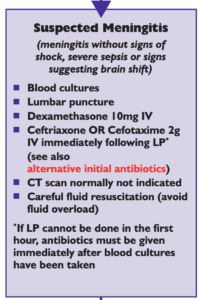
National (UK) guidelines on meningitis (community acquired meningitis in the immunocompetent host) available from meningitis.org. , including this box:

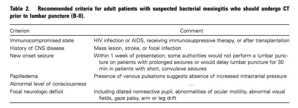
Practice Guidelines for the Management of Bacterial Meningitis These 2004 guidelines from the Infectious Diseases Society of America provide the following table listing the recommended criteria for adult patients with suspected bacterial meningitis who should undergo CT prior to lumbar puncture:

Clin Infect Dis. (2004) 39 (9): 1267-1284 Full text

4. This is potentially even more of an issue with paediatric patients

Fatal Lumbar Puncture: Fact Versus Fiction—An Approach to a Clinical Dilemma

An excellent summary of the above mentioned issues presented in a paediatric context, including the following:

On initial consideration a cranial CT would seem to be an appropriate and potentially useful diagnostic study for confirming the diagnosis of cerebral herniataion. The fallacy in this assessment has been emphasized by the finding that no clinically significant CT abnormalities are found that are not suspected on clinical assessments. Further, as previously noted, a normal CT examination may be found at about the time of a fatal herniation. Thus, the practical usefulness of a cranial CT in the majority of pediatric patients is limited to those rare patients whose increased ICP is secondary to mass lesions, not in the initial approach to acute meningitis.

Pediatrics. 2003 Sep;112(3 Pt 1):e174-6 Full Text

The last words should go to Dr Brad Spellberg, who in response to the IDSA’s guidelines wrote an excellent letter summarising much of the evidence at the time, confessed:

Why do we persist in using the CT scan for this purpose, despite the lack of supportive data? I am as guilty of this practice as anyone else, and the reason is simple: I am a chicken.

Clin Infect Dis. (2005) 40 (7): 1061 Full Text

Pre-hospital CPAP for pulmonary oedema

The physician-staffed mobile intensive care units of SAMU (Service d’Aide Médicale Urgente) in France provided the location for this randomised controlled trial of CPAP for acute cardiogenic pulmonary oedema.


STUDY OBJECTIVE: The purpose of this randomized controlled trial was to determine the immediate and delayed effects of noninvasive ventilation for patients in acute cardiogenic pulmonary edema (ACPE) in addition to aggressive usual care in a medical prehospital setting.

METHODS: Out-of-hospital patients in severe ACPE were eligible for the study. Patients were randomized to receive either usual care, including conventional optimal treatment with furosemide, oxygen, and high-dose boluses of isosorbide dinitrate plus oxygen, or conventional medications plus out-of-hospital continuous positive airway pressure (CPAP). The primary outcome was the treatment success defined as all of respiratory rate less than 25 breaths per minute and oxygen saturation of greater than 90% at the end of 1-hour study. Secondary end points included death during 30 days after inclusion. Lengths of intensive care unit and hospital stays were also recorded.

RESULTS: In total, 124 patients were enrolled into the study. The 2 groups had similar baseline characteristics. For the primary outcome analysis, 22 (35.5%) of 62 patients were considered as experiencing a treatment success in the usual care group vs 19 (31.7%) of 60 in the CPAP group (P = .65). Seven patients died within 30 days in the usual care group vs 6 in the CPAP group (P = .52). There were no statistically significant differences between the treatment groups for length of stay either in hospital or in the intensive care unit.

CONCLUSION: In the prehospital setting, in spite of its potential advantages for patients in ACPE, CPAP may not be preferred to a strict optimal intravenous treatment.

Continuous positive airway pressure for cardiogenic pulmonary edema: a randomized study
Am J Emerg Med. 2011 Sep;29(7):775-81

Pre-hospital thoracotomy

The London Helicopter Emergency Medical Service provides a physician / paramedic team to victims of trauma. One of the interventions performed by their physicians is pre-hospital resuscitative thoracotomy to patients with cardiac arrest due to penetrating thoracic trauma. They have published the outcomes from this procedure over a 15 year period which show an 18% survival to discharge rate, with a high rate of neurologically intact survivors1.
The article was submitted for publication on February 1, 2010, and in the discussion mentions a further two survivors from the procedure performed after conducting the study. It is likely therefore in the year and a half since submission still more patients have been saved. It will be interesting to read future reports from this team as the numbers accumulate; penetrating trauma missions are sadly increasing in frequency.
Having worked for these guys and performed this procedure in the field a few times myself, I can attest to the training and governance surrounding this system. The technique of clamshell thoracotomy is well described 2 and one I would recommend for the non-surgeon.

BACKGROUND: Prehospital cardiac arrest associated with trauma almost always results in death. A case of survival after prehospital thoracotomy was published in 1994 and several others have followed. This article describes the result of prehospital thoracotomy in a physician-led system for patients with stab wounds to the chest who suffered cardiac arrest on scene.
METHODS: A 15-year retrospective prehospital trauma database review identified victims of stab wounds to the chest who suffered cardiac arrest on scene and had thoracotomy performed according to local standard operating procedures.
RESULTS: Overall, 71 patients met inclusion criteria. Thirteen patients (18%) survived to hospital discharge. Neurologic outcome was good in 11 patients and poor in 2. Presenting cardiac rhythm was asystole in four patients, pulseless electrical activity in five, and unrecorded in the remaining four. All survivors had cardiac tamponade. The medical team was present at the time of cardiac arrest for six survivors (good neurologic outcome): arrived in the first 5 minutes after arrest in three patients (all good neurologic outcome), arrived 5 minutes to 10 minutes after arrest in two patients (one poor neurologic outcome), and in one patient (poor neurologic outcome) the period was unknown. Of the survivors, seven thoracotomies were performed by emergency physicians and six by anesthesiologists.
CONCLUSIONS: Prehospital thoracotomy is a well-established procedure in this physician-led prehospital service. Results from this and other similar systems suggest that when performed for the subgroup of patients described, significant numbers of survivors with good neurologic outcome can be expected.

1. Thirteen Survivors of Prehospital Thoracotomy for Penetrating Trauma: A Prehospital Physician-Performed Resuscitation Procedure That Can Yield Good Results
J Trauma. 2011 May;70(5):E75-8
2. Emergency thoracotomy: “how to do it”
Emerg Med J. 2005 January; 22(1):22–24
Full text available here

Exsanguinating cardiac arrest not always fatal

The British Military has developed a reputation for aggressive pre-hospital critical care including (but not limited to) the use of blood products and tourniquets, and coordinated field hospital trauma care. They now report the outcomes for patients with traumatic cardiac arrest, mainly from improvised explosive devices. Of 52 patients, 14 (27%) demonstrated return of spontaneous circulation (ROSC), of whom four (8%) survived to hospital discharge with a neurologically good recovery. Resuscitative thoracotomy (RT) was performed on 12 patients (8 in the ED), including all four survivors. RT enabled open-chest CPR, release of pericardial tamponade, lung resection and compression of the descending thoracic aorta for haemorrhage control.
No patients who arrested in the field survived, although one of the neurologically well-recovered survivors arrested during transport to hospital and was in cardiac arrest for 24 minutes. The authors propose this individual’s survival was in part due to ‘the high level of care that he received during retrieval, including haemorrhage control, tracheal intubation and transfusion of blood products‘.
Asystole was universally associated with death but agonal / bradycardic rhythms were not. In keeping with other studies, cardiac activity on ultrasound was associated with ROSC.


AIM: To determine the characteristics of military traumatic cardiorespiratory arrest (TCRA), and to identify factors associated with successful resuscitation.

METHODS: Data was collected prospectively for adult casualties suffering TCRA presenting to a military field hospital in Helmand Province, Afghanistan between 29 November 2009 and 13 June 2010.

RESULTS: Data was available for 52 patients meeting the inclusion criteria. The mean age (range) was 25 (18-36) years. The principal mechanism of injury was improvised explosive device (IED) explosion, the lower limbs were the most common sites of injury and exsanguination was the most common cause of arrest. Fourteen (27%) patients exhibited ROSC and four (8%) survived to discharge. All survivors achieved a good neurological recovery by Glasgow Outcome Scale. Three survivors had arrested due to exsanguination and one had arrested due to pericardial tamponade. All survivors had arrested after commencing transport to hospital and the longest duration of arrest associated with survival was 24min. All survivors demonstrated PEA rhythms on ECG during arrest. When performed, 6/24 patients had ultrasound evidence of cardiac activity during arrest; all six with cardiac activity subsequently exhibited ROSC and two survived to hospital discharge.

CONCLUSION: Overall rates of survival from military TCRA were similar to published civilian data, despite military TCRA victims presenting with high Injury Severity Scores and exsanguination due to blast and fragmentation injuries. Factors associated with successful resuscitation included arrest beginning after transport to hospital, the presence of electrical activity on ECG, and the presence of cardiac movement on ultrasound examination.

Outcomes following military traumatic cardiorespiratory arrest: A prospective observational study
Resuscitation. 2011 Sep;82(9):1194-7

Trauma mortality and systolic BP

Here’s some further evidence that a ‘lowish’ – as opposed to a low – systolic blood pressure is a reason to be vigilant in trauma. In this study, it was BP measurement in the ED (rather than pre-hospital) that was assessed:


Introduction: Non-invasive systolic blood pressure (SBP) measurement is often used in triaging trauma patients. Traditionally, SBP < 90 mmHg has represented the threshold for hypotension, but recent studies have suggested redefining hypotension as SBP < 110 mmHg. This study aims to examine the association of SBP with mortality in blunt trauma patients.
Methods: This is an analysis of prospectively recorded data from adult (≥16 years) blunt trauma patients. Included patients presented to hospitals belonging to the Trauma Audit and Research Network (TARN) between 2000 and 2009. The primary outcome was the association of SBP and mortality rates at 30 days. Multivariate logistic regression models were used to adjust for the influence of age, gender, Injury Severity Score (ISS) and Glasgow Coma Score (GCS) on mortality.

Results: 47,927 eligible patients presented to TARN hospitals during the study period. Sample demographics were: median age: 51.1 years (IQR=32.8–67.4); male 60% (n=28,694); median ISS 9 (IQR = 8–10); median GCS 15 (IQR = 15–15); and median SBP 135 mmHg (IQR = 120–152). We identified SBP < 110 mmHg as a cut off for hypotension, where a significant increase in mortality was observed. Mor- tality rates doubled at <100 mmHg, tripled at <90 mmHg and were 5- to 6-fold at <70 mmHg, irrespective of age.
Conclusion: We recommend triaging adult blunt trauma patients with a SBP < 110 mmHg to resuscitation areas within dedicated trauma units for close monitoring and appropriate management.

Systolic blood pressure below 110mmHg is associated with increased mortality in blunt major trauma patients: Multicentre cohort study
Resuscitation. 2011 Sep;82(9):1202-7

Score to predict traumatic coagulopathy

Acute traumatic coagulopathy (ATC) is present in up to 25% of major trauma patients by the time they arrive in hospital. A predictive tool called the coagulopathy of severe trauma (COAST) score was retrospectively derived and then prospectively validated in major trauma patients in the state of Victoria, Australia. The definition of ATC was INR > 1.5 (1.0–1.3) or aPTT of > 60 s (25–38 s) on hospital presentation.
The study claims that a subgroup of patients with acute traumatic coagulopathy can be accurately identified based on simple observations in the pre-hospital phase or immediately on presentation to the ED, and that this could improve the feasibility of prospective interventional studies. Perhaps this will lead on to evaluation of pre-hospital tranexamic acid or even blood products?
At the cutoff score of ≥3, 40 coagulopathic patients would have been missed with 60 patients correctly predicted. The authors argue that while the low sensitivity of the score missed these coagulopathic patients, they had significantly better outcomes (and contained a significantly higher proportion of patients with isolated severe head injury).


Introduction: The inability to accurately predict acute traumatic coagulopathy (ATC) has been a key factor in the low level of evidence guiding its management. The aim of this study was to develop a tool to accurately identify patients with ATC using pre-hospital variables without the use of pathology or radiological testing.

Methods: Retrospective data from the trauma registry on major trauma patients were used to identify vari- ables independently associated with coagulopathy. These variables were clinically evaluated to develop a scoring system to predict ATC, which was prospectively validated in the same setting.

Results: There were 1680 major trauma patients in the derivation dataset, with 151 patients being coagulopathic. Pre-hospital variables independently associated with ATC were entrapment (OR 1.85; 95% CI: 1.12–3.06), temperature (OR 0.60; 95% CI: 0.60–0.72), systolic blood pressure (OR 0.99; 95% CI: 0.98–0.99), abdominal or pelvic content injury (OR 2.0; 95% CI: 1.27–3.12) and pre-hospital chest decompression (OR 4.99; 2.77–8.99). The COAST score was developed, scoring points for entrapment, temperature <35 ◦ C, systolic blood pressure <100 mm Hg, abdominal or pelvic content injury and chest decompression. Prospectively validated using 1225 major trauma patients, a COAST score of ≥3 had a specificity of 96.4% with a sensitivity of 60.0%, with an area under the receiver operating characteristic curve of 0.83 (0.78–0.88).
Conclusions: The COAST score accurately identified a group of patients with ATC using pre-hospital obser- vations. This predictive tool can be used to select patients for inclusion into prospective studies examining management options for ATC. Mortality in these patients is high, potentially improving feasibility of outcome studies.

Glutamine in ICU

Of interest to intensivists….


Background: Low plasma glutamine concentration is an independent prognostic factor for an unfavourable outcome in the intensive care unit (ICU). Intravenous (i.v.) supplementation with glutamine is reported to improve outcome. In a multi-centric, double-blinded, controlled, randomised, pragmatic clinical trial of i.v. glutamine supplementation for ICU patients, we investigated outcomes regarding sequential organ failure assessment (SOFA) scores and mortality. The hypothesis was that the change in the SOFA score would be improved by glutamine supplementation.

Methods: Patients (n=413) given nutrition by an enteral and/or a parenteral route with the aim of providing full nutrition were included within 72 h after ICU admission. Glutamine was supplemented as i.v. l-alanyl-l-glutamine, 0.283 g glutamine/kg body weight/24 h for the entire ICU stay. Placebo was saline in identical bottles. All included patients were considered as intention-to-treat patients. Patients given supplementation for >3 days were considered as predetermined per protocol (PP) patients.

Results: There was a lower ICU mortality in the treatment arm as compared with the controls in the PP group, but not at 6 months. For change in the SOFA scores, no differences were seen, 1 (0,3) vs. 2 (0.4), P=0.792, for the glutamine group and the controls, respectively.

Conclusion: In summary, a reduced ICU mortality was observed during i.v. glutamine supplementation in the PP group. The pragmatic design of the study makes the results representative for a broad range of ICU patients.

Scandinavian glutamine trial: a pragmatic multi-centre randomised clinical trial of intensive care unit patients
Acta Anaesthesiol Scand. 2011 Aug;55(7):812-818