
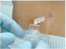
Here ultrasound was used to ascertain the best position for doing a lumbar puncture in kids, where the interspinous space was maximised:
BACKGROUND Lumbar punctures are commonly performed in the pediatric emergency department. There is no standard, recommended, optimal position for children who are undergoing the procedure.
OBJECTIVE To determine a position for lumbar punctures where the interspinous space is maximized, as measured by bedside ultrasound.
METHODS A prospective convenience sample of children under age 12 was performed. Using a portable ultrasound device, the L3-L4 or L4-L5 interspinous space was measured with the subject in 5 different positions. The primary outcome was the interspinous distance between 2 adjacent vertebrae. The interspinous space was measured with the subject sitting with and without hip flexion. In the lateral recumbent position, the interspinous space wasmeasured with the hips in a neutral position as well as in flexion, both with and without neck flexion. Data were analyzed by comparing pairwise differences.
RESULTS There were 28 subjects enrolled (13 girls and 15 boys) at a median age of 5 years. The sitting-flexed position provided a significantly increased interspinous space (P < .05). Flexion of the hips increased the interspinous space in both the sitting and lateral recumbent positions (P < .05). Flexion of the neck, did not significantly change the interspinous space (P = .998).
CONCLUSIONS The interspinous space of the lumbar spine was maximally increased with children in the sitting position with flexed hips; therefore we recommend this position for lumbar punctures. In the lateral recumbent position, neck flexion does not increase the interspinous space and may increase morbidity; therefore, it is recommended to hold patients at the level of the shoulders as to avoid neckflexion.
Positioning for lumbar puncture in children evaluated by bedside ultrasound
Pediatrics. 2010 May;125(5):e1149-53
A more recent study also used ultrasound in infants to investigate the anatomic necessity and advantage derived from a tight flexed lateral recumbent position, since hypoxia has been observed in that position:
Objectives: Hypoxia has been observed when infants undergo lumbar puncture in a tight flexed lateral recumbent position. This study used sonographic measurements of lumbar interspinous spaces to investigate the anatomic necessity and advantage derived from this tight flexed positioning in infants.
Methods: This was a brief, prospective, observational study of a convenience sample of patients. Twenty-one healthy infants under 1 month of age were scanned in two positions: prone in a spine-neutral position and lateral recumbent with their knees bent into their chest and their neck flexed. In each position, a 5- to 10-MHz linear array transducer was used to scan midline along the lumbar spinous processes in the sagittal plane. The distances between the spinous processes were measured near the ligamentum flavum using the ultrasound machine’s calipers. Pulse oximetry was monitored on all infants during flexed positioning.
Results: In the spine-neutral position, all studied interspinous spaces were much wider than a 22-gauge spinal needle (diameter 0.072 cm). The mean (±SD) interspinous spaces for L3-4, L4-5, and L5-S1 in a spine-neutral position were 0.42 (±0.07), 0.37 (±0.06), and 0.36 (±0.11) cm, respectively. Flexing the infants increased the mean lumbar interspinous spaces at L3-4, L4-5, and L5-S1 by 31, 51, and 44%, respectively.
Conclusions: This study verified that tight, lateral flexed positioning substantially enhances the space between the lumbar spinous processes and that a spine-neutral position also allows for a large enough anatomic interspinous space to perform lumbar puncture. However, further clinical research is required to establish the feasibility of lumbar puncture in a spine-neutral position.
Evaluating infant positioning for lumbar puncture using sonographic measurements
Acad Emerg Med. 2011 Feb;18(2):215-8
Tag Archives: paediatric
Therapeutic hypothermia for newborns
More evidence that cooling the hypoxic neonatal brain improves outcomes….
OBJECTIVE Mild hypothermia after perinatal hypoxic-ischemic encephalopathy (HIE) reduces neurologic sequelae without significant adverse effects, but studies are needed to determine the most-efficacious methods.
METHODS In the neo.nEURO.network trial, term neonates with clinical and electrophysiological evidence of HIE were assigned randomly to either a control group, with a rectal temperature of 37°C (range: 36.5–37.5°C), or a hypothermia group, cooled and maintained at a rectal temperature of 33.5°C (range: 33–34°C) with a cooling blanket for 72 hours, followed by slow rewarming. All infants received morphine (0.1 mg/kg) every 4 hours or an equivalent dose of fentanyl. Neurodevelopmental outcomes were assessed at the age of 18 to 21 months. The primary outcome was death or severe disability.
RESULTS A total of 129 newborn infants were enrolled, and 111 infants were evaluated at 18 to 21 months (53 in the hypothermia group and 58 in the normothermia group). The rates of death or severe disability were 51% in the hypothermia group and 83% in the normothermia group (P = .001; odds ratio: 0.21 [95% confidence interval [CI]: 0.09–0.54]; number needed to treat: 4 [95% CI: 3–9]). Hypothermia also had a statistically significant protective effect in the group with severe HIE (n = 77; P = .005; odds ratio: 0.17 [95% CI: 0.05–0.57]). Rates of adverse events during the intervention were similar in the 2 groups except for fewer clinical seizures in the hypothermia group.
CONCLUSION Systemic hypothermia in the neo.nEURO.network trial showed a strong neuroprotective effect and was effective in the severe HIE group.
Systemic Hypothermia After Neonatal Encephalopathy: Outcomes of neo.nEURO.network RCT
Pediatrics. 2010 Oct;126(4):e771-8
Update Dec 2014:
An RCT to determine if longer duration cooling (120 hours), deeper cooling (32.0°C), or both are superior to cooling at 33.5°C for 72 hours in neonates who are full-term with moderate or severe hypoxic ischemic encephalopathy.
Longer cooling, deeper cooling, or both compared with hypothermia at 33.5°C for 72 hours did not reduce NICU death. Small study.
Effect of depth and duration of cooling on deaths in the NICU among neonates with hypoxic ischemic encephalopathy: a randomized clinical trial
JAMA. 2014 Dec 24;312(24):2629-39
2J or 4J/kg in Paediatric Defibrillation?
Should we shock with 2J/kg or 4J/kg in Paediatric Defibrillation? The answer seems to be ‘we still don’t know’. Don’t worry – just follow the guidelines (reproduced for you at the bottom)
OBJECTIVE To examine the effectiveness of initial defibrillation attempts. We hypothesized that (1) an initial shock dose of 2 ± 10 J/kg would be less effective for terminating fibrillation than suggested in published historical data and (2) a 4 J/kg shock dose would be more effective.
PATIENTS AND METHODS This was a National Registry of Cardiopulmonary Resuscitation prospective, multisite, observational study of in-hospital pediatric (aged 18 years) ventricular fibrillation or pulseless ventricular tachycardia cardiac arrests from 2000–2008. Termination of ventricular fibrillation or pulseless ventricular tachycardia and event survival after initial shocks of 2 J/kg were compared with historic controls and a 4 J/kg shock dose.

RESULTS Of 266 children with 285 events, 173 of 285 (61%) survived the event and 61 of 266 (23%) survived to discharge. Termination of fibrillation after initial shock was achieved for 152 of 285 (53%) events. Termination of fibrillation with 2 ± 10 J/kg was much less frequent than that seen among historic control subjects (56% vs 91%; P < .001), but not different than 4 J/kg. Compared with 2 J/kg, an initial shock dose of 4 J/kg was associated with lower rates of return of spontaneous circulation (odds ratio: 0.41 [95% confidence interval: 0.21–0.81]) and event survival (odds ratio: 0.42 [95% confidence interval: 0.18–0.98]).
CONCLUSIONS The currently recommended 2 J/kg initial shock dose for in-hospital cardiac arrest was substantially less effective than previously published. A higher initial shock dose (4 J/kg) was not associated with superior termination of ventricular fibrillation or pulseless ventricular tachycardia or improved survival rates. The optimal pediatric defibrillation dose remains unknown.
Effect of defibrillation energy dose during in-hospital pediatric cardiac arrest
Pediatrics. 2011 Jan;127(1):e16-23
Here’s what the guidelines say:
Many AEDs have high specificity in recognizing pediatric shockable rhythms, and some are equipped to decrease (or attenuate) the delivered energy to make them suitable for infants and children <8 years of age. For infants a manual defibrillator is preferred when a shockable rhythm is identified by a trained healthcare provider (Class IIb, LOE C). The recommended first energy dose for defibrillation is 2 J/kg. If a second dose is required, it should be doubled to 4 J/kg. If a manual defibrillator is not available, an AED equipped with a pediatric attenuator is preferred for infants. An AED with a pediatric attenuator is also preferred for children <8 year of age. If neither is available, an AED without a dose attenuator may be used (Class IIb, LOE C). AEDs that deliver relatively high energy doses have been successfully used in infants with minimal myocardial damage and good neurological outcomes
Pediatric Basic Life Support: 2010 American Heart Association Guidelines for Cardiopulmonary Resuscitation and Emergency Cardiovascular Care
Full text document
Out of hospital monitoring in kids
I don’t have full text access to the Journal Pediatrics, so I’m not sure what I make of this small randomised trial comparing two types of blood pressure monitoring during paediatric transport:
BACKGROUND The “golden-hour” concept has led to emphasis on speed of patient delivery during pediatric interfacility transport. Timely intervention, in addition to enhanced monitoring during transport, is the key to improved outcomes in critically ill patients. Taking the ICU to the patient may be more beneficial than rapid delivery to a tertiary care center.

METHODS The Improved Monitoring During Pediatric Interfacility Transport trial was the first randomized controlled trial in the out-of-hospital pediatric transport environment. It was designed to determine the impact of improved blood pressure monitoring during pediatric interfacility transport and the effect on clinical outcomes in patients with systemic inflammatory response syndrome and moderate-to-severe head trauma. Patients in the control group had their blood pressure monitored intermittently with an oscillometric device; those in the intervention group had their blood pressure monitored every 12 to 15 cardiac contractions with a near-continuous, noninvasive device.
RESULTS Between May 2006 and June 2007, 1995, consecutive transport patients were screened, and 94 were enrolled (48 control, 46 intervention). Patients in the intervention group received more intravenous fluid (19.8 ± 22.2 vs 9.9 ± 9.9 mL/kg; P = .01), had a shorter hospital stay (6.8 ± 7.8 vs 10.9 ± 13.4 days; P = .04), and had less organ dysfunction (18 of 206 vs 32 of 202 PICU days; P = .03).
CONCLUSIONS Improved monitoring during pediatric transport has the potential to improve outcomes of critically ill children. Clinical trials, including randomized controlled trials, can be accomplished during pediatric transport. Future studies should evaluate optimal equipment, protocols, procedures, and interventions during pediatric transport, aimed at improving the clinical and functional outcomes of critically ill patients.
Enhanced Monitoring Improves Pediatric Transport Outcomes: A Randomized Controlled Trial
Pediatrics. 2011 Jan;127(1):42-8
Estimating child weight in Hong Kong
We know that the ‘APLS formula’ is inaccurate as a tool for estimating weight in Western children, and British and Australian researchers have devised more fitting formulae for their local populations as described here.

The emergency medicine team at the Accident and Emergency Medicine Academic Unit, Chinese University of Hong Kong have now provided a solution for Chinese children:
weight (kg) = (3 x age) + 5.
This was most accurate and precise in children <7 years old.
Age-based formulae to estimate children’s weight in the emergency department
Emerg Med J. 2010 Oct 13. [Epub ahead of print]

UK children sedation guideline
 Despite the huge number of articles in the literature on paediatric sedation, one still encounters acrimonious debates about the appropriateness of non-anaesthetists doing it. How refreshing then, to see that the UK’s National Institute for Health & Clinical Excellence (“NICE”) has tackled this subject and come up with some reasonable recommendations. I’ve as yet only read the summary, but some of the good things are:
- No unachievable ‘two doctors present’ rule: ‘Two trained healthcare professionals should be available during sedation‘
- Differentiating painless imaging from painful procedures
- Monitoring standards that are appropriate for the age of child and depth of sedation (no mandatory blood pressure or ECG monitoring unless deep sedation; end-tidal capnography in deep sedation).
- Acknowledgement of the special features of ketamine: ‘Ketamine is a dissociative agent: the state of dissociative sedation cannot be readily categorised as either moderate or deep sedation; the drug is considered to have a wide margin of safety.’
- Recognition that specialists other than anaesthetists may have specialist sedation and airway skills
There are some rather conservative recommendations on fasting, although the wording of the guideline in my interpretation allows some flexibility if ketamine is used for an emergency procedure.
Sedation in children and young people
National Institute for Health & Clinical Excellence
Better outcome with paediatric retrieval teams
Data from the England and Wales Paediatric Intensive Care Audit Network on children (aged 16 years or younger) admitted to 29 regional paediatric intensive care units (PICUs) between 1 January 2005 and 31 December 2008 were analysed in a retrospective cohort study to assess the effectiveness of the specialist retrieval teams.

The type of transferring team (specialist or non-specialist) was known for 16 875 cases and was specialist in 13 729 (81%). Compared with children transferred to PICUs from within the same hospital, children transferred from other hospitals were younger (median age 10 months vs 18 months), more acutely ill (mortality risk 6% vs 4% using the Paediatric Index of Mortality), needed more resources (such as invasive ventilation, vasoactive drugs, renal replacement therapy, extracorporeal membrane oxygenation and/or multiple-organ support), had longer stays in the PICU (median 75 h vs 43 h) and had a higher crude mortality (8% vs 6%). On multivariable analysis after adjustment for case mix and organisational factors, the risk of death among interhospital transfers was significantly (35%) lower than among intrahospital transfers. With similar analysis, the times spent in PICU did not differ significantly between these two groups. When the type of transferring team was considered, crude mortality was similar with specialist and non-specialist teams, although the children transferred by the specialist teams were more severely ill. On multivariable analysis, the risk of death was 42% lower with specialist team transfer.
These findings appear to confirm the value of specialist retrieval teams. Why children transferred from other hospitals did better than children transferred to the PICU in the same hospital is not explained.
Effect of specialist retrieval teams on outcomes in children admitted to paediatric intensive care units in England and Wales: a retrospective cohort study
Lancet. 2010 Aug 28;376(9742):698-704
Swimming the Channelopathy
Drowning is one of the leading causes of accidental death in children. Some apparent drownings may be related to sudden cardiac death, in particular to unidentified channelopathies, which are known to precipitate fatal arrhythmias during swimming-related events.
The majority of cases of sudden cardiac death in children and adolescents are secondary to either hypertrophic or right ventricular cardiomyopathy with coronary artery abnormalities also prevalent, and reports have demonstrated these cardiac abnormalities on autopsy following sudden swimming-related deaths.
However, the majority of autopsies in swimming-related sudden deaths are normal suggesting causation at molecular level, in particular ion channel defects such as type 1 long-QT syndrome (LQT1) and catecholaminergic polymorphic ventricular tachycardia (CPVT).


Some recommendations are made in an article in Archives of Disease in Childhood:
Proposed implementations to improve detection and appropriate management of apparent drownings secondary to cardiac channelopathies
- Improving awareness in the coronial service of the possibility of a cardiac cause for poorly explained drownings.
- Education of lifeguards and provision of automated defibrillators in swimming pools.
- Molecular autopsy for non-survivors to look for potential channelopathies.
- Screening for survivors and family members of non-survivors to identify those with a channelopathy.
- Proper counselling for those identified to have a channelopathy on family screening.
Drowning and sudden cardiac death
Arch Dis Child 2011;96:5-8
Tracheobronchial Foreign Bodies in Children
Asphyxiation by an inhaled foreign body is a leading cause of accidental death among children younger than 4 years. A review article examining 12,979 paediatric bronchoscopies made the following observations:
Epidemiology
- Most aspirated foreign bodies are organic materials (81%, confidence interval [CI] = 77%-86%), nuts and seeds being the most common.
- The majority of foreign bodies (88%, CI = 85%-91%) lodge in the bronchial tree, with the remainder catching in the larynx or trachea.
- The incidence of right-sided foreign bodies (52%, CI = 48%-55%) is higher than that of left-sided foreign bodies (33%, CI = 30%-37%). A small number of objects fragment and lodge in different parts of the airways.
- A history of a witnessed choking event is highly suggestive of an acute aspiration.
- A history of cough is highly sensitive for foreign body aspiration but is not very specific. On the other hand, a history of cyanosis or stridor is very specific for foreign body aspiration but is not very sensitive.
- Signs and symptoms typical in delayed presentations include unilateral decreased breath sounds and rhonchi, persistent cough or wheezing, recurrent or nonresolving pneumonia, or rarely, pneumothorax.
- Only 11% (CI = 8%-16%) of the foreign bodies were radio-opaque on radiograph, with chest radiographs being normal in 17% of children (CI = 13%-22%).
- The common radiographic abnormalities included localized emphysema and air trapping, atelectasis, infiltrate, and mediastinal shift.
- Although rigid bronchoscopy is the traditional diagnostic “gold standard,” the use of computerized tomography, virtual bronchoscopy, and flexible bronchoscopy is increasing.
- Reported mortality during bronchoscopy is 0.42%.
- Although asphyxia at presentation or initial emergency bronchoscopy causes some deaths, hypoxic cardiac arrest during retrieval of the object, bronchial rupture, and unspecified intraoperative complications in previously stable patients constitute the majority of in-hospital fatalities.
- Major complications include severe laryngeal edema or bronchospasm requiring tracheotomy or reintubation, pneumothorax, pneumomediastinum, cardiac arrest, tracheal or bronchial laceration, and hypoxic brain damage (0.96%).
- Aspiration of gastric contents is not reported.

Anaesthetic considerations
- Preoperative assessment should determine where the aspirated foreign body has lodged, what was aspirated, and when the aspiration occurred (“what, where, when”).
- The choices of inhaled or IV induction, spontaneous or controlled ventilation, and inhaled or IV maintenance may be individualized to the circumstances. Although several anesthetic techniques are effective for managing children with foreign body aspiration, there is no consensus from the literature as to which technique is optimal.
- An induction that maintains spontaneous ventilation is commonly practiced to minimize the risk of converting a partial proximal obstruction to a complete obstruction.
- Controlled ventilation combined with IV drugs and paralysis allows for suitable rigid bronchoscopy conditions and a consistent level of anesthesia.
- Close communication between the anesthesiologist, bronchoscopist, and assistants is essential.
The Anesthetic Considerations of Tracheobronchial Foreign Bodies in Children: A Literature Review of 12,979 Cases
Anesth Analg. 2010 Oct;111(4):1016-25
Hypertonic saline in bronchiolitis
A Canadian randomised controlled trial compared nebulised 3% saline with 0.9% saline in 81 infants under 2 years of age with bronchiolitis. The short-term use of nebulised 3% saline did not result in any statistically significant benefits, although a non-significant trend toward a decrease in hospital admission and improvement in respiratory distress was found. A larger study would be required to determine whether these trends arise from a clinically relevant treatment effect.

There’s really not much that’s been shown to make a difference in this disease, as this review article reminds us.
Effect of inhaled hypertonic saline on hospital admission rate in children with viral bronchiolitis: a randomized trial.
CJEM. 2010 Nov;12(6):477-84