Category Archives: PHARM

Prehospital and Retrieval Medicine

Compression-only CPR vs standard CPR

Two studies comparing compression-only CPR with conventional CPR:

BACKGROUND: The role of rescue breathing in cardiopulmonary resuscitation (CPR) performed by a layperson is uncertain. We hypothesized that the dispatcher instructions to bystanders to provide chest compression alone would result in improved survival as compared with instructions to provide chest compression plus rescue breathing.
METHODS: We conducted a multicenter, randomized trial of dispatcher instructions to bystanders for performing CPR. The patients were persons 18 years of age or older with out-of-hospital cardiac arrest for whom dispatchers initiated CPR instruction to bystanders. Patients were randomly assigned to receive chest compression alone or chest compression plus rescue breathing. The primary outcome was survival to hospital discharge. Secondary outcomes included a favorable neurologic outcome at discharge.
RESULTS: Of the 1941 patients who met the inclusion criteria, 981 were randomly assigned to receive chest compression alone and 960 to receive chest compression plus rescue breathing. We observed no significant difference between the two groups in the proportion of patients who survived to hospital discharge (12.5% with chest compression alone and 11.0% with chest compression plus rescue breathing, P=0.31) or in the proportion who survived with a favorable neurologic outcome in the two sites that assessed this secondary outcome (14.4% and 11.5%, respectively; P=0.13). Prespecified subgroup analyses showed a trend toward a higher proportion of patients surviving to hospital discharge with chest compression alone as compared with chest compression plus rescue breathing for patients with a cardiac cause of arrest (15.5% vs. 12.3%, P=0.09) and for those with shockable rhythms (31.9% vs. 25.7%, P=0.09).
CONCLUSIONS: Dispatcher instruction consisting of chest compression alone did not increase the survival rate overall, although there was a trend toward better outcomes in key clinical subgroups. The results support a strategy for CPR performed by laypersons that emphasizes chest compression and minimizes the role of rescue breathing. (Funded in part by the Laerdal Foundation for Acute Medicine and the Medic One Foundation; ClinicalTrials.gov number, NCT00219687.)

CPR with chest compression alone or with rescue breathing
N Engl J Med. 2010 Jul 29;363(5):423-3

BACKGROUND: Emergency medical dispatchers give instructions on how to perform cardiopulmonary resuscitation (CPR) over the telephone to callers requesting help for a patient with suspected cardiac arrest, before the arrival of emergency medical services (EMS) personnel. A previous study indicated that instructions to perform CPR consisting of only chest compression result in a treatment efficacy that is similar or even superior to that associated with instructions given to perform standard CPR, which consists of both compression and ventilation. That study, however, was not powered to assess a possible difference in survival. The aim of this prospective, randomized study was to evaluate the possible superiority of compression-only CPR over standard CPR with respect to survival.
METHODS: Patients with suspected, witnessed, out-of-hospital cardiac arrest were randomly assigned to undergo either compression-only CPR or standard CPR. The primary end point was 30-day survival.
RESULTS: Data for the primary analysis were collected from February 2005 through January 2009 for a total of 1276 patients. Of these, 620 patients had been assigned to receive compression-only CPR and 656 patients had been assigned to receive standard CPR. The rate of 30-day survival was similar in the two groups: 8.7% (54 of 620 patients) in the group receiving compression-only CPR and 7.0% (46 of 656 patients) in the group receiving standard CPR (absolute difference for compression-only vs. standard CPR, 1.7 percentage points; 95% confidence interval, -1.2 to 4.6; P=0.29).
CONCLUSIONS: This prospective, randomized study showed no significant difference with respect to survival at 30 days between instructions given by an emergency medical dispatcher, before the arrival of EMS personnel, for compression-only CPR and instructions for standard CPR in patients with suspected, witnessed, out-of-hospital cardiac arrest. (Funded by the Swedish Heart–Lung Foundation and others; Karolinska Clinical Trial Registration number, CT20080012.)

Compression-Only CPR or Standard CPR in Out-of-Hospital Cardiac Arrest
N Engl J Med. 2010 Jul 29;363(5):434-42

Weight formula for kids

The traditional ‘APLS formula’ for weight estimation in children based on age (wt in kg = [age+4] x 2) is recognised as underestimating weight in ‘developed’ countries, with the degree of underestimation increasing with increasing age.
Several authors have attempted to derive a more accurate formula.
In the UK, the measured weights of over 93 000 children aged 1-16 who attended a paediatric emergency department were used to compare a previously derived formula (wt=3[age]+7) with the APLS formula.
The formula ‘Weight=2(age+4)’ underestimated children’s weights by a mean of 33.4% (95% CI 33.2% to 33.6%) over the age range 1–16 years whereas the formula ‘Weight=3(age)+7’ provided a mean underestimate of 6.9% (95% CI 6.8% to 7.1%); this latter formula remained applicable from 1 to 13 years inclusive.
The authors state: ‘The APLS formula has clearly become a victim of better nourished children. With a mean underestimate of more than 20% (nearly 40% at age 10 years), its place as a weight estimation tool is questionable…. To continue with an inaccurate formula with no evidence base cannot be considered good medical practice.’
Weight estimation in paediatrics: a comparison of the APLS formula and the formula ‘Weight=3(age)+7’
Emerg Med J. 2010 Jul 20. [Epub ahead of print]
A previous retrospective Australian study on over 70 000 paediatric ED attendances derived formulae for three different age ranges:

  • For Infants < 12 months: Weight (kg) = (age in months + 9)/2
  • For Children aged 1-5 years: Weight (kg) = 2 x (age in years + 5)
  • For Children aged 5-14 years: Weight (kg) = 4 x age in years.

Make your Best Guess: An updated method for paediatric weight estimation in emergencies
Emerg Med Australas. 2007 Dec;19(6):528-34

ETCO2 and ROSC

One for the ‘hardly surprising’ category….
A study of end-tidal CO2 during out-of-hospital adult and child cardiac arrest resuscitation showed a sudden rise in CO2 was associated with return of spontaneous circulation (ROSC), suggesting that witnessing this would be a good time for a pulse check. Data from the 59 patients who achieved ROSC are shown below, time zero being time of ROSC. There was no such observed rise in the 49 patients who did not achieve ROSC.

A Sudden Increase in Partial Pressure End-Tidal Carbon Dioxide (PETCO2) at the Moment of Return of Spontaneous Circulation
The Journal of Emergency Medicine, Vol. 38, No. 5, pp. 614–621, 2010

Etomidate in RSI – systematic review

A systematic review of 20 included studies comparing a bolus dose of etomidate for rapid sequence induction with other induction agents resulted in the following conclusion:
“The available evidence suggests that etomidate suppresses adrenal function transiently without demonstrating a significant effect on mortality. However, no studies to date have been powered to detect a difference in hospital, ventilator, or ICU length of stay or in mortality”
The Effect of a Bolus Dose of Etomidate on Cortisol Levels, Mortality, and Health Services Utilization: A Systematic Review
Ann Emerg Med. 2010 Aug;56(2):105-13

Tracheal tube cuff pressure in flight

Tracheal tube cuff pressures increased from a mean 28.7 cm H2O pre-flight to 62.6 cm H2O in flight (mean altitude increase 2260 feet) in a Swiss helicopter-based study.
At cruising altitude, 98% of patients had intracuff pressure >30 cm H2O, 72% had intracuff pressure>50 cm H2O, and 20% even had intracuff pressure>80 cm H2O.
Multiple different referring hospitals meant the type of tracheal tube was not controlled for.

Endotracheal Tube Intracuff Pressure During Helicopter Transport
Ann Emerg Med. 2010 Aug;56(2):89-93

Tactical Combat Casualty Care

The brave men and women of the military not only risk their lives for us – they also provide a wealth of trauma experience and publish interesting stuff.
This month’s Journal of Trauma contains a military trauma supplement. One of the articles describes the latest guidelines on Tactical Combat Casualty Care. These include:

  • tourniquet use
  • Quikclot Combat Gauze as the haemostatic agent which has replaced Quikclot powder and HemCon. This preference is based on field experience that powder and granular agents do not work well in wounds in which the bleeding vessel is at the bottom of a narrow wound tract or in windy environments. WoundStat was a backup agent but this has been removed because of concerns over possible embolic and thrombotic complications.
  • longer catheters for decompression of tension pneumothorax (Harcke et al. found a mean chest wall thickness of 5.36 cm in 100 autopsy computed tomography studies of military fatalities. Several of the cases in their autopsy series were noted to have had unsuccessful attempts at needle thoracostomy because the needle/catheter units used for the procedure were too short to reach the pleural space*.)
  • close open chest wounds immediately with an occlusive material, such as Vaseline gauze, plastic wrap, foil, or defibrillator pads
  • a rigid eye shield and antibiotics for penetrating eye injury

Tactical Combat Casualty Care: Update 2009
The Journal of TRAUMA 2010;69(1):S10-13 (no abstract available)
Full text of guidelines in PDF at itstactical.com
*Harcke HT, Pearse LA, Levy AD, Getz JM, Robinson SR. Chest wall thickness in military personnel: implications for needle thoracentesis in tension pneumothorax. Mil Med. 2007;172:1260 –1263

Less RSI desaturation with Roc

Some of my pre-hospital critical care colleagues in the UK exclusively use rocuronium in preference to suxamethonium for rapid sequence induction (RSI) of anaesthesia in critically ill patients. I couldn’t see a good reason to switch although now there’s some evidence that adds to the argument.
The muscle fasciculations caused by the depolarising effect of suxamethonium may increase oxygen consumption, which may shorten the apnoea time before desaturation. Non-depolarising neuromuscular blockers such as rocuronium should allow a longer apnoea time after RSI. In addition, drugs which reduce fasciculations (such as lidocaine and fentanyl) should delay the the onset of desaturation when given prior to suxamethonium.

A large dose of Roc

These hypotheses were tested in a blinded, randomised controlled trial in 60 ASA-1 or -2 patients, who were scheduled for elective surgery under general anaesthesia. All patients received 2mg/kg propofol. One group was randomised to receive suxamethonium 1.5 mg/kg, a second group received rocuronium 1mg/kg plus lidocaine 1.5mg/kg and fentanyl 2mcg/kg, and a third group was given suxamethonium 1.5 mg/kg plus lidocaine 1.5mg/kg and fentanyl 2mcg/kg. The facemask was removed 50 seconds after the neuromuscular blocker was given and patients were intubated; the tube was then left open to air until desaturation to 95% occurred, which was timed.
Desaturation occurred significantly sooner in the suxamethonium-only group, followed by the sux/lido/fentanyl group, followed by the roc/lido/fentanyl group.
Of course these results are not necessarily directly applicable to the critically ill patient, and in this study there was no direct comparison between induction agent + rocuronium only and induction agent + suxamethonium only. Nevertheless the argument that suxamethonium-induced muscle fasciculations contribute to an avoidable increase in oxygen consumption is persuasive.
Effect of suxamethonium vs rocuronium on onset of oxygen desaturation during apnoea following rapid sequence induction
Anaesthesia. 2010 Apr;65(4):358-61

Pre-hospital RSI successes

A couple of papers in Prehospital Emergency Care this month contribute to the pre-hospital airway management / rapid sequence intubation (RSI) literature.
Intensive physician oversight of a pre-hospital RSI program increased the prescription of post-intubation morphine and midazolam, and decreased vecuronium use, although did not significantly increase the successful intubation rate in a before-and-after study. There was also an improvement in patient selection for RSI.
Effect of intensive physician oversight on a prehospital rapid-sequence intubation program
Prehosp Emerg Care. 2010 Jul-Sep;14(3):310-6
A prospective study examined intubation success rates and peri-intubation hypoxaemia in critical care transport (CCT) services in North America, whose services are mainly crewed by registered nurses (RNs) and emergency medical technicians–paramedic (EMT-Ps).
There was a mixture of pre-hospital and interhospital work: 51.9% of the 603 patients studied were intubated at the trauma scene, 27% were intubated inside a hospital, and interestingly 21.1% were intubated inside a vehicle (most of which were helicopters).
Neuromuscular blockade was used to facilitate intubation in only 428 patients (71%). Endotracheal intubation (ETI) was successful in 582 patients (96.5% of 603, 95% CI 94.7-97.8%). There was a greater need (p < 0.001) for multiple attempts at ETI when CCT crews performed the procedure in transport (37.3%) as compared with rate of requirement for multiple ETI attempts while in hospital (16.6%) or on scene (19.4%). Logistic regression identified a three-fold increase in the odds of requiring multiple attempts for intratransport ETI as compared with in-hospital ETI (OR 3.0, 95 CI 1.7–5.2, p < 0.001). 21 patients (3.5%) could not be intubated by the CCT crews resulting in a number of different rescue modalities including 3 cricothyroidotomies. At least there were no unrecognised oesophageal intubations. There were low rates of new hypoxaemia but peri-ETI SpO2 was only recorded for 494 patients (82%).
Airway management success and hypoxemia rates in air and ground critical care transport: a prospective multicenter study
Prehosp Emerg Care. 2010 Jul-Sep;14(3):283

Pre-hospital chest escharotomy

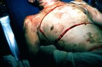
Two cases are described in Pre-hospital Emergency Care of severely burned patients who were impossible to adequately ventilate after tracheal intubation until they underwent escharotomy by a pre-hospital physician.
The review that follows reminds us of some intersting escharotomy facts:

  • circumferential extremity burns can cause limb ischaemia
  • abdominal burns can cause elevated intra-abdominal pressure and ischemic bowel
  • neck burns can cause tracheal and jugular venous compression
  • chest burns can cause respiratory compromise
  • one previous study showed that chest and abdominal escharotomies significantly decreased intra-abdominal pressure, retention of carbon dioxide, and central venous and inferior vena caval pressures while significantly increasing serum oxygen concentration and systolic blood pressure.
  • escharotomies may be performed on multiple body parts, including the extremities, the digits, the chest, the abdomen, the neck, and the penis
  • neck escharotomy is a relatively simple procedure that involves an incision of the skin eschar longitudinally in the anterior midline from the chin to the sternal notch
  • although different ways of doing chest escharotomies have been described, in the two reported cases in this article the procedure only involved longitudinal incisions, with good immediate effect.


Of note, neither of the physicians concerned had seen or done an escharotomy before. I’m adding this to my list of life-saving surgical interventions that are technically straightforward to perform, cannot always wait for another specialist to do, and happen too rarely to train for in the traditional way (ie being taught on a patient under supervision prior to the first time you do one).
Out-of-hospital chest escharotomy: a case series and procedure review
Prehosp Emerg Care. 2010 Jul-Sep;14(3):349-54

Military pre-hospital thoracotomy

Military doctors in Afghanistan reviewed their experience of thoracotomy done within 24 hours of admission to their hospital. The ballistic nature of thoracic penetrating trauma (mainly Afghan civilians without body armour) differs from the typical knife-wound related injury seen in survivors of thoracotomy reported in the pre-hospital literature.
Six of the patients presented in cardiac arrest – four PEA and two asystole. One of the PEA patients survived; this patient had sustained a thoracoabdominal GSW and had arrested 8 minutes from hospital. Following emergency thoracotomy, aortic control, and concomitant massive transfusion, return of spontaneous circulation (ROSC) was achieved and damage control surgery undertaken in both chest and abdomen.
The two patients in asystole had sustained substantial pulmonary and hilar injuries, and ROSC was never achieved. The patients in PEA all had arrested as a consequence of hypovolaemia from solid intra-abdominal visceral haemorrhage. All patients in PEA had ROSC achieved, albeit temporarily.
Following thoracotomy, patients required surgical manoeuvres such as pulmonary hilar clamping, packing and temporary aortic occlusion; hypovolaemia was the leading underlying cause of the cardiac arrest. These factors lead the authors to conclude that although isolated cardiac wounds do feature in war, they are unusual and the injury pattern of casualties in conflict zones are often complex and multifactorial.
Is pre-hospital thoracotomy necessary in the military environment?
Injury. 2010 Jul;41(7):1008-12