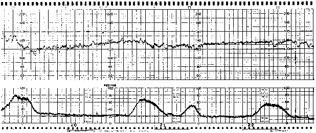
Can cardiotocography be applied in the pre-hospital setting? French physicians assessed its feasibility in 145 patients enrolled during 119 interhospital transfers and 26 primary prehospital missions.
Their physician-staffed ambulance teams included 19 emergency physicians and one anaesthetist.

Interpretable tracings were obtained for 81% of the patients during the initial examination, but this rate decreased to 66% during handling and transfer procedures. Only ground EMS transportations were included in the study. For 17 patients (12%), the monitoring led to a change in the patient’s management: an acceleration of chronology of prehospital management in 5 cases, a decision to directly admit the patient to the operating room for immediate cesarean section in three cases, a change in hospital admission in three cases, an adaptation or implementation of tocolytic treatment in six cases, and placing the patient in the left lateral decubitus position or oxygen administration in three cases.
Fetal monitoring in the prehospital setting
J Emerg Med. 2010 Nov;39(5):623-8
Tag Archives: monitoring
Evidence refutes ATLS shock classification
I have always had a problem with the ATLS classification of hypovolaemic shock, and omit it from teaching as any clinical applicability and reproducibility seem to be entirely lost on me. I was therefore reassured to read that real physiological data from the extensive national trauma registry in the UK (TARN) of 107,649 adult blunt trauma patients do not strongly support this classification. A key observation we regularly make in trauma patients is the frequent presence of normo- or bradycardia in hypovolaemic patients, which is well documented in the literature.

An excellent discussion section in this paper states: ‘it is clear that the ATLS classification of shock that associates increasing blood loss with an increasing heart rate, is too simplistic. In addition, blunt injury, which forms the majority of trauma in the UK, is usually a combination of haemorrhage and tissue injury and the classification fails to consider the effect of tissue injury‘
Testing the validity of the ATLS classification of hypovolaemic shock
Resuscitation. 2010 Sep;81(9):1142-7
capnometry versus pulse oximetry during procedural sedation
During emergency department procedural sedation, some clinicians (myself included) advocate non-invasive capnography for the early detection of apnoea. Others argue against routine administration of oxygen so that if desaturation occurs it provides an earlier more correctable warning of respiratory depression than if it occurs on supplemental oxygen. A Canadian study using prospective data from research on propofol with either ketamine or fentanyl compared changes in capnography with desaturation in sedated patients breathing only room air. Desaturation detectable by pulse oximeter occurred before overt changes in capnometry were identified.

It’s hard to ascertain the relevance of this finding. The authors wisely state ‘these findings should not be extrapolated to patients administered supplemental oxygen where it is possible capnometry may be helpful’. Since I use capnography in the hope that it will assist in the earlier detection of ketamine-associated laryngospasm in children, I’m not going to discard it in favour of waiting for the saturation to fall. Perhaps we just need to be clear that capnography may be more useful at detecting apnoea than hypoventilation.
A comparative evaluation of capnometry versus pulse oximetry during procedural sedation and analgesia on room air
CJEM. 2010 Sep;12(5):397-404
ETCO2 and ROSC
One for the ‘hardly surprising’ category….
A study of end-tidal CO2 during out-of-hospital adult and child cardiac arrest resuscitation showed a sudden rise in CO2 was associated with return of spontaneous circulation (ROSC), suggesting that witnessing this would be a good time for a pulse check. Data from the 59 patients who achieved ROSC are shown below, time zero being time of ROSC. There was no such observed rise in the 49 patients who did not achieve ROSC.

A Sudden Increase in Partial Pressure End-Tidal Carbon Dioxide (PETCO2) at the Moment of Return of Spontaneous Circulation
The Journal of Emergency Medicine, Vol. 38, No. 5, pp. 614–621, 2010
Tracheal tube cuff pressure in flight
Tracheal tube cuff pressures increased from a mean 28.7 cm H2O pre-flight to 62.6 cm H2O in flight (mean altitude increase 2260 feet) in a Swiss helicopter-based study.
At cruising altitude, 98% of patients had intracuff pressure >30 cm H2O, 72% had intracuff pressure>50 cm H2O, and 20% even had intracuff pressure>80 cm H2O.
Multiple different referring hospitals meant the type of tracheal tube was not controlled for.
_7093.jpg)
Endotracheal Tube Intracuff Pressure During Helicopter Transport
Ann Emerg Med. 2010 Aug;56(2):89-93
Femoral SvO2 not so useful
Bloods sampled from both femoral vein and SVC-sited catheters in critically ill patients showed good correlation in lactate levels but the oxygen saturation was not so reliable, with >5% variation in more than 50% and >15% variation in some patients. The authors suggest one reason is that the femoral catheter tip usually sits in the iliac vein and samples blood prior to the mixing of blood returning from intra-abdominal organs. They advise caution in using SfvO2 to guide resuscitation when narrow end points are used, as this may lead to inappropriate vasoactive drug or blood component therapy.

Femoral-Based Central Venous Oxygen Saturation Is Not a Reliable Substitute for Subclavian/Internal Jugular-Based Central Venous Oxygen Saturation in Patients Who Are Critically Ill
Chest. 2010 Jul;138(1):76-83
Thoracic electrical bioimpedance in dyspnoea
Thoracic electrical bioimpedance (TEB) was used in ED patients presenting with dyspnoea to differentiate between cardiac and non-cardiac causes.
The fundamental principle behind TEB is based on Ohm’s law. If a constant electrical current is applied to the thorax, changes in impedance (ΔZ) to flow are equal to changes in voltage drop across the circuit. As a current will always seek the path of lowest resistivity, which in the human body is blood, ΔZ of the thorax will primarily reflect the dynamic changes of blood volume in the thoracic aorta. Changes in thoracic electrical impedance are continuously recorded and processed using a computer algorithm to calculate a number of cardiohaemodynamic parameters such as stroke volume, CO, CI, SVR and systemic vascular resistance index (SVRi).

A cardiac index cut-off of 3.2 l/m/m2 had a 86.7% sensitive (95% CI 59.5% to 98.0%) and 88.9% specific (95% CI 73.9% to 96.8%) for cardiac dyspnoea in the 52 patients studies, of which 15 had cardiac-related dyspnoea.
The study has several limitations including small numbers and using the gold standard of discharge diagnosis.
Thoracic electrical bioimpedance: a tool to determine cardiac versus non-cardiac causes of acute dyspnoea in the emergency department
Emerg Med J. 2010 May;27(5):359-63
Free Full Text
Target Oxygen Saturation in Extreme Prems
1316 infants who were born between 24 weeks 0 days and 27 weeks 6 days of gestation were randomised to one of two different target ranges of oxygen saturation: 85 – 89% vs. 91 – 95%. The primary outcome was a composite of severe retinopathy of prematurity (defined as the presence of threshold retinopathy, the need for surgical ophthalmologic intervention, or the use of bevacizumab), death before discharge from the hospital, or both.
All infants were also randomly assigned to continuous positive airway pressure or intubation and surfactant in a 2-by-2 factorial design.
The rates of severe retinopathy or death did not differ significantly between the lower-oxygen-saturation group and the higher-oxygen-saturation group (28.3% and 32.1%, respectively; relative risk with lower oxygen saturation, 0.90; 95% confidence interval [CI], 0.76 to 1.06; P=0.21). Death before discharge occurred more frequently in the lower-oxygen-saturation group (in 19.9% of infants vs. 16.2%; relative risk, 1.27; 95% CI, 1.01 to 1.60; P=0.04), whereas severe retinopathy among survivors occurred less often in this group (8.6% vs. 17.9%; relative risk, 0.52; 95% CI, 0.37 to 0.73; P<0.001). There were no significant differences in the rates of other adverse events.
An editorial notes that the unmasked trial data showed that the distribution of oxygen saturation levels was within or above the target range in the higher-oxygen-saturation group, but in the lower-oxygen-saturation group, it was about 90 to 95% (i.e., above the target range). The difference in oxygen saturation levels between the groups was about 3 percentage points instead of the 6 percentage points that had been planned. Therefore, this study actually compared saturation levels of about 89 to 97% with saturation levels of 91 to 97%; the results should be ascribed to these higher ranges.
Targeting oxygen saturation levels is difficult, and a recommended oxygen saturation range that is effective yet safe remains elusive. A lower oxygen saturation level significantly reduces the incidence of severe retinopathy of prematurity but may increase the rate of death.
Target Ranges of Oxygen Saturation in Extremely Preterm Infants
N Engl J Med. 2010 May 16. [Epub ahead of print]
Protected: Positive capnography in cadavers
Low PPV can still be fluid responsive
Pulse pressure variation with respiration (PPV) predicts fluid responsiveness in mechanically ventilated patients. Because this is due to transmission of airway pressures to the vasculature, it is hypothesised that low tidal volume ventilation (or non compliant lungs, or both) results in less PPV even in fluid-responsive patients. This was confirmed in a study looking at the effect of airway driving pressure (Pplat – PEEP) on PPV. The study confirmed the positive predictive value of a high PPV, but some of those patients with a ‘low’ PPV (below a commonly accepted cut-off of 13%) were still fluid responsive, which was defined as a 15% or more increase in stroke index after a fluid challenge. In fluid responders with a low PPV, (Pplat – PEEP) was less than or equal to 20 cmH20.
Take home message: In mechanically ventilated patients, PPV values <13% do not rule out fluid responsiveness, especially when (Pplat – PEEP) was less than or equal to 20
The influence of the airway driving pressure on pulsed pressure variation as a predictor of fluid responsiveness
Intensive Care Med. 2010 Mar;36(3):496-503
