Category Archives: Kids

Acute Paediatrics

Hyperosmolar therapy

A great review article from the New England Journal of Medicine summaries the current knowledge base regarding the use of hypertonic saline and mannitol for raised intracranial pressure.

Hyperosmolar Therapy for Raised Intracranial Pressure 
N Engl J Med. 2012 Aug 23;367(8):746-52
Full text access is only available to New England Journal subscribers, but I’ve summarised some of the interesting bits in a short quiz you can take to test your knowledge. Just 13 True/False questions.

If you liked the quiz and want to use it at your local teaching sessions, here’s a Keynote Version and a PowerPoint Version

Don't ignore the diastolic

Most of us are pretty good at spotting hypotension and activating help or initiating therapy.
But ‘hypotension’ in many practitioners’ minds refers to a low systolic blood pressure. Who pays serious attention to the diastolic blood pressure? A low diastolic in a sick patient to me is a warning sign that their mean arterial pressure (MAP) is – or will be – low. After all, we spend about twice as long in diastole as in systole, so the diastolic pressure contributes more to the MAP than does the systolic.
A recent study showed that a low diastolic BP was one of several factors predictive of cardiac arrest on hospital wards: the most accurate predictors were maximum respiratory rate, heart rate, pulse pressure index, and minimum diastolic BP. These factors were more predictive than some of the variables included in the commonly used Early Warning Scores that trigger an emergency review.
The ‘pulse pressure index’ examined in the study is the pulse pressure divided by the systolic blood pressure (ie. [SBP-DBP]/SBP) which of course will be higher with lower diastolic blood pressures.
Importantly, the authors point out:


“In addition, our findings suggest that for many patients there is ample time prior to cardiac arrest to provide potentially life-saving interventions.”

…suggesting that there is still room for improvement in the identification and management of patients at risk for cardiac arrest, as the NCEPOD report ‘Cardiac Arrest Procedures: Time to Intervene?’ also showed.
They also recommend:


“…although systolic BP is commonly used in rapid response team activation criteria, incorporation of pulse pressure, pulse pressure index, or diastolic BP in place of systolic BP into the predictive model may be superior.”

Perhaps this may remind all of us to keep an eye on the diastolic as well as systolic BP when patients first present to us, and to include the importance of recognising diastolic hypotension in the teaching we provide our medical, paramedical and nursing students.

Predicting Cardiac Arrest on the Wards: A Nested Case-Control Study
Chest. 2012 May;141(5):1170-6 Free Full Text here
[EXPAND Click to read abstract]


Background: Current rapid response team activation criteria were not statistically derived using ward vital signs, and the best vital sign predictors of cardiac arrest (CA) have not been determined. In addition, it is unknown when vital signs begin to accurately detect this event prior to CA.

Methods: We conducted a nested case-control study of 88 patients experiencing CA on the wards of a university hospital between November 2008 and January 2011, matched 1:4 to 352 control subjects residing on the same ward at the same time as the case CA. Vital signs and Modified Early Warning Scores (MEWS) were compared on admission and during the 48 h preceding CA.

Results: Case patients were older (64 ± 16 years vs 58 ± 18 years; P = .002) and more likely to have had a prior ICU admission than control subjects (41% vs 24%; P = .001), but had similar admission MEWS (2.2 ± 1.3 vs 2.0 ± 1.3; P = .28). In the 48 h preceding CA, maximum MEWS was the best predictor (area under the receiver operating characteristic curve [AUC] 0.77; 95% CI, 0.71-0.82), followed by maximum respiratory rate (AUC 0.72; 95% CI, 0.65-0.78), maximum heart rate (AUC 0.68; 95% CI, 0.61-0.74), maximum pulse pressure index (AUC 0.61; 95% CI, 0.54-0.68), and minimum diastolic BP (AUC 0.60; 95% CI, 0.53-0.67). By 48 h prior to CA, the MEWS was higher in cases (P = .005), with increasing disparity leading up to the event.

Conclusions: The MEWS was significantly different between patients experiencing CA and control patients by 48 h prior to the event, but includes poor predictors of CA such as temperature and omits significant predictors such as diastolic BP and pulse pressure index.

[/EXPAND]

Life, limb and sight-saving procedures

The challenge of competence in the face of rarity

by Dr Cliff Reid FCEM, and Dr Mike Clancy FCEM
This article is to be published in Emergency Medicine Journal (EMJ), and is reproduced here with permission of the BMJ Group.
Emergency physicians require competence in procedures which are required to preserve life, limb viability, or sight, and whose urgency cannot await referral to another specialist.
Some procedures that fit this description, such as tracheal intubation after neuromuscular blockade in a hypoxaemic patient with trismus, or placement of an intercostal catheter in a patient with a tension pneumothorax, are required sufficiently frequently in elective clinical practice that competence can be acquired simply by training in emergency department, intensive care, or operating room environments.
Other procedures, such as resuscitative thoracotomy, may be required so infrequently that the first time a clinician encounters a patient requiring such an intervention may be after the completion of specialist training, or in the absence of colleagues with prior experience in the technique.
Some techniques that might be considered limb or life saving may be too technically complex to acquire outside specialist surgical training programs. Examples are damage control laparotomy and limb fasciotomy. One could however argue that these are rarely too urgent to await arrival of the appropriate specialist.
The procedures which might fit the description of a time­‐critical life, limb, or sight saving procedure in which it is technically feasible to acquire competence within or alongside an emergency medicine residency, and that cannot await another specialist, include:

  • limb amputation for the entrapped casualty with life-­threatening injuries;
  • escharotomy for a burns patient with compromised ventilation or limb perfusion;

 
Defining competence for emergency physicians
A major challenge is the acquisition of competence in the face of such clinical rarity. One medical definition of competence is ‘the knowledge, skill, attitude or combination of these, that enables one to effectively perform the activities of a particular occupation or role to the standards expected’[1]; in essence the ability to perform to a standard, but where are these standards defined?
If we look to the curricula which are used to assess specialist emergency physicians in several English-­speaking nations, all the procedures in the short list above are included, although no one single nation’s curriculum includes the entire list (Table 1).

 
So an emergency physician is expected to be able to conduct these procedures, and a competent emergency physician effectively performs them to the ‘standards’ expected. It appears then that the question is not whether emergency physicians should perform them, but to what standard should they be trained? Only then can the optimal approach to training be decided.
There are convincing arguments that even after minimal training the performance of these procedures by emergency physicians is justifiable:

  • All the abovementioned interventions could be considered to carry 100% morbidity or mortality if not performed, with some chance of benefit whose magnitude depends on the timeliness of intervention. In some cases that risk is quantifiable: cardiac arrest due to penetrating thoracic trauma has 100% mortality if untreated, but an 18% survival to discharge rate, with a high rate of neurologically intact survivors, if performed by prehospital emergency medicine doctors in the field according to defined indications[2] and using a simple operative procedure[3]. In this extreme clinical example, no further harm to the patient can result from the procedure but a chance of supreme benefit exists. Thus, the ethical requirements of beneficence and non-­maleficence are both met even in the circumstance of very limited training for the procedure. It is hard to conceive of many other circumstances in medicine where the benefit:harm ratio approaches infinity.
  • The procedures in question are technically straightforward and can be executed without specialist equipment in non-­operating room environments. These factors appear to be underappreciated by non-­emergency specialist opponents of emergency physician-­provided thoracotomy whose practice and experience is likely to be predominantly operating room-­based[4].
  • Some of the procedures are recommended or mandated by official guidelines[5], raising the possibility of medicolegal consequences of failure to perform them.
  • The procedures are time-­critical and cannot await the arrival of an alternative specialist not already present. Simple pragmatism dictates that emergency physicians be trained to provide the necessary interventions.

 
The challenge of training
So how does one best train for these procedures? High volume trauma experience provided by a registrar term with the London Helicopter Emergency Medical Service or at a South African trauma centre will be an option for a very limited subset of trainees. Alternative training can be provided using simulation, animal labs, and cadaver labs, without risk to patients or requiring dedicated surgical specialty attachments.
Simulation manikins are not yet available for all the procedures mentioned, and lack realistic operable tissue. Human cadaver labs and live animal training bring administrative, legal, ethical and financial challenges that may be prohibitive to time and cash‐limited training schemes, or be less available to the ‘already trained’ providers in existing consultant posts. Even excellent focused cadaver-­based courses such as the Royal College of Surgeons’ Definitive Surgical Trauma Skills course[6] may not be appropriate for the emergency medicine environment: on such a course one of the authors (CR) was publicly castigated by a cardiothoracic surgeon instructor for inexpert suture technique during the resuscitative thoracotomy workshop, despite the former having successfully performed the procedure on several occasions ‘in the field’ without need of elaborate needlework.
An additional training challenge is that of metacompetence: the decision and ability to apply the competence at the right time. In the light of the relative technical simplicity of the practical procedures under discussion, this may indeed be the greatest challenge. Both authors can recount sad tales of colleagues failing to provide indicated life-­saving interventions despite being technically capable of intervening. Reasons for reticence include ‘I haven’t been properly trained’, and ‘I wouldn’t feel supported if it went wrong’.
 
Where do we go from here?
We have presented clinical, ethical, practical, and medicolegal arguments in favour of emergency physicians providing these procedures. Collectively, the emergency medicine curricula of English-­speaking nations mandate competence in them. The relative technical simplicity and overwhelming benefit:harm equation obviate the need to match the competence of a surgical subspecialist; these factors suggest training can be limited in time and cost as long as the metacompetences of ‘decision to act and knowing when to act’ are taught, simulated, and tested.
While we should capitalise on the technical expertise of surgical colleagues in the training situation, it is imperative that emergency physicians appreciative of the emergency department environment and equipment are directly involved in translating this training to emergency medicine practice. The rarity of the situations requiring these procedures requires that training should be revisited on a regular basis, preferably in the context of local departmental simulation in order to optimise equipment and teamwork preparation.
Finally, the College of Emergency Medicine needs to make it clear to its members and fellows that these procedures lie unquestionably within the domain of emergency medicine, and that emergency physicians are supported in performing them to the best of their abilities with limited training when circumstances dictate that this in the best interests of preserving a patient’s life, limb, or sight.
 
 
References
1. British Medical Association. Competency-­based assessment discussion paper for consultants, May 2008. http://www.bma.org.uk/employmentandcontracts/doctors_performance/1_app raisal/CompetencyBasedAssessment.jsp Accessed 22nd March 2012
2. Davies GE, Lockey DJ. Thirteen Survivors of Prehospital Thoracotomy for Penetrating Trauma: A Prehospital Physician‐Performed Resuscitation Procedure That Can Yield Good Results. J Trauma. 2011;70(5):E75-­8
3. Wise D, Davies G, Coats T, et al. Emergency thoracotomy: “how to do it”. Emerg Med J. 2005; 22(1):22–24 Free full text
4. Civil I. Emergency room thoracotomy: has availability triumphed over advisability in the care of trauma patients in Australasia? Emerg Med Australas. 2010;22(4):257­‐9
5. Soar J, Perkins GD, Abbas G, et al. European Resuscitation Council Guidelines for Resuscitation 2010 Section 8. Cardiac arrest in special circumstances: Electrolyte abnormalities, poisoning, drowning, accidental hypothermia, hyperthermia, asthma, anaphylaxis, cardiac surgery, trauma, pregnancy, electrocution. Resuscitation. 2010;81(10):1400-­33 Full text
6. Definitive Surgical Trauma Skills course. http://www.rcseng.ac.uk/courses/course-search/dsts.html Accessed 22nd March 2012
7. http://www.collemergencymed.ac.uk/Training-Exams/Curriculum/Curriculum%20from%20August%202010/ Accessed 22nd March 2012
8. http://www.eusem.org/cms/assets/1/pdf/european_curriculum_for_em-aug09-djw.pdf accessed 17 May 2012
9. The Model of the Clinical Practice of Emergency Medicine http://www.abem.org/PUBLIC/portal/alias__Rainbow/lang__en-%C2%AD%20US/tabID__4223/DesktopDefault.aspx Accessed 22nd March 2012
10. http://rcpsc.medical.org/residency/certification/objectives/emergmed_e.pdf Accessed 22nd March 2012
11. http://www.acem.org.au/media/publications/15_Fellowship_Curriculum.pdf accessed 17 May 2012
12. http://www.collegemedsa.ac.za/Documents/doc_173.pdf accessed 17 May 2012
Life, limb and sight-saving procedures-the challenge of competence in the face of rarity
Emerg Med J. 2012 Jul 16. [Epub ahead of print]

Hypotonic Versus Isotonic Fluids After Surgery for Children

Kids in hospital with injury, infection or other illness, and those undergoing the physiological stress of surgery, produce (appropriately) elevated antidiuretic hormone levels which contribute to the risk of hyponatraemia by impairing free water excretion in the kidney.
Deaths have occurred on general paediatric and surgery wards when fluid regimens containing low concentrations of sodium (classically 0.18% or 0.225% NaCl) have resulted in hyponatraemia in children without adequate electrolyte monitoring, leading some bodies to recommend at least 0.45% NaCl solutions for maintenance fluid therapy in children.
However two recent studies1,2 on postoperative children show an increased risk of hyponatraemia even with 0.45% saline, when compared with 0.9% saline or Hartmann’s solution (Hartmann’s is similar – almost identical – to Ringer’s lactate).
I like the fact that paediatricians used Hartmann’s in one of these studies1. I have worked with several paediatricians who never use Hartmann’s, either from lack of experience or because of concern about its lactate content (not appreciating the lactate is metabolised by the liver to bicarbonate).
This is ironic, since Alexis Hartmann (1898–1964) was a paediatrician.
Want more fluid therapy irony? The ‘balanced salt solution’ used by Brits and Australasians is Hartmann’s solution – named after an American. The one used by Americans is Lactated Ringer’s solution – named after the British physician Sydney Ringer (1834-1910).
Medical history enthusiasts can read more about Hartmann and Ringer here.
1. A randomised controlled trial of Hartmann’s solution versus half normal saline in postoperative paediatric spinal instrumentation and craniotomy patients.
Arch Dis Child. 2012 Jun;97(6):491-6
[EXPAND Click to read abstract]

OBJECTIVE: To compare the difference in plasma sodium at 16-18 h following major surgery in children who were prescribed either Hartmann’s and 5% dextrose or 0.45% saline and 5% dextrose.
DESIGN: A prospective, randomised, open label study.
SETTING: The paediatric intensive care unit (650 admissions per annum) in a tertiary children’s hospital in Brisbane, Australia.
PATIENTS: The study group comprised 82 children undergoing spinal instrumentation, craniotomy for brain tumour resection, or cranial vault remodelling.
INTERVENTIONS: Patients received either Hartmann’s and 5% dextrose at full maintenance rate or 0.45% saline and 5% dextrose at two-thirds maintenance rate.
MAIN OUTCOMES MEASURES: Primary outcome measure: plasma sodium at 16-18 h postoperatively; secondary outcome measure: number of fluid boluses administered.
RESULTS: Mean postoperative plasma sodium levels of children receiving 0.45% saline and 5% dextrose were 1.4 mmol/l (95% CI 0.4 to 2.5) lower than those receiving Hartmann’s and 5% dextrose (p=0.008). In the 0.45% saline group, seven patients (18%) became hyponatraemic (Na <135 mmol/l) at 16-18 h postoperatively; in the Hartmann’s group no patient became hyponatraemic (p=0.01). No child in either fluid group became hypernatraemic.
CONCLUSIONS: The postoperative fall in plasma sodium was smaller in children who received Hartmann’s and 5% dextrose compared to those who received 0.45% saline and 5% dextrose. It is suggested that Hartmann’s and 5% dextrose should be administered at full maintenance rate postoperatively to children who have undergone major surgery in preference to hypotonic fluids.

[/EXPAND]
2. Hypotonic versus isotonic maintenance fluids after surgery for children: a randomized controlled trial
Pediatrics. 2011 Nov;128(5):857-66.
[EXPAND Click to read abstract]

OBJECTIVE: The objective of this randomized controlled trial was to evaluate the risk of hyponatremia following administration of a isotonic (0.9% saline) compared to a hypotonic (0.45% saline) parenteral maintenance solution (PMS) for 48 hours to postoperative pediatric patients.
METHODS: Surgical patients 6 months to 16 years of age with an expected postoperative stay of >24 hours were eligible. Patients with an uncorrected baseline plasma sodium level abnormality, hemodynamic instability, chronic diuretic use, previous enrollment, and those for whom either hypotonic PMS or isotonic PMS was considered contraindicated or necessary, were excluded. A fully blinded randomized controlled trial was performed. The primary outcome was acute hyponatremia. Secondary outcomes included severe hyponatremia, hypernatremia, adverse events attributable to acute plasma sodium level changes, and antidiuretic hormone levels.
RESULTS: A total of 258 patients were enrolled and assigned randomly to receive hypotonic PMS (N = 130) or isotonic PMS (N = 128). Baseline characteristics were similar for the 2 groups. Hypotonic PMS significantly increased the risk of hyponatremia, compared with isotonic PMS (40.8% vs 22.7%; relative risk: 1.82 [95% confidence interval: 1.21-2.74]; P = .004). Admission to the pediatric critical care unit was not an independent risk factor for the development of hyponatremia. Isotonic PMS did not increase the risk of hypernatremia (relative risk: 1.30 [95% confidence interval: 0.30-5.59]; P = .722). Antidiuretic hormone levels and adverse events were not significantly different between the groups.
CONCLUSION: Hypotonic Versus Isotonic Maintenance Fluids After Surgery for Children: A Randomized Controlled Trial.

[/EXPAND]

Not a pin cushion

This is the daughter of my friend. Avery is only seven months old and has survived a critical illness and is thankfully now fully recovered. Her Dad has nothing but praise for the medical and nursing staff who cared for her. But one thing could have been better. Avery endured multiple attempts at vascular access without ultrasound guidance.

If you were her parent, and you were an emergency physician with galaxy-class expertise in emergency ultrasound, how would you react? Complaints? Incident forms? Outrage?
How about education? For free. Accompanied by lavish praise for the experts who treated Avery and made her better.
Avery’s Dad is ultrasound podcaster and gentleman Dr Matt Dawson. He is offering FREE ultrasound training to anyone who wants to improve their vascular access skills.
Are there nurses, physicians, or technicians in your ED or ICU that could improve their care with this training? Please consider sending them for this training. To register for the course, and to read Avery’s full story, go to notapincushion.com.
And if you’re already comfortable with ultrasound-guided vascular access, then visit the site anyway, as there is some education here for all of us: how to turn a gut-wrenchingly distressing experience into something positive that will benefit countless others. I am thoroughly inspired.
Best wishes to an amazing family.
Cliff

Laryngospasm after Ketamine

A case is reported in Prehospital Emergency Care1 in which an agitated patient (due to mania and alcohol intoxication) received 5 mg/kg (500 mg) of ketamine intramuscularly by an EMS crew which dissociated him within a few minutes. He subsequently developed episodes of laryngospasm in the emergency department which were unrelieved by head tilt, chin lift and simple airway adjuncts but responded to bag-mask ventilation (BMV). The patient was intubated because the laryngospasm recurred, although it had again responded to BMV.
The authors make the point that because of the response of laryngospasm to simple manoeuvres, and because in the prehospital environment a patient will not be left without an EMS provider present, ‘restricting ketamine to EMS units capable of rapid-sequence intubation therefore seems unnecessary.
This is one for EMS directors to consider seriously. Personally, I think practicing prehospital care without access to ketamine is like having a hand tied behind my back. Ketamine opens up a world of possibilities in controlling combative patients, optimising scene safety, providing sedation for painful procedures including extrication, and enabling severe pain to be controlled definitively.
I’ve been using ketamine regularly for prehospital analgesia and emergency department procedural sedation in both adults and kids for more than a decade. I’ve seen significant laryngospasm 5 times (twice in kids). On one of those occasions, a 3 year old child desaturated to around 50% twice during two episodes of laryngospasm. We weren’t slow to pick it up – that was just her showing us how quickly kids can desaturate which continued while we went through a stepwise approach until BMV resolved it. It was however an eye opener for the registrar (senior resident) assisting me, who became extremely respectful of ketamine after that. Our ED sedation policy (that I wrote) required that suxamethonium was ready and available and that an appropriate dose had been calculated before anyone got ketamine. Paralysis may extremely rarely be required, but when it’s needed you need to be ready.

The best monitor for laryngospasm – noninvasive capnography

Laryngospasm is rare but most regular prescribers of ketamine will have seen it; the literature says it occurs in about 1-2% of sedations, although anecdotally I think it’s a bit less frequent. Importantly for those weighing the risks of allowing non-RSI competent prescribers, the requirement for intubation is exceptionally rare (2 of 11,589 reported cases in one review). Anyone interested should read this excellent review of ketamine-related adverse effects provided by Chris Nickson at Life in The Fast Lane. Chris reminds us of the Larson manouevre, which is digital pressure in the notch behind and below the ear, described by Larson2 as follows:

The technique involves placing the middle finger of each hand in what I term the laryngospasm notch. This notch is behind the lobule of the pinna of each ear. It is bounded anteriorly by the ascending ramus of the mandible adjacent to the condyle, posteriorly by the mastoid process of the temporal bone, and cephalad by the base of the skull. The therapist presses very firmly inward toward the base of the skull with both fingers, while at the same time lifting the mandible at a right angle to the plane of the body (i.e., forward displacement of the mandible or “jaw thrust”). Properly performed, it will convert laryngospasm within one or two breaths to laryngeal stridor and in another few breaths to unobstructed respirations.

I use this point most often to provide painful stimuli when assessing GCS in a patient, particular those I think may be feigning unconsciousness (I’ve done this for a number of years since learning how painful it can be when I was shown it by a jujitsu instructor). Dr Larson says he was taught the technique by Dr Guadagni, so perhaps we should be calling it the ‘Guadagni manouevre’. The lack of published evidence has led to some appropriate skepticism3, but as it can be combined with a jaw thrust it needn’t delay more aggressive interventions should they become necessary, it may work, and it’s likely to be harmless.
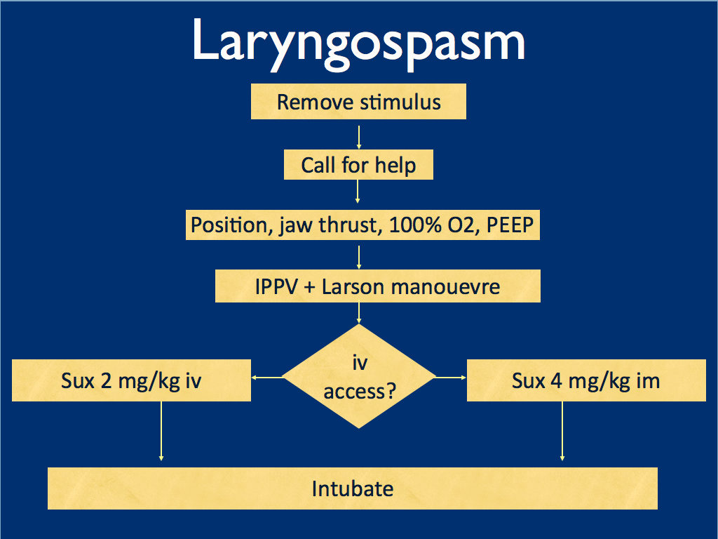
I presented the following suggested algorithm for management of laryngospasm during ketamine procedural sedation at a regional emergency medicine ‘Fellows Forum’ meeting in November 2007 in the UK. Since many paediatric procedural sedations were done using intramuscular (im) ketamine, it gives guidance based on whether or not vascular access has been obtained:

Some things I considered were:

    • Neuromuscular blockade (NMB) isn’t always necessary – laryngospasm may be managed with other sedatives such as propofol. However, titrating further sedatives in a desaturating child in my view is inferior to definitive airway management and laryngeal relaxation with suxamethonium and a tube.
    • Laryngospasm may be managed with much smaller doses of suxamethonium than are required for intubation – as little as 0.1 mg/kg may be effective. However, I think once we go down the NMB route we’re committed to intubation and therefore we should use a dose guaranteed to be effective in achieving intubating conditions.
    • In the child without vascular access, I considered intraosseous and intralingual sux. However, intramuscular suxamethonium is likely to have a relaxant effect on the laryngeal muscles within 30-45 seconds, which has to be compared with time taken to insert and confirm intraosseous needle placement. I do not think the traditionally recommended intralingual injection has any role to play in modern airway management.
  • At the time I wrote this most paediatric resuscitation bays in my area in the United Kingdom had breathing circuits capable of delivering PEEP – usually the Ayr’s T-Piece (specifically the Mapleson F system), which is why PEEP was included early in in the algorithm prior to BMV.
I have since modified it for two reasons: firstly, we might as well do the Larson manoeuvre during the jaw thrust; secondly, many Australasian and US EDs will only be able to deliver PEEP with a PEEP valve attached to a BVM, so PEEP has been moved to the BVM stage.
I would love to hear what people are doing in their prehospital and inhospital practice. Should ketamine only be administered by providers who can offer RSI? Do you have a laryngospasm protocol? If so, I’d love to see it. If not, feel free to use or adapt my unvalidated one at your own risk.

ABSTRACT An advanced life support emergency medical services (EMS) unit was dispatched with law enforcement to a report of a male patient with a possible overdose and psychiatric emergency. Police restrained the patient and cleared EMS into the scene. The patient was identified as having excited delirium, and ketamine was administered intramuscularly. Sedation was achieved and the patient was transported to the closest hospital. While in the emergency department, the patient developed laryngospasm and hypoxia. The airway obstruction was overcome with bag–valve–mask ventilation. Several minutes later, a second episode of laryngospasm occurred, which again responded to positive-pressure ventilation. At this point the airway was secured with an endotracheal tube. The patient was uneventfully extubated several hours later. This is the first report of laryngospam and hypoxia associated with prehospital administration of intramuscular ketamine to a patient with excited delirium.

1. Laryngospasm and Hypoxia After Intramuscular Administration of Ketamine to a Patient in Excited Delirium
Prehosp Emerg Care. 2012 Jul;16(3):412-4
2. Laryngospasm-The best treatment
Anesthesiology 1998; 89:1293-4
3. Management of Laryngospasm
http://www.respond2articles.com/ANA/forums/thread/1096.aspx

Out-of hospital traumatic paediatric cardiac arrest

This small study on traumatic arrests in children1 refutes the “100% mortality from traumatic arrest” dogma that people still spout and gives information on the mechanisms associated with survival: drowning and strangulation were associated with greater rates of survival to hospital admission compared with blunt, penetrating, and other traumas. Overall, drowning had the greatest rate of survival to discharge (19.1%).
I would like to know the injuries sustained in non-survivors, to determine whether they were potentially treatable. Strikingly, in the list of prehospital procedures performed, there were NO attempts at pleural decompression, something that is standard in traumatic arrest protocols in prehospital services were I have worked.
It is interesting to compare these results with those of the London HEMS team2, who for traumatic paediatric arrest achieved 19/80 (23.8%) survival to discharged from the emergency department and 7/80 (8.75%) survival to hospital discharge. They also noted a large proportion of the survivors suffered hypoxic or asphyxial injuries, whereas those patients with hypovolaemic cardiac arrest did not survive.


OBJECTIVE:To determine the epidemiology and survival of pediatric out-of-hospital cardiac arrest (OHCA) secondary to trauma.

METHODS:The CanAm Pediatric Cardiac Arrest Study Group is a collaboration of researchers in the United States and Canada sharing a common goal to improve survival outcomes for pediatric cardiac arrest. This was a prospective, multicenter, observational study. Twelve months of consecutive data were collected from emergency medical services (EMS), fire, and inpatient records from 2000 to 2003 for all OHCAs secondary to trauma in patients aged ≤18 years in 36 urban and suburban communities supporting advanced life support (ALS) programs. Eligible patients were apneic and pulseless and received chest compressions in the field. The primary outcome was survival to discharge. Secondary measures included return of spontaneous circulation (ROSC), survival to hospital admission, and 24-hour survival.

RESULTS:The study included 123 patients. The median patient age was 7.3 years (interquartile range [IQR] 6.0-17.0). The patient population was 78.1% male and 59.0% African American, 20.5% Hispanic, and 15.7% white. Most cardiac arrests occurred in residential (47.1%) or street/highway (37.2%) locations. Initial recorded rhythms were asystole (59.3%), pulseless electrical activity (29.1%), and ventricular fibrillation/tachycardia (3.5%). The majority of cardiac arrests were unwitnessed (49.5%), and less than 20% of patients received chest compressions by bystanders. The median (IQR) call-to-arrival interval was 4.9 (3.1-6.5) minutes and the on-scene interval was 12.3 (8.4-18.3) minutes. Blunt and penetrating traumas were the most common mechanisms (34.2% and 25.2%, respectively) and were associated with poor survival to discharge (2.4% and 6.5%, respectively). For all OHCA patients, 19.5% experienced ROSC in the field, 9.8% survived the first 24 hours, and 5.7% survived to discharge. Survivors had triple the rate of bystander cardiopulmonary resuscitation (CPR) than nonsurvivors (42.9% vs. 15.2%). Unlike patients sustaining blunt trauma or strangulation/hanging, most post-cardiac arrest patients who survived the first 24 hours after penetrating trauma or drowning were discharged alive. Drowning (17.1% of cardiac arrests) had the highest survival-to-discharge rate (19.1%).

CONCLUSIONS:The overall survival rate for OHCA in children after trauma was low, but some trauma mechanisms are associated with better survival rates than others. Most OHCA in children is preventable, and education and prevention strategies should focus on those overrepresented populations and high-risk mechanisms to improve mortality.

1. Epidemiology of out-of hospital pediatric cardiac arrest due to trauma
Prehosp Emerg Care, 2012 vol. 16 (2) pp. 230-236
2. Outcome from paediatric cardiac arrest associated with trauma
Resuscitation. 2007 Oct;75(1):29-34

Passive leg raise predicted fluid responsiveness in kids

Passive leg raising (PLR) is a great ‘free reversible fluid challenge’ to see if a shocked or hypotensive patient is likely to respond to volume therapy. A new study assesses its applicability in children.
PLR predicted fluid responders with 85% specificity but a lack of response did not rule out fluid responsiveness. Also, the effect of the PLR on cardiac index measured by echocardiography was the only way of predicting response – there was no relation to the more easily monitored effects of PLR on systolic blood pressure or heart rate.
Want to learn how to measure cardiac output using ultrasound? Mike Mallin from the Emergency Ultrasound Podcast shows you how here


OBJECTIVE: Fluid challenge is often used to predict fluid responsiveness in critically ill patients. Inappropriate fluid expansion can lead to some unwanted side effects; therefore, we need a noninvasive predictive parameter to assess fluid responsiveness. We want to assess the hemodynamic parameter changes after passive leg raising, which can mimic fluid expansion, to predict fluid responsiveness in pediatric intensive care unit patients and to get a cutoff value of cardiac index in predicting fluid responsiveness in pediatric patients.

DESIGN: Nonrandomized experimental study.

SETTING: Tertiary academic pediatric intensive care.

PATIENTS: Children admitted to pediatric intensive care.

INTERVENTION: Hemodynamic parameters were assessed at baseline, after passive leg raising, at second baseline, and after volume expansion (10 mL/kg normal saline infusion over 15 mins).

MEASUREMENTS AND MAIN RESULTS: We measured the heart rate, systolic blood pressure, and stroke volume and cardiac index using Doppler echocardiography. The hemodynamic parameter changes induced by passive leg raising were monitored. Among 40 patients included in the study, 20 patients had a cardiac index increase of ≥10% after volume expansion (responders). Changes in heart rate, systolic blood pressure, and stroke volume after passive leg raising did not significantly relate to the response to volume expansion. There was significant relation between changes in cardiac index to predict fluid responsiveness (p = .012, r = .22, 95% confidence interval 1.529 to 31.37). A cardiac index increase by ≥10% induced by passive leg raising predicted preload-dependent status with sensitivity of 55% and specificity of 85% (area under the curve 0.71 ± 0.084, 95% confidence interval 0.546-0.874).

CONCLUSION: The concomitant measurements in cardiac index changes after the passive leg raising maneuver can be helpful in predicting who might have an increase in cardiac index with subsequent fluid resuscitation.

The role of passive leg raising to predict fluid responsiveness in pediatric intensive care unit patients.
Pediatric Critical Care Medicine. 13(3):e155-e160, May 2012

Clotbusting wisdom on tap – your questions answered

The prevention and management of venous thromboembolic disease is a huge topic, which generates questions for emergency, critical care, and acute physicians during many shifts:

  • How long should someone requiring cardioversion for atrial fibrillation be anticoagulated for?
  • How should I provide thromboprophylaxis for this intubated patient?
  • This patient with submassive pulmonary embolism isn’t hypotensive yet. Can I thrombolyse them? Can I?
  • There’s a large superficial vein thrombosis in that limb – is anticoagulation indicated?
  • This asymptomatic patient on warfarin has an INR of 9.0 – should I reverse them?
  • Do I need to add Vitamin K if I’ve reversed warfarin with prothrombin complex concentrate?

The answers to these – and many, many more – questions are provided in one of the most comprehensive guidelines I’ve ever come across. I can see myself clicking on the link below in future when on duty in the ED.
Antithrombotic Therapy and Prevention of Thrombosis, 9th ed: American College of Chest Physicians Evidence-Based Clinical Practice Guidelines
Chest. 2012 Feb;141(2 Suppl) Full Text

Severe Traumatic Brain Injury in Children

The Brain Trauma Foundation has released updated guidelines on traumatic brain injury in children.
Most of the recommendations are Grade C and therefore based on limited evidence.

Indications for ICP monitoring

Use of intracranial pressure (ICP) monitoring may be considered in infants and children with severe traumatic brain injury (TBI) (Grade C).
Four lines of evidence support the use of ICP monitoring in children with severe TBI:

  • a frequently reported high incidence of intracranial hypertension in children with severe TBI
  • a widely reported association of intracranial hypertension and poor neurologic outcome
  • the concordance of protocol-based intracranial hypertension therapy and best-reported clinical outcomes
  • and improved outcomes associated with successful ICP-lowering therapies.

Threshold for treatment of intracranial hypertension
Treatment of intracranial pressure (ICP) may be considered at a threshold of 20 mm Hg (Grade C).
Sustained elevations in ICP (>20 mm Hg) are associated with poor outcome in children after severe TBI.
Normal values of blood pressure and ICP are age-dependent (lower at younger ages), so it is anticipated that the optimal ICP treatment threshold may be age-dependent.
Cerebral perfusion pressure thresholds
A CPP threshold 40–50 mm Hg may be considered. There may be age-specific thresholds with infants at the lower end and adolescents at the upper end of this range (Grade C).
Survivors of severe pediatric TBI undergoing ICP monitoring consistently have higher CPP values vs. nonsurvivors, but no study demonstrates that active maintenance of CPP above any target threshold in pediatric TBI reduces mortality or morbidity.
CPP should be determined in a standard fashion with ICP zeroed to the tragus (as an indicator of the foramen of Monro and midventricular level) and MAP zeroed to the right atrium with the head of the bed elevated 30°.
Advanced neuromonitoring
If brain oxygenation monitoring is used, maintenance of partial pressure of brain tissue oxygen (PbtO2) >10 mm Hg may be considered.
Neuroimaging
In the absence of neurologic deterioration or increasing intracranial pressure (ICP), obtaining a routine repeat computed tomography (CT) scan >24 hrs after the admission and initial follow-up study may not be indicated for decisions about neurosurgical intervention (Grade C).
Hyperosmolar therapy
Hypertonic saline should be considered for the treatment of severe pediatric traumatic brain injury (TBI) associated with intracranial hypertension. Effective doses for acute use range between 6.5 and 10 mL/kg (of 3%) (Grade B).
Hypertonic saline should be considered for the treatment of severe pediatric TBI associated with intracranial hypertension. Effective doses as a continuous infusion of 3% saline range between 0.1 and 1.0 mL/kg of body weight per hour administered on a sliding scale. The minimum dose needed to maintain intracranial pressure (ICP)
Temperature control
Moderate hypothermia (32–33°C) beginning early after severe traumatic brain injury (TBI) for only 24 hrs’ duration should be avoided.
Moderate hypothermia (32–33°C) be- ginning within 8 hrs after severe TBI for up to 48 hrs’ duration should be considered to reduce intracranial hypertension.
If hypothermia is induced for any indication, rewarming at a rate of >0.5°C/hr should be avoided (Grade B).
Moderate hypothermia (32–33°C) be- ginning early after severe TBI for 48 hrs, duration may be considered (Grade C).
Note: after completion of these guidelines, the committee became aware that the Cool Kids trial of hypothermia in pediatric TBI was stopped because of futility. The implications of this development on the recommendations in this section may need to be considered by the treating physician when details of the study are published.
Cerebrospinal fluid drainage
Cerebrospinal fluid (CSF) drainage through an external ventricular drain may be considered in the management of increased intracranial pressure (ICP) in children with severe traumatic brain injury (TBI).
The addition of a lumbar drain may be considered in the case of refractory intracranial hypertension with a functioning external ventricular drain, open basal cis- terns, and no evidence of a mass lesion or shift on imaging studies (Grade C).
Barbiturates
High-dose barbiturate therapy may be considered in hemodynamically stable patients with refractory intracranial hypertension despite maximal medical and surgical management.
When high-dose barbiturate therapy is used to treat refractory intracranial hy- pertension, continuous arterial blood pressure monitoring and cardiovascular support to maintain adequate cerebral perfusion pressure are required (Grade C).
Decompressive craniectomy for the treatment of intracranial hypertension
Decompressive craniectomy (DC) with duraplasty, leaving the bone flap out, may be considered for pediatric patients with traumatic brain injury (TBI) who are showing early signs of neurologic deterioration or herniation or are developing intracranial hypertension refractory to medical management during the early stages of their treatment (Grade C).
Hyperventilation
Avoidance of prophylactic severe hyperventilation to a PaCO2 If hyperventilation is used in the management of refractory intracranial hypertension, advanced neuromonitoring for evaluation of cerebral ischemia may be considered (Grade C).
Corticosteroids
The use of corticosteroids is not recommended to improve outcome or reduce intracranial pressure (ICP) for children with severe traumatic brain injury (TBI) (Grade B).
Analgesics, sedatives, and neuromuscular blockade
Etomidate may be considered to control severe intracranial hypertension; however, the risks resulting from adrenal suppression must be considered.
Thiopental may be considered to control intracranial hypertension.
In the absence of outcome data, the specific indications, choice and dosing of analgesics, sedatives, and neuromuscular-blocking agents used in the management of infants and children with severe traumatic brain injury (TBI) should be left to the treating physician.
*As stated by the Food and Drug Administration, continuous infusion of propofol for either sedation or the management of refractory intracranial hypertension in infants and children with severe TBI is not recommended (Grade C).
The availability of other sedatives and analgesics that do not suppress adrenal function, small sample size and single-dose administration in the study discussed previously, and limited safety profile in pediatric TBI limit the ability to endorse the general use of etomidate as a sedative other than as an option for single-dose administration in the setting of raised ICP.
Glucose and nutrition
The evidence does not support the use of an immune-modulating diet for the treatment of severe traumatic brain injury (TBI) to improve outcome (Grade B).
In the absence of outcome data, the specific approach to glycemic control in the management of infants and children with severe TBI should be left to the treating physician (Grade C).
Antiseizure prophylaxis
Prophylactic treatment with phenytoin may be considered to reduce the incidence of early posttraumatic seizures (PTS) in pediatric patients with severe traumatic brain injury (TBI) (Grade C).
The incidence of early PTS in pediatric patients with TBI is approximately 10% given the limitations of the available data. Based on a single class III study (4), prophylactic anticonvulsant therapy with phenytoin may be considered to reduce the incidence of early posttraumatic seizures in pediatric patients with severe TBI. Concomitant monitoring of drug levels is appropriate given the potential alterations in drug metabolism described in the context of TBI. Stronger class II evidence is available supporting the use of prophylactic anticonvulsant treatment to reduce the risk of early PTS in adults. There are no compelling data in the pediatric TBI literature to show that such treatment reduces the long-term risk of PTS or improves long-term neurologic outcome.
Guidelines for the Acute Medical Management of Severe Traumatic Brain Injury in Infants, Children, and Adolescents-Second Edition
Pediatr Crit Care Med 2012 Vol. 13, No. 1 (Suppl.)
Read online
Download PDF (617k)
Other Brain Trauma Foundation Guidelines