Category Archives: Resus

Life-saving medicine

Swallow a camera in GI bleed

Two recent studies evaluate the use of a novel ingestable camera to diagnose upper gastrointestinal bleeding in emergency department patients.
The potential advantages of video capsule endoscopy over traditional endoscopy could include immediate availability, avoidance of sedation, patient tolerance, and the ability to rule out active bleeding in the emergency department.
The device used was the PillCam ESO2 – shown here in this animation:

Further research is needed. These small interesting studies demonstrate the potential for this imaging technology to be used in stable patients presenting to emergency departments. Since it can only diagnose rather than treat, it would not appear to have any role in unstable patients.
Video capsule endoscopy in the emergency department: a prospective study of acute upper gastrointestinal hemorrhage.
Ann Emerg Med. 2013 Apr;61(4):438-443
[EXPAND Abstract]


STUDY OBJECTIVE: Video capsule endoscopy has been used to diagnose gastrointestinal hemorrhage and other small bowel diseases but has not been tested in an emergency department (ED) setting. The objectives in this pilot study are to demonstrate the ability of emergency physicians to detect blood in the upper gastrointestinal tract with capsule endoscopy after a short training period, measure ED patient acceptance of capsule endoscopy, and estimate the test characteristics of capsule endoscopy to detect acute upper gastrointestinal hemorrhage.

METHODS: During a 6-month period at a single academic hospital, eligible patients underwent video capsule endoscopy (Pillcam Eso2; Given Imaging) in the ED. Video images were reviewed by 4 blinded physicians (2 emergency physicians with brief training in capsule endoscopy interpretation and 2 gastroenterologists with capsule endoscopy experience).

RESULTS: A total of 25 subjects with acute upper gastrointestinal hemorrhage were enrolled. There was excellent agreement between gastroenterologists and emergency physicians for the presence of fresh or coffee-ground blood (0.96 overall agreement; κ=0.90). Capsule endoscopy was well tolerated by 96% of patients and showed an 88% sensitivity (95% confidence interval 65% to 100%) and 64% specificity (95% confidence interval 35% to 92%) for the detection of fresh blood. Capsule endoscopy missed 1 bleeding lesion located in the postpyloric region, which was not imaged because of expired battery life.

CONCLUSION: Video capsule endoscopy is a sensitive way to identify upper gastrointestinal hemorrhage in the ED. It is well tolerated and there is excellent agreement in interpretation between gastroenterologists and emergency physicians.

[/EXPAND]
Capsule endoscopy in acute upper gastrointestinal hemorrhage: a prospective cohort study
Endoscopy. 2013 Jan;45(1):12-9
[EXPAND Abstract]


BACKGROUND AND STUDY AIMS: Capsule endoscopy may play a role in the evaluation of patients presenting with acute upper gastrointestinal hemorrhage in the emergency department.

METHODS: We evaluated adults with acute upper gastrointestinal hemorrhage presenting to the emergency departments of two academic centers. Patients ingested a wireless video capsule, which was followed immediately by a nasogastric tube aspiration and later by esophagogastroduodenoscopy (EGD). We compared capsule endoscopy with nasogastric tube aspiration for determination of the presence of blood, and with EGD for discrimination of the source of bleeding, identification of peptic/inflammatory lesions, safety, and patient satisfaction.

RESULTS:The study enrolled 49 patients (32 men, 17 women; mean age 58.3 ± 19 years), but three patients did not complete the capsule endoscopy and five were intolerant of the nasogastric tube. Blood was detected in the upper gastrointestinal tract significantly more often by capsule endoscopy (15 /18 [83.3 %]) than by nasogastric tube aspiration (6 /18 [33.3 %]; P = 0.035). There was no significant difference in the identification of peptic/inflammatory lesions between capsule endoscopy (27 /40 [67.5 %]) and EGD (35 /40 [87.5 %]; P = 0.10, OR 0.39 95 %CI 0.11 - 1.15). Capsule endoscopy reached the duodenum in 45 /46 patients (98 %). One patient (2.2 %) had self-limited shortness of breath and one (2.2 %) had coughing on capsule ingestion.

CONCLUSION:In an emergency department setting, capsule endoscopy appears feasible and safe in people presenting with acute upper gastrointestinal hemorrhage. Capsule endoscopy identifies gross blood in the upper gastrointestinal tract, including the duodenum, significantly more often than nasogastric tube aspiration and identifies inflammatory lesions, as well as EGD. Capsule endoscopy may facilitate patient triage and earlier endoscopy, but should not be considered a substitute for EGD.

[/EXPAND]

Identifying the febrile kid who's too tachypnoeic

Body temperature raises heart rate and respiratory rate in kids, potentially affecting our interpretation of these clinical signs.
Dutch investigators developed centile charts of respiratory rates for specific body temperatures (derivation study), so that abnormally high rates could be identified as a means of predicting lower respiratory infection (validation set).
Respiratory rate increased overall by 2.2 breaths/min per 1°C rise (standard error 0.2) after accounting for age and temperature in the model, which is similar to a previous UK study that suggested a rise in respiratory rate of around 0.5-2 breaths per minute and an increase in heart rate of about 10 beats per minute for every 1 degree celcius above normal.
Cut-off values at the 97th centile were more useful in detecting the presence of LRTI than existing (Advanced Paediatric Life Support) respiratory rate thresholds.
The respiratory rate charts are available here.
Derivation and validation of age and temperature specific reference values and centile charts to predict lower respiratory tract infection in children with fever: prospective observational study
BMJ. 2012 Jul 3;345:e4224
Free Full Text Link

Hypothermia as an inotrope

This small study supports the hypothesis that therapeutic hypothermia can have positive inotropic effects in patients with cardiogenic shock of ischaemic or non-ischaemic origin.
Cooling resulted in a temperature-dependent decrease in heart rate and temperature-dependent increases in stroke volume index, cardiac index, mean arterial pressure, and cardiac power output. These changes reversed when the patients were rewarmed.
The authors summarise as follows:


In summary, our studies demonstrate that moderate hypothermia is feasible and safe also for patients in cardiogenic shock.

Improved cardiac performance may contribute to the considerable decrease of mortality for survivors of cardiac arrest, and the use of hypothermia can be recommended for patients with a clear indication for cooling and poor cardiac performance.

Moreover, hypothermia might be considered as a positive inotropic intervention during cardiogenic shock.

Moderate hypothermia for severe cardiogenic shock (COOL Shock Study I & II)
Resuscitation. 2013 Mar;84(3):319-25.
[EXPAND Abstract]


AIM OF THE STUDY: Hypothermia exerts profound protection from neurological damage and death after resuscitation from circulatory arrest. Its application during concomitant cardiogenic shock has been discussed controversially, and still hypothermia is used with reserve when haemodynamic parameters are impaired. On the other hand hypothermia improves force development in isolated human myocardium. Thus, we hypothesized that hypothermia could beneficially affect cardiac function in patients during cardiogenic shock.

METHODS: 14 Patients, admitted to Intensive Care Unit for cardiogenic shock under inotropic support, were enrolled and moderate hypothermia (33°C) was induced for either one (n=5, short-term) or twenty-four (n=9, mid-term) hours.

RESULTS: 12 patients suffered from ischaemic cardiomyopathy, 2 were female, and 6 were included after cardiac arrest and resuscitation. Body temperature was controlled by an intravascular cooling device. Short-term hypothermia consistently decreased heart rate, and increased stroke volume, cardiac index and cardiac power output. Metabolic and electrocardiographic parameters remained constant during cooling. Improved cardiac function persisted during mid-term hypothermia, but was reversed during re-warming. No severe or persistent adverse effects of hypothermia were observed.

CONCLUSION: Moderate Hypothermia is safe and feasable in patients during cardiogenic shock. Moreover, hypothermia improved parameters of cardiac function, suggesting that hypothermia might be considered as a positive inotropic intervention rather than a risk for patients during cardiogenic shock.

[/EXPAND]

Beherrsche die Reanimation

TLsm-icon The whole purpose behind my career and this blog is to save life. Like most emergency physicians I don’t see a huge number of resuscitation patients myself in a given week, so my best hope in making a difference is to develop my teaching skills so that I can motivate and inspire others to improve their ability to manage resuscitation.
The highlight of my week therefore has been the receipt of some email feedback from a colleague in Germany. An intensivist, internist, and prehospital doctor (I like him already) who tells me he found my ‘Own the Resus‘ talk helpful:


Dear Dr. Reid,

Few days ago, too tired too sleep after a long shift on my ICU (18 beds internal medicine ICU, I am specialist in internal medicine specialized in intensive care and prehospital emergency medicine in a major German city) I watched your talk via emcrit podcast. I was immediately caught, I soaked in every word, I was fascinated, watched it twice in the middle of the night and next afternoon I listened to it in my car driving to work.

At this very day I did some overdue crap beyond the end of my shift when I heard the ominous shuffling of feet and rolling of the emergency cart from the other end of the ward… “I think we need your help….”

There it was, difficult airway situation. Patient crashing.

Then what followed was a kind of “out of body experience”. I did what was necessary, made things happen like calling anesthesia difficult airway code, calling the surgeons, organizing fiber optics and meanwhile trying to secure that airway myself until i could dispatch anesthesia to the head and surgeons to the neck. Within few minutes there were 6 doctors and 5 nurses shuffling on 9 square meters…

I found myself 1 meter behind the foot end of the pts bed and with your talk in my head I found me consciously controlling the crowd. There was suddenly the messages of your talk and there was me. I don’t know how to put it into words, I wouldn’t have done something else in medical terms but thanks to your talk I had the vocabulary, the tools to reflect myself as the leader to be in charge of the situation somehow with more distance, and after a successful resus the 10 people involved in this code went off with a good feeling that everybody contributed in what they could and all for the pts benefit.

Your talk was a kind of transition to the next level for me: from the colleague who asks how to get out of trouble in many situations because he was often deeply in trouble, to the one who leads out of trouble.

With your talk many things suddenly became clear and I am looking forward to be able to work harder on this role of leading.

Thank you very much.

D

High flow nasal cannula oxygen

Where I work high flow humidified nasal cannula oxygen (HFNC) is used for infants with bronchiolitis and our ICU also employs it for selected adult patients. This is a relatively recent addition to our choice of oxygen delivery systems, and many emergency physicians may still be unfamiliar with it.
A recent review outlines the (scant) evidence for its use in neonates, infants, and adults, and proposes some mechanisms for its effect.
It’s a bit like the traditional delivery of oxygen via nasal cannulae. However, it is recommended that flow rates above 6 l/min are heated and humidified, so the review referred to heated, humidified, high flow nasal cannulae (HFNC).
Neonates
HFNC began as an alternative to nasal CPAP for premature infants. There are as yet no definitive studies showing its superiority over CPAP.
Infants
HFNC may decrease the need for intubation when compared to standard nasal cannula in infants with bronchiolitis.
Adults
No hard outcome data yet exist. It has mainly been used for hypoxemic respiratory failure rather than patients with hypercarbia such as COPD patients.
How it works
The following are proposed mechanisms for improvements in gas exchange / oxygenation:

1. A high FiO2 is maintained because flow rates are higher than spontaneous inspiratory demand, compared with standard facemasks and low flow nasal cannulae which entrain a significant amount of room air.

2. Nasopharyngeal dead space ‘washout’. The additional gas flow within the nasopharyngeal space may  reduce dead space: tidal volume ratio. There are some animal neonatal data to show improved CO2 clearance with flows up to 8 l/min.

3. Stenting of the upper airway by positive pressure may decrease upper airways resistance and reduce work of breathing.

4. Some positive pressure (akin to CPAP) may be generated, which can help recruit lung and decrease ventilation–perfusion mismatch; however this is not consistently present in all studies, and high flows are needed to generate even modest pressures. For example, in a study on postoperative cardiac surgery patients, HFNC at 35 l/min generated a nasopharyngeal pressure of only 2.7 ± 1 cmH2O.

 
Drawbacks and things to know

Studies suggest that if benefit is going to be seen in adult or paediatric patients, this should be evident in the first 30-60 minutes.

Any modest positive pressure generated will be reduced by an open mouth or when there is a significant leak between the cannulae and the nares.

HFNC maintain a fixed flow and generate variable pressures, and the pressures may be more inconsistent in patients with respiratory distress with high respiratory rates and mouth breathing. Compare this with non-invasive ventilation (CPAP and or BiPAP) in which variable flow is used to generate a fixed pressure.

 
The authors’ summary is helpful:


We postulate that the predominant benefit of HFNC is the ability to match the inspiratory demands of the distressed patient while washing out the nasopharyngeal dead space. Generation of positive airway pressure is dependent on the absence of significant leak around the nares and mouth and seems less likely to be a predominant factor in relieving respiratory distress for most patients.

NIV such as CPAP and bilevel positive airway pressure should still be considered first line therapy in moderately distressed patients in whom supplementation oxygen is insufficient and when a consistent positive pressure is indicated.

There are numerous ongoing trials which should hopefully clarify indications for HFNC and the mechanisms by which it may be beneficial.

An earlier summary of the evidence was written by my Scandinavian chums. And Reuben Strayer uses it to optimise oxygenation during RSI as a modification of the NODESAT technique.
Use of high flow nasal cannula in critically ill infants, children, and adults: a critical review of the literature
Intensive Care Med. 2013 Feb;39(2):247-57
[EXPAND Abstract]

BACKGROUND: High flow nasal cannula (HFNC) systems utilize higher gas flow rates than standard nasal cannulae. The use of HFNC as a respiratory support modality is increasing in the infant, pediatric, and adult populations as an alternative to non-invasive positive pressure ventilation.
OBJECTIVES: This critical review aims to: (1) appraise available evidence with regard to the utility of HFNC in neonatal, pediatric, and adult patients; (2) review the physiology of HFNC; (3) describe available HFNC systems (online supplement); and (4) review ongoing and planned trials studying the utility of HFNC in various clinical settings.
RESULTS: Clinical neonatal studies are limited to premature infants. Only a few pediatric studies have examined the use of HFNC, with most focusing on this modality for viral bronchiolitis. In critically ill adults, most studies have focused on acute respiratory parameters and short-term physiologic outcomes with limited investigations focusing on clinical outcomes such as duration of therapy and need for escalation of ventilatory support. Current evidence demonstrates that HFNC generates positive airway pressure in most circumstances; however, the predominant mechanism of action in relieving respiratory distress is not well established.
CONCLUSION: Current evidence suggests that HFNC is well tolerated and may be feasible in a subset of patients who require ventilatory support with non-invasive ventilation. However, HFNC has not been demonstrated to be equivalent or superior to non-invasive positive pressure ventilation, and further studies are needed to identify clinical indications for HFNC in patients with moderate to severe respiratory distress.

[/EXPAND]

Lateral chest thrusts for choking

An interesting animal study examined the techniques recommended in basic choking management algorithms for foreign body airway obstruction (chest and abdominal thrusts). In terms of the pressures generated, lateral chest thrusts were the most effective, although they are not recommended in current guidelines.
The technique described (on intubated pigs) was:


The animals were placed on the floor and on their side. The lower (dependent) side of the chest was braced by the ground and thrust was applied to the upper part of the upper side by two hands side by side with the higher one just below the axilla.

Interestingly – and I didn’t know this (although perhaps should have!) – the Australian Resuscitation Council (ARC) recommended lateral chest thrusts instead of abdominal thrusts for over 20 years.
While we should always exercise extreme caution in extrapolating animal studies to humans, this makes me want to consider lateral thrusts in the first aid (ie. no equipment) situation if other measures are failing.
Lateral versus anterior thoracic thrusts in the generation of airway pressure in anaesthetised pigs
Resuscitation. 2013 Apr;84(4):515-9
[EXPAND Abstract]


Objective Anterior chest thrusts (with the subject sitting or standing and thrusts applied to the lower sternum) are recommended by the Australian Resuscitation Council as part of the sequence for clearing upper airway obstruction by a foreign body. Lateral chest thrusts (with the victim lying on their side) are no longer recommended due to a lack of evidence. We compared anterior, lateral chest and abdominal thrusts in the generation of airway pressures using a suitable animal model.

Methods This was a repeated-measures, cross-over, clinical trial of eight anaesthetised, intubated, adult pigs. For each animal, ten trials of each technique were undertaken with the upper airway obstructed. A chest/abdominal pressure transducer, a pneumotachograph and an intra-oesophageal balloon catheter recorded chest/abdominal thrust, expiratory air flows, airway and intrapleural pressures, respectively.

Results The mean (SD) thrust pressures generated for the anterior, lateral and abdominal techniques were 120.9 (11.0), 135.2 (20.0), and 142.4 (27.3) cmH2O, respectively (p < 0.0001). The mean (SD) peak expiratory airway pressures were 6.5 (3.0), 18.0 (5.5) and 13.8 (6.7) cmH2O, respectively (p < 0.0001). The mean (SD) peak expiratory intrapleural pressures were 5.4 (2.7), 13.5 (6.2) and 10.3 (8.5) cmH2O, respectively (p < 0.0001). At autopsy, no rib, intra-abdominal or intra-thoracic injury was observed.
Conclusion Lateral chest and abdominal thrust techniques generated significantly greater airway and pleural pressures than the anterior thrust technique. We recommend further research to provide additional evidence that may inform management guidelines for clearing foreign body upper airway obstruction.

[/EXPAND]

Save a life by watching telly?

BB2.055If you’re in the United Kingdom on Thursday 21st March please consider watching BBC’s Horizon program at 9pm on BBC2.
I’m in Australia so I’ll miss it, but I’m moved by the whole background to this endeavour and really want you to help me spread the word.
Many of you will be familiar with the tragic case of Mrs Elaine Bromiley, who died from hypoxic brain injury after clinicians lost control of her airway during an anaesthetic for elective surgery. Her husband Martin has heroically campaigned for a greater awareness of the need to understand human factors in healthcare so such disasters can be prevented in the future.
Mr Bromiley describes the program, which is hosted by intensivist and space medicine expert Dr Kevin Fong:


Kevin and the Horizon team have produced something inspirational yet scientific, and – just as importantly – it’s by a clinician, for clinicians. It’s written in a way that will appeal to both those in healthcare and the public. It uses a tragic death to highlight human factors that all of us are prone to, and looks at how we can learn from others both in and outside healthcare to make a real difference in the future.

The lessons of this programme are for everyone in healthcare.

It would be wonderful if you could pass on details of the programme to anyone you know who works in healthcare. My goal is that by the end of this week, every one of the 1 million or so people who work in healthcare in the UK will be able to watch it (whether on Thursday or on iPlayer).


From the Health Foundation blog

Please help us reach this 1000 000 viewer target by watching on Thursday or later on iPlayer. Tweet about it or forward this message to as many healthcare providers you know. Help Martin help the rest of us avoid the kind of tragedy that he and his children have so bravely endured.
For more information on Mrs Bromiley’s case, watch ‘Just a Routine Operation’:


Cliff

Traumatic cardiac arrest outcomes

simEver heard anyone spout dogma along the lines of: “it’s a traumatic cardiac arrest – resuscitation is futile as the outcome is hopeless: survival is close to zero per cent”?
I have. Less frequently in recent years, I’ll admit, but you still hear it spout forth from the anus of some muppet in the trauma team. Here’s some recent data to add to the existing literature that challenges the ‘zero per cent survival’ proponents. A Spanish study retrospectively analysed 167 traumatic cardiac arrests (TCAs). 6.6% achieved a complete neurological recovery (CNR), which increased to 9.4% if the first ambulance to arrive contained an advanced team including a physician. Rhythm and age were important: CNR was achieved in 36.4% of VFs, 7% of PEAs, and 2.7% of those in asystole; survival rate by age groups was 23.1% in children, 5.7% in adults, and 3.7% in the elderly.
Since traumatic arrest tends to affect a younger age group than medical arrests, the authors suggest:
Avoiding the potential decrease in life expectancy in this kind of patient justifies using medical resources to their utmost potential to achieve their survival
Since 2.7% of the asystolic patients achieved a CNR, the authors challenge the practice proposed by some authors that Advanced Life Support be withheld in TCA patients with asystole as the initial rhythm:
had that indication been followed, three of our patients who survived neurologically intact would have been declared dead on-scene.”
I’d like to know what interventions were making the difference in these patients. They describe what’s on offer as:


In our EMS, all TCA patients receive ALS on-scene, which includes intubation, intravenous access, fluid and drug therapy, point-of-care blood analysis, and procedures such as chest drain insertion, pericardiocentesis, or Focused Assessment with Sonography for Trauma ultrasonography to improve the treatment of the cause of the TCA.

It appears that crystalloids and colloids are their fluid therapy of choice; unlike many British and Australian physician-based prehospital services they made no mention of the administration of prehospital blood products.
Traumatic cardiac arrest: Should advanced life support be initiated?
J Trauma Acute Care Surg. 2013 Feb;74(2):634-8
[EXPAND Abstract]


BACKGROUND: Several studies recommend not initiating advanced life support in traumatic cardiac arrest (TCA), mainly owing to the poor prognosis in several series that have been published. This study aimed to analyze the survival of the TCA in our series and to determine which factors are more frequently associated with recovery of spontaneous circulation (ROSC) and complete neurologic recovery (CNR).

METHODS: This is a cohort study (2006-2009) of treatment benefits.

RESULTS: A total of 167 TCAs were analyzed. ROSC was obtained in 49.1%, and 6.6% achieved a CNR. Survival rate by age groups was 23.1% in children, 5.7% in adults, and 3.7% in the elderly (p < 0.05). There was no significant difference in ROSC according to which type of ambulance arrived first, but if the advanced ambulance first, 9.41% achieved a CNR, whereas only 3.7% if the basic ambulance first. We found significant differences between the response time and survival with a CNR (response time was 6.9 minutes for those who achieved a CNR and 9.2 minutes for those who died). Of the patients, 67.5% were in asystole, 25.9% in pulseless electrical activity (PEA), and 6.6% in VF. ROSC was achieved in 90.9% of VFs, 60.5% of PEAs, and 40.2% of those in asystole (p < 0.05), and CNR was achieved in 36.4% of VFs, 7% of PEAs, and 2.7% of those in asystole (p < 0.05). The mean (SD) quantity of fluid replacement was greater in ROSC (1,188.8 [786.7] mL of crystalloids and 487.7 [688.9] mL of colloids) than in those without ROSC (890.4 [622.4] mL of crystalloids and 184.2 [359.3] mL of colloids) (p < 0.05).

CONCLUSION: In our series, 6.6% of the patients survived with a CNR. Our data allow us to state beyond any doubt that advanced life support should be initiated in TCA patients regardless of the initial rhythm, especially in children and those with VF or PEA as the initial rhythm and that a rapid response time and aggressive fluid replacement are the keys to the survival of these patients.

[/EXPAND]

On chicken bombs and muppets

I want to clarify some terminology I use on a day-to-day basis, which is now so ingrained in my vocabulary that I forget that its meaning may not be obvious to all.

“You go in there and it looks like a chicken bomb has gone off…”

“..external muppet factors can delay preparation for transport”

Muppets

The first is ‘muppet’. This does not refer to the much loved and trademarked invention of Jim Henson, (and now property of Disney) – a word originally thought to be a synthesis of ‘marionette’ and ‘puppet’. If I were referring to these wonderful icons of children’s televisual theatre I would capitalise the ’m’. Nope. I refer to the British meaning, which the Oxford English Dictionary lists as: ‘an incompetent or foolish person’. However I apply it in the context of behaviour rather than character. A wealth of evidence has proven that good people can do bad things given the circumstances, and situational factors can lead us to behave in a way that we would not normally consider to be correct.
Certain situations can therefore lead our behaviour to at least appear to be incompetent or foolish. So perfectly good clinicians can appear to act like muppets during a resuscitation, given the circumstances. Various environmental and psychological factors contribute to this. Those factors generated within our own brains or bodies that influence our personal behaviour and performance have been called ‘internal muppet factors’. These include various cognitive errors such as inattention or fixation, or simple physiological stresses like fatigue or hunger. Those that relate to external forces such as environmental pressures or interaction with other team members are grouped under ‘external muppet factors’. These are most often a consequence of poor leadership and communication, and a lack of a shared mental model and agreed mission trajectory.
I had the privilege of working with Norwegian critical care doctor Per Bredmose, aka Viking One. He and I coined the terms internal and external muppet factors as a framework for debriefing resuscitation cases when attempting to understand the human factors involved. This was when we worked together in the UK in Basingstoke, where for the duration of my tenure we had a sign up on the wall in the resus room saying ‘No muppets’ (this now lives in my office in Sydney).

Chicken Bombs

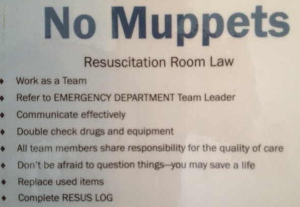
When the external muppet factor is allowed to escalate unchecked, the end result is frenetic activity and noise from the staff without coordinated meaningful intervention for the patient. Comparisons with ‘headless chickens’ are often drawn. In particularly challenging scenarios, it can appear that the panic has swelled to such magnitude that it goes nova, as though the headless chickens have actually exploded, metaphorically filling the room with a gruesome blanket of giblets and a snowstorm of feathers, clouding ones ability to assess and manage the patient effectively. This high-point of group anxiety and ineffectiveness is what I mean by the term ’chicken bomb’, and I bet most readers of this blog will have witnessed the detonation of one.
I credit the invention of this term to emergency and prehospital physician James French, a master resuscitationist and human factors wizard who also introduced the idea of clinical logistics to us.
So, next time you encounter muppets and chicken bombs, feel free to use the terminology, although preferably not during an actual resus with those who might take it personally.

The importance of first pass success

mv-vl-iconA large single-centre study in an academic tertiary care center emergency department (where residents perform most of the intubations) examined 1,828 orotracheal intubations, of which 1,333 were intubated successfully on the first attempt (72.9%).
Adverse events (AE) captured were oesophageal intubation, oxygen desaturation, witnessed aspiration, mainstem intubation, accidental extubation, cuff leak, dental trauma, laryngospasm, pneumothorax, hypotension, dysrhythmia, and cardiac arrest.
When the first pass was successful, the incidence of AEs was 14.2%. More than one attempt was associated with significantly more AEs. Patients requiring two attempts had 33% more AEs (47.2%) and as the number of attempts increased, so did the risk of AEs, with the largest increase in AEs occurring between an unsuccessful first attempt and the second intubation attempt.
This is a powerful argument in favour of optimising first pass success. In the prehospital service I work for, We like to include this in a ‘first pass, no desat, no hypotension’ package that includes team simulation training, pre-intubation briefing, checklist use, optimisation of position, ketamine induction (and avoidance of propofol), apnoeic oxygenation, bougie use, bimanual laryngoscopy, and waveform capnography.
The Importance of First Pass Success When Performing Orotracheal Intubation in the Emergency Department
Academic Emergency Medicine 2013;20(1):71–78, Free Full Text
[EXPAND Abstract]


Objectives The goal of this study was to determine the association of first pass success with the incidence of adverse events (AEs) during emergency department (ED) intubations.

Methods This was a retrospective analysis of prospectively collected continuous quality improvement data based on orotracheal intubations performed in an academic ED over a 4-year period. Following each intubation, the operator completed a data form regarding multiple aspects of the intubation, including patient and operator characteristics, method of intubation, device used, the number of attempts required, and AEs. Numerous AEs were tracked and included events such as witnessed aspiration, oxygen desaturation, esophageal intubation, hypotension, dysrhythmia, and cardiac arrest. Multivariable logistic regression was used to assess the relationship between the primary predictor variable of interest, first pass success, and the outcome variable, the presence of one or more AEs, after controlling for various other potential risk factors and confounders.

Results Over the 4-year study period, there were 1,828 orotracheal intubations. If the intubation was successful on the first attempt, the incidence of one or more AEs was 14.2% (95% confidence interval [CI] = 12.4% to 16.2%). In cases requiring two attempts, the incidence of one or more AEs was 47.2% (95% CI = 41.8% to 52.7%); in cases requiring three attempts, the incidence of one or more AEs was 63.6% (95% CI = 53.7% to 72.6%); and in cases requiring four or more attempts, the incidence of one or more AEs was 70.6% (95% CI = 56.2.3% to 82.5%). Multivariable logistic regression showed that more than one attempt at tracheal intubation was a significant predictor of one or more AEs (adjusted odds ratio [aOR] = 7.52, 95% CI = 5.86 to 9.63).

Conclusions When performing orotracheal intubation in the ED, first pass success is associated with a relatively small incidence of AEs. As the number of attempts increases, the incidence of AEs increases substantially.

[/EXPAND]